Vertebrate photoreceptors are unusual because they hyperpolarize in response to light.
In photoreceptors of the retina, opsin molecules are arranged in cylindrical groups in the plasma membrane of most photosensitive cells. Each opsin protein contains a light-
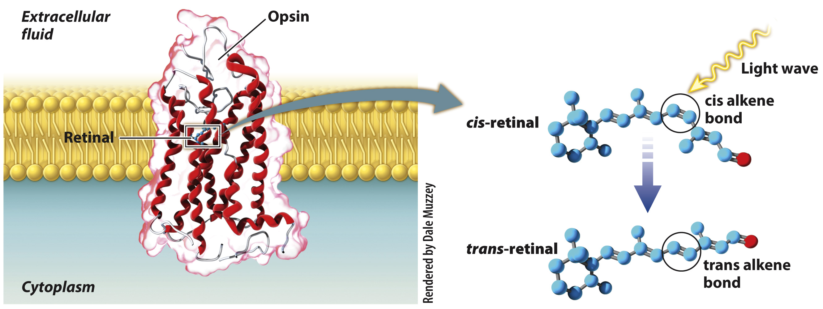
Vertebrate photoreceptor cells have leaky Na+ channels that let some Na+ ions into the cell even at rest. As a result, their resting membrane potential (in the dark) is less negative (about –35 mV) than that of nerve cells. When a photoreceptor is in the dark and its resting potential is –35 mV, it continuously releases the neurotransmitter glutamate. However, when retinal absorbs a photon of light, it undergoes a conformational change from a cis to a trans configuration, causing Na+ channels to close completely. Without Na+ ions entering the cell, the cell membrane becomes hyperpolarized (rather than depolarized) and the cell’s release of glutamate is reduced. Photoreceptors themselves do not fire action potentials, but when stimulated by light they increase or decrease the firing rate of neurons in the retina of the eye. That is, the reduction in neurotransmitter triggers ESPNs in the dendrites of some connecting neurons, and ISPNs in others. Differences in the firing rates of these neurons provide information about the intensity and location of light.