Skeletal and cardiac muscle fibers are organized into repeating contractile units called sarcomeres.
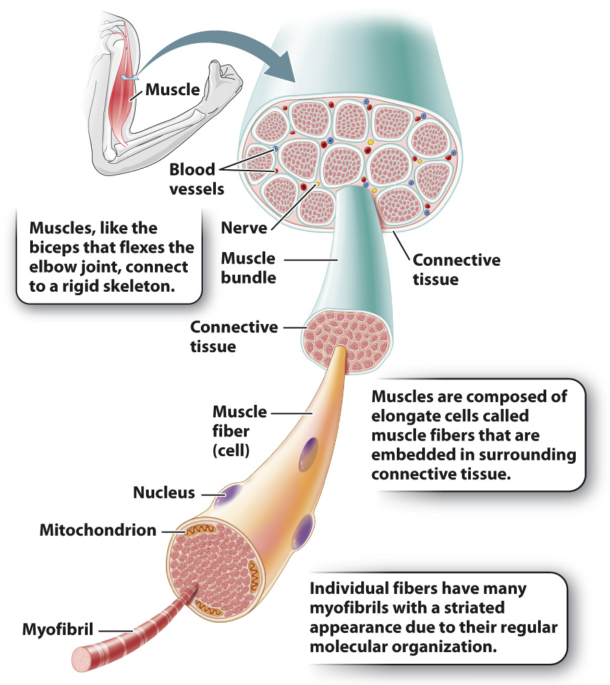
The visible bands in skeletal and cardiac muscles are an important clue to the mechanism that produces muscle contractions. Whole muscles are made up of parallel bundles of individual muscle fibers (Fig. 37.3). Recall that a muscle fiber is a muscle cell. Each muscle fiber in turn contains hundreds of long, rodlike structures called myofibrils. Myofibrils contain parallel arrays of actin and myosin filaments that cause a muscle to contract.
788
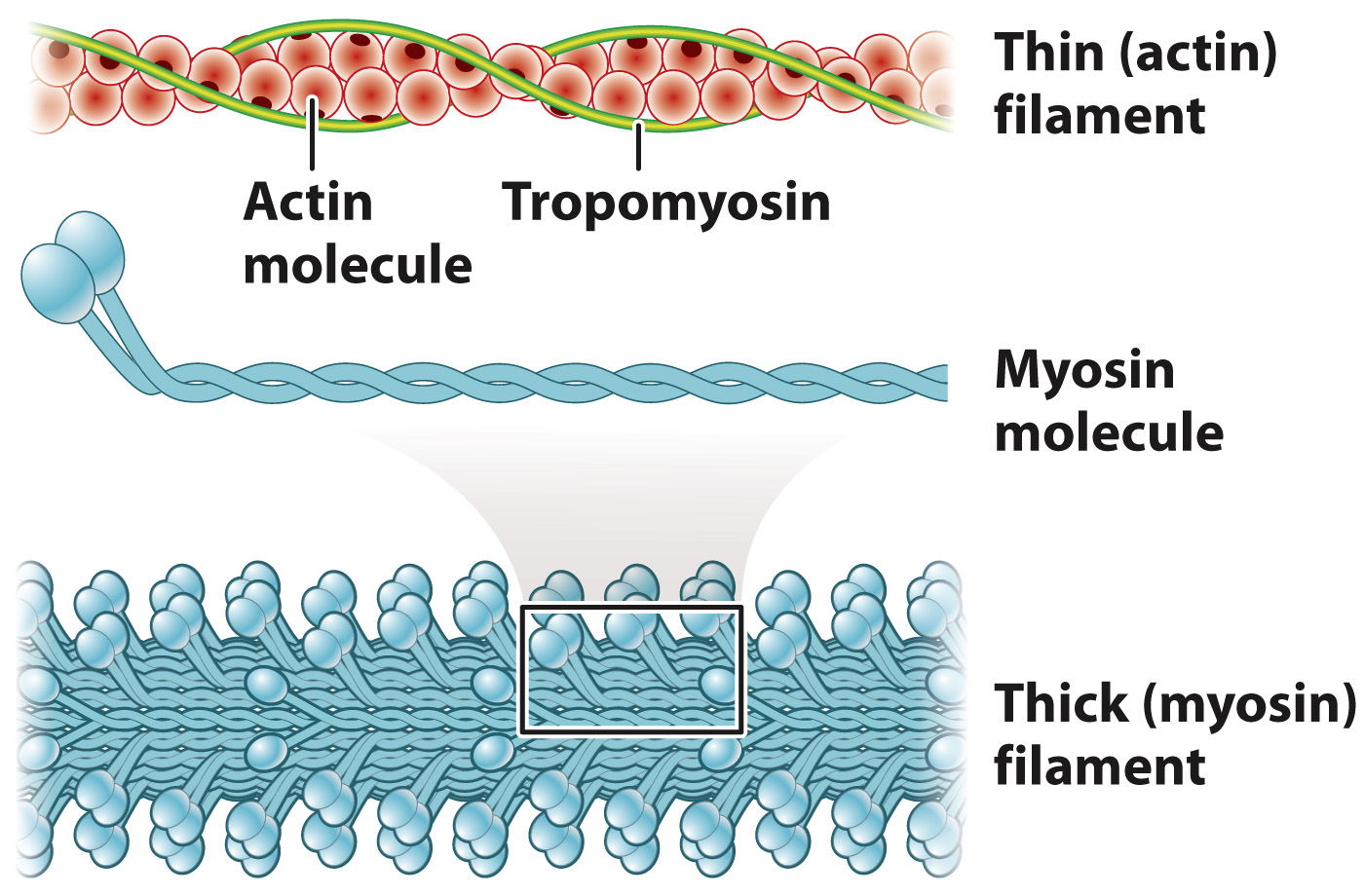
Let’s consider the structural organization of myosin and actin filaments in skeletal muscles in more detail (Fig. 37.4). Each myosin molecule consists of two long polypeptide chains coiled together, each ending with a globular head. Consequently, the myosin molecule resembles a double-
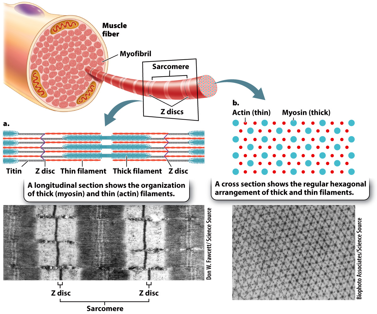
The thin filaments are attached to protein backbones called Z discs that are regularly spaced along the length of the myofibril (Fig. 37.5a). The region from one Z disc to the next is called the sarcomere. Sarcomeres are the functional units of muscles. In other words, the shortening (contraction) of a muscle is ultimately the result of the shortening of thousands of sarcomeres along a myofibril.
789
Sarcomeres are arranged in series along the length of the myofibril. Sets of actin thin filaments extend from both sides of the Z discs toward the midline of the sarcomere (Fig. 37.5a). In the middle of the sarcomere, not directly contacting the Z discs, are myosin thick filaments. The thin filaments overlap with the myosin thick filaments, forming two regions of overlap within a sarcomere. Toward either end of the sarcomere, as well as in the middle, are regions where the actin and myosin filaments do not overlap. The regions that overlap appear dark under an electron microscope, while the regions of non-
As we have seen, the regular pattern of actin and myosin filaments within sarcomeres along the length of the fiber gives skeletal muscles their striated appearance (Fig. 37.5a). In cross section, thick and thin filaments are arranged in a hexagonal lattice. This arrangement allows each thick filament to interact with six adjacent thin filaments (Fig. 37.5b). The precise geometry of thin and thick filaments is critical to their interaction, as we discuss next.