Calcium regulates actin–
We have discussed what makes muscles contract. We next consider when they contract. Skeletal and smooth muscle fibers are both activated by the nervous system. Whereas vertebrate skeletal muscles are innervated by the somatic nervous system, smooth muscles are innervated by the autonomic nervous system, as are cardiac muscles (Chapter 35).
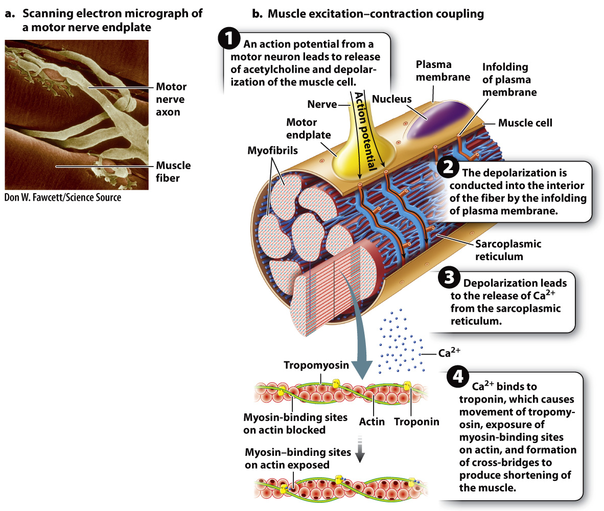
Like nerve cells, muscle fibers are electrically excitable. Skeletal muscle fibers are activated by impulses transmitted by motor nerves to synaptic junctions (Fig. 37.8). Motor neuron axons have branches that allow them to synapse with multiple muscle fibers. When action potentials traveling down a motor neuron arrive at the neuromuscular junction, the neurotransmitter acetylcholine is released into the synaptic cleft. The neurotransmitter binds with postsynaptic receptors on the muscle cell at a region called the motor endplate, triggering the opening of Na+ channels. The resulting influx of Na+ ions in turn initiates a wave of depolarization that passes from the neuromuscular junction toward both ends of the muscle fiber (Chapter 35).
How does membrane depolarization lead to the cross-
Let’s look at the chain of events that concludes with the exposure of the myosin-
A muscular contraction is initiated when depolarization of the muscle fiber causes the SR to release Ca2+. The Ca2+ diffuses into the myofibrils and binds to a protein called troponin, causing the troponin molecule to change shape. This conformational change of troponin, in turn, causes tropomyosin to move, exposing myosin-
792
The process by which membrane depolarization leads to Ca2+ release from the SR and the formation of myosin-
Quick Check 2 Curare is a paralyzing compound that blocks the action of the neurotransmitter acetylcholine at the muscle fiber’s motor endplate. What effect do you think curare has on the release of calcium ions from the sarcoplasmic reticulum of the muscle cell?
Quick Check 2 Answer
Because acetylcholine is blocked and cannot depolarize the muscle cell membrane, calcium is not released from the sarcoplasmic reticulum, and the muscle is unable to contract.