The pituitary gland integrates diverse bodily functions by secreting hormones in response to signals from the hypothalamus.
The hypothalamus is the main route by which nervous system signals are transmitted to the vertebrate endocrine system. The function of the hypothalamus is to transmit these signals to the pituitary gland, the endocrine gland that acts as a control center for many other endocrine glands in the body.
The pituitary gland is divided into anterior and posterior regions (Fig. 38.11). This division is not arbitrary—
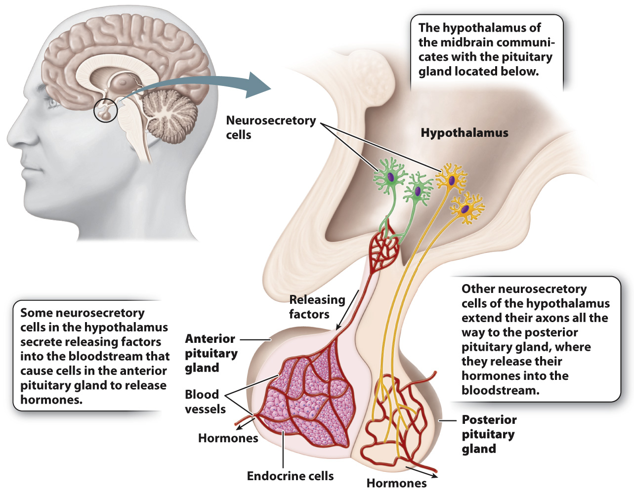
Because of these developmental differences, the anterior and posterior pituitary glands receive input from the hypothalamus in different ways. As does the brain of arthropods, the vertebrate hypothalamus contains neurosecretory cells, which are part of the brain and therefore are neurons. As with invertebrate neurosecretory cells, these cells release hormones into the bloodstream. Some of these neurosecretory cells communicate with the anterior pituitary gland. In this case, they secrete hormones called releasing factors into small blood vessels that travel to and supply the anterior pituitary gland (Fig. 38.11). In response, cells of the anterior pituitary gland release hormones into the bloodstream. These hormones circulate throughout the body and bind to receptors on target cells, tissues, and organs.
In contrast, communication between the hypothalamus and the posterior pituitary gland does not involve hypothalamic releasing factors. Instead, the posterior pituitary gland contains the axons of neurosecretory cells whose cell bodies are located in the hypothalamus. These axons release hormones directly into the bloodstream, and these hormones are transported in the blood to distant sites (Fig. 38.11). Consequently, the posterior pituitary is part of the nervous system itself.
In response to signals from the hypothalamus, distinct hormones are secreted by the anterior and posterior pituitary glands (Table 38.2). The hormones released by the anterior pituitary hormone act on an endocrine gland to cause release of other hormones. Hormones that control the release of other hormones are called tropic hormones. In response to thyroid-
820
The anterior pituitary gland also secretes the hormones growth hormone (GH) and prolactin. Growth hormone acts generally on the muscles, bones, and other body tissues to stimulate their growth, and prolactin stimulates milk production in the breasts of female mammals in response to an infant’s suckling at the mother’s nipple.
The hormones released by the posterior pituitary gland include oxytocin and antidiuretic hormone (ADH; also called vasopressin). These are two evolutionarily related peptide hormones of similar structure. As we discussed earlier, oxytocin plays several roles related to female reproduction: It causes uterine contraction during labor and stimulates the release of milk during breastfeeding. Antidiuretic hormone acts on the kidneys (Chapter 41), regulating the concentration of urine that an animal excretes, which is critical to maintaining water and solute balance in the body.
In addition to their roles in reproduction and kidney function, oxytocin and antidiuretic hormone are released by cells in the brain and may play roles in social behaviors. For instance, recent evidence indicates that oxytocin may be important in regulating maternal behavior toward infants. Recent research suggests that oxytocin may also have a role in behaviors such as trust as a prelude to mating in vertebrate animals. These functions reflect the long evolutionary history of oxytocin, as it plays a role in the social behavior of insects.
Recent evidence also suggests that antidiuretic hormone plays a parallel role in regulating male social behavior and parental behavior in mammals. This is not surprising because oxytocin and antidiuretic hormone have very similar chemical structures (see Fig. 38.7). The integration of reproductive function with parental and social behavior for the care of young is likely favored strongly by natural selection. Receptors for these hormones are expressed within the brain. Thus, in addition to acting on reproductive and renal organs of the body, oxytocin and antidiuretic hormone affect mammalian behavior by acting directly on the brain.