Mammals and birds have four-
Mammals and birds evolved four-
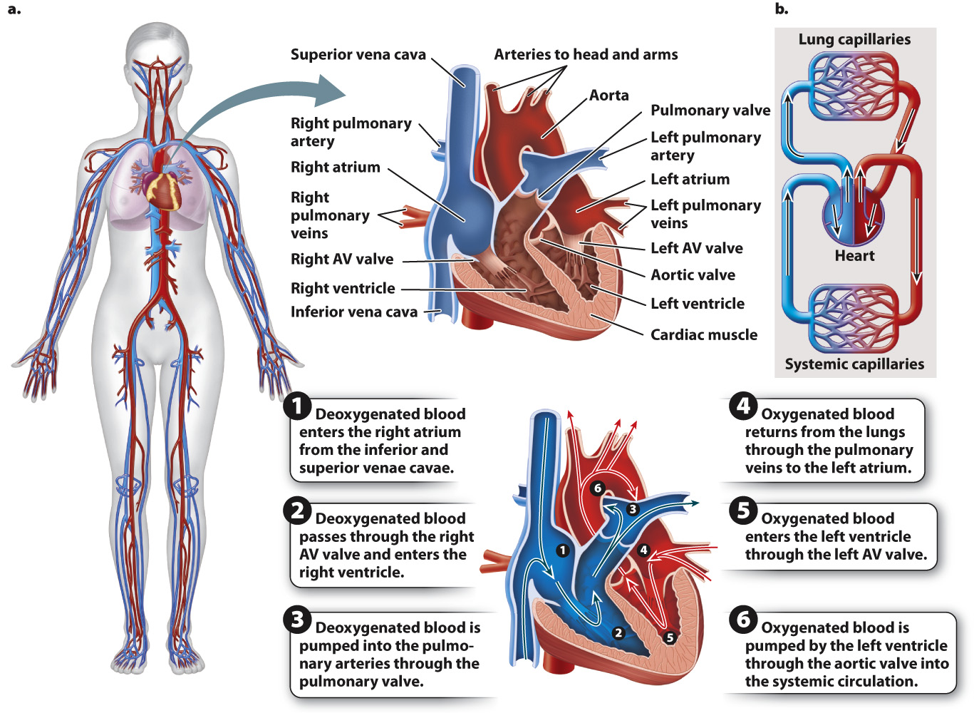
Following the pattern established in amphibians and reptiles, deoxygenated blood enters the right atrium from the venae cavae and, when the atrium contracts, moves through an atrioventricular (AV) valve into the right ventricle (Fig. 39.22). When the right ventricle contracts, blood is pumped through the pulmonary valve into the pulmonary trunk, which divides into the left and right pulmonary arteries, and then to the lungs for oxygenation. The atrioventricular and pulmonary valves ensure that blood flows in one direction into and then out of the right ventricle. The walls of the right ventricle are thinner than those of the left ventricle, and its weaker contractions eject blood at a lower pressure. As a result, the blood of the pulmonary circulation moves at a slower rate, allowing greater time for gases to diffuse into and out of the lungs.
847
The oxygenated blood returns from the lungs through the pulmonary veins and enters the left atrium of the heart. When the left atrium contracts, blood is pumped through a second atrioventricular valve into the left ventricle. The thick muscular walls of the left ventricle eject the blood under high pressure to the body. Oxygenated blood leaving the left ventricle passes through the aortic valve and then flows to the head and the rest of the body through the large artery called the aorta. The one-
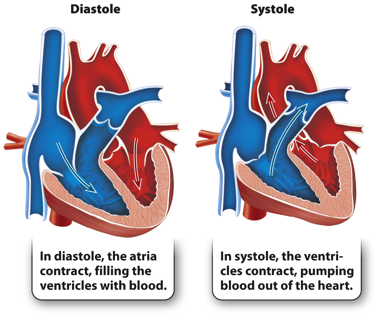
The contraction of the two atria followed by the contraction of the two ventricles makes up the cardiac cycle (Fig. 39.23). The cardiac cycle is divided into two main phases. Diastole is the relaxation of the ventricles, and systole is the contraction of the ventricles. Systole, therefore, is the phase of the cardiac cycle in which blood is pumped from the heart into the pulmonary and systemic circulations, and diastole is the phase in which the atria contract and the ventricles fill with blood.
Quick Check 6 What pressure difference between the ventricle and the atrium causes the atrioventricular valve to close?
Quick Check 6 Answer
The atrioventricular valve closes when pressure in the ventricle exceeds the pressure in the atrium.