Mammalian lungs are well adapted for gas exchange.
Despite the limitations of tidal respiration, mammalian lungs are well adapted for breathing air. They have an enormous surface area and a short diffusion distance for gas exchange. Consequently, the lungs of mammals supply O2 quickly enough to support high metabolic rates.
Except in the case of birds (discussed in next section), the lungs of air-breathing vertebrates are blind-ended sacs located within the thoracic cavity. Air is taken in through the mouth and nasal passages and then passes through the larynx, an organ in the throat made of muscle and cartilage that helps to isolate breathing from swallowing and within which the vocal cords are located. (The vocal cords enable speech, song, and sound production.) Air then enters the trachea, the central airway leading to the lungs (Fig. 39.8). The trachea divides into two airways, called the primary bronchi, one of which supplies each lung. The trachea and bronchi are supported by rings of cartilage that prevent them from collapsing during respiration, ensuring that airflow meets with minimal resistance. You can feel some of these cartilage rings at the front of your throat below your larynx (the part of the larynx you can feel is your Adam’s apple). The primary bronchi divide into smaller secondary bronchi and again into finer bronchioles. This branching continues until the terminal bronchioles have a diameter of less than 1 mm. The very fine bronchioles end with clusters of tiny thin-walled sacs, the alveoli (singular, alveolus), where gas exchange by diffusion takes place. As a result of this branching pattern, each lung consists of several million alveoli, providing a large surface area. Together, human lungs consist of 300 to 500 million alveoli with a combined surface area as large as a tennis court—about 100 m2!
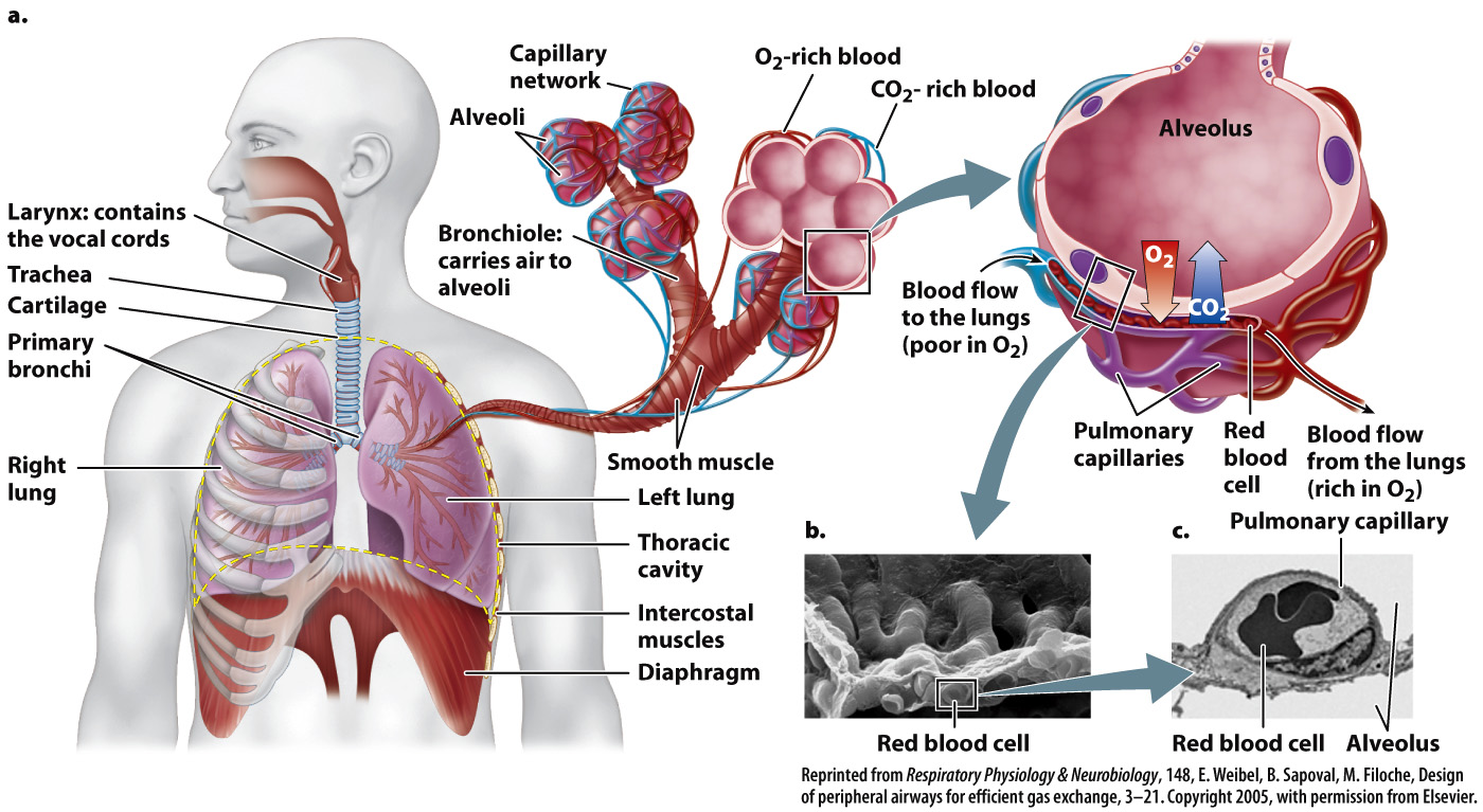
FIG. 39.8 Human lung anatomy. (a) Millions of alveoli provide a vast surface for gas exchange. (b) A scanning electron micrograph of the lung alveolar surface. (c) A transmission electron micrograph showing a cross section of the alveolar wall and lung capillary.
Small blood vessels, the pulmonary capillaries, supply the alveolar wall. Each alveolus is lined with thin epithelial cells in intimate contact with the endothelium of these small blood vessels (Fig. 39.8). As a result, the diffusion distance from the alveolus into the capillary is extremely short (about 2 µm), similar to that of gill lamellae and the terminal airways of bird lungs.
Keeping the surfaces of the alveoli moist is critical because moisture helps move O2 molecules from the air into solution and thus helps them diffuse across the alveolar wall. Certain alveolar epithelial cells produce a surfactant, a compound that acts to reduce the surface tension of the fluid film (soap, for example, is a surfactant). Surface tension is a cohesive force that holds the molecules of a liquid together, causing the surface to act like an elastic membrane. Because of surface tension, it requires greater pressure to inflate a small balloon than a large one. The surfactant allows the lungs to be inflated easily at low volumes when the alveoli are partially collapsed and small. It also ensures that alveoli of different sizes inflate with similar ease, enabling more nearly uniform ventilation of the lung.
Mucus-secreting cells line the airways of the lung not only to keep them moist, but also to trap and remove foreign particles and microorganisms that an animal may breathe in with air. Beating cilia on the surface of these cells move the mucus and foreign debris out of the lungs and into the throat, where it is swallowed and digested. Smoking even one cigarette can stop the cilia’s beating for several hours or even destroy them. This is one reason why smokers cough—they must clear the mucus that builds up.