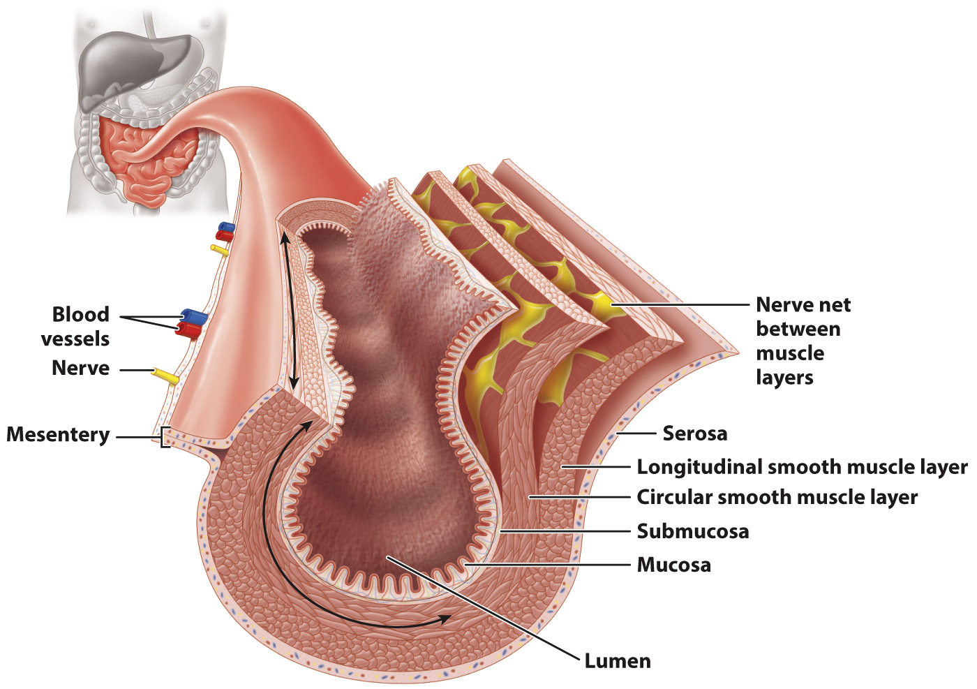
The lining of the digestive tract is composed of distinct layers.
As we have seen, the digestive tract is not a passive tube. It secretes enzymes and other chemical compounds for digestion, absorbs nutrients, and actively moves food through the body. How does a series of tubes accomplish all of this? As shown in Fig. 40.18, the digestive tract is made up of several layers of tissue, each with a specialized function. The central space, or lumen, through which the gut contents travel is surrounded by an inner tissue layer, the mucosa, which both secretes enzymes and absorbs nutrients. The cells of the mucosa secrete mucus to protect the gut wall from digestive enzymes, and, in the stomach, hydrochloric acid. Surrounding the mucosa is the submucosa, a layer containing blood vessels, lymph vessels, and nerves.
870
Outside these layers are two smooth muscle layers (Fig. 40.18). An inner circular muscle layer contracts to reduce the size of the lumen. An outer longitudinal muscle layer contracts to shorten small sections of the gut. These two muscle layers contract alternately to mix gut contents, producing a traveling wave during peristalsis that moves the contents along the digestive tract from compartment to compartment. Between the two muscle layers are autonomic nerves that control the contractions of the two sets of smooth muscle.
An outer layer of cells and connective tissue called the serosa covers and protects the gut (Fig. 40.18). The gut is supported in the abdominal cavity by a membrane called the mesentery, through which blood vessels, nerves, and lymph travel to supply the gut.