Animals have diverse excretory organs.
As we have seen, the processes of filtration, reabsorption, and secretion serve two important and complementary functions: They allow wastes to be isolated and subsequently removed from the body, and they allow the organism to adjust the amounts of water and electrolytes required to meet its osmoregulatory needs. These two functions are carried out by diverse excretory organs.
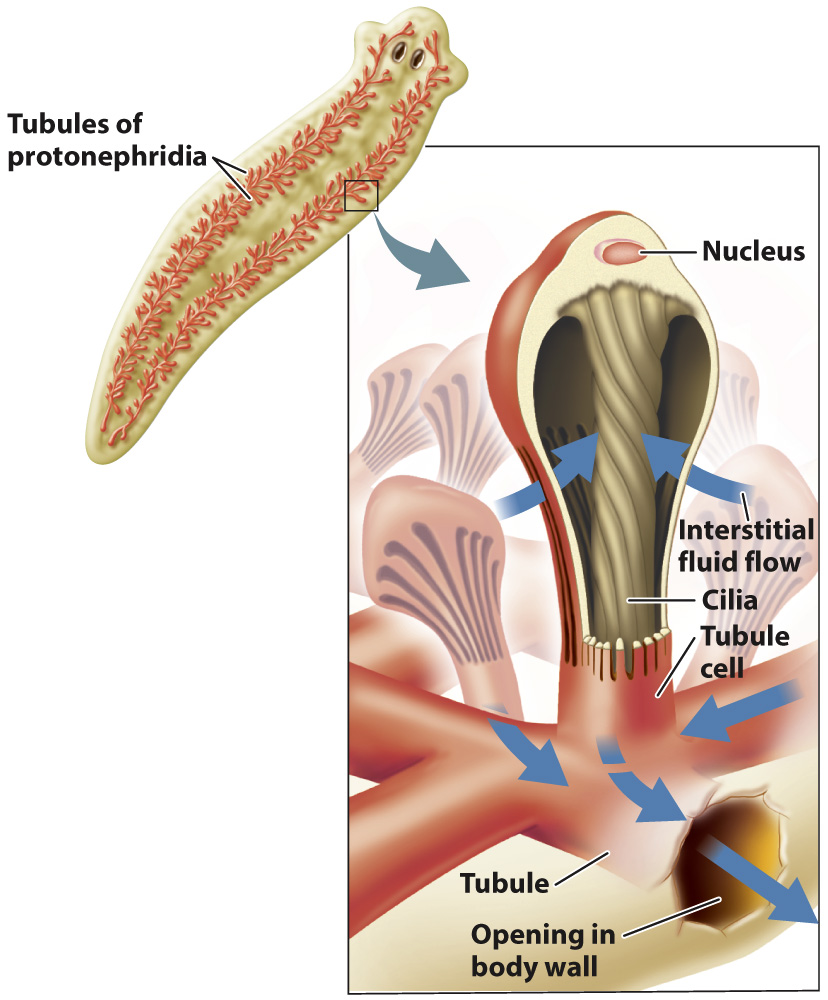
For example, consider waste excretion in flatworms, small flattened animals that glide along the streambed floor. In flatworms, waste is excreted without being filtered: The fluid from the body cavity is isolated in excretory organs called protonephridia (Fig. 41.9). Cells with beating cilia move the fluid down the lumen of a tubule to an excretory pore. Freshwater flatworms, such as Planaria, have an extensive network of tubules that eliminate excess fluid along with waste products. As the fluid passes down the tubule, cells lining the tubule modify the fluid’s contents through reabsorption and secretion. The urine leaving a freshwater flatworm contains nitrogenous waste and is less concentrated (that is, more dilute) than its internal body fluids to balance the water it ingests as it feeds, as is typical of freshwater animals, as we saw earlier.
884
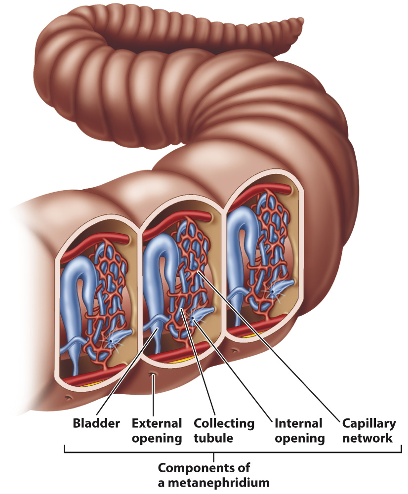
In segmented annelid worms such as earthworms, the blood is filtered through small capillaries into a pair of excretory organs called metanephridia within each body segment (Fig. 41.10). The fluid enters a metanephridium at a funnel-
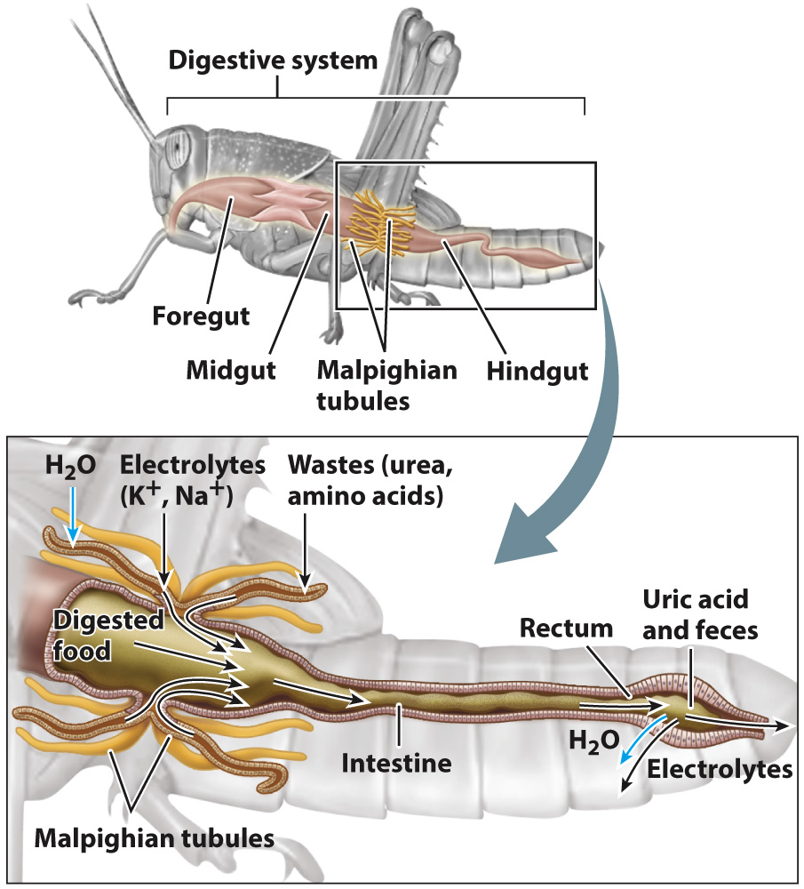
In insects and other terrestrial arthropods, body fluid called hemolymph passes from the main body cavity into a series of tubes called Malpighian tubules that empty into the hindgut (Fig. 41.11). The cells lining the tubules actively secrete uric acid and excess electrolytes into the lumen of the tubules while retaining key solutes. Muscle fibers in the walls of the tubules help push the fluid to the hindgut. In the hindgut, the fluid becomes increasingly acidic because of the buildup of uric acid, causing uric acid to precipitate from solution when it enters the rectum. Because uric acid is removed from the fluid, it does not influence the movement of water. Water is therefore free to diffuse back into the tissues as electrolytes are reabsorbed from the rectum. The uric acid and remaining dried digestive wastes are then eliminated. Malpighian tubules represent one of the most successful adaptations for excreting nitrogenous wastes with minimal water loss, enabling insects to live in diverse and arid terrestrial environments.