Sponges are simple and widespread in the oceans.
As we discussed in Chapters 27 and 28, the unicellular ancestors of animals carried out all functions from metabolism to reproduction as individual cells. The cells making up sponges retain much of this independence, while also having some benefits of multicellularity. Coordination among cells means that there can be distinct kinds of cell specialized for different functions—
949
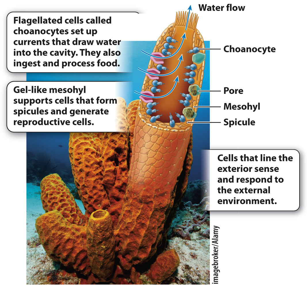
The sponge body plan resembles a flower vase, with many small pores along its sides and a larger opening at the top (Fig. 44.6). The cells on the outer surface of the sponge are tough and act as the sponge’s skin. The interior surface is lined by cells called choanocytes, which have flagella and function in nutrition and gas exchange. Choanocyte cells have a collar of small cilia around their flagellum, much like the cells of animals’ closest unicellular relatives, the choanoflagellates (Chapter 27). Between the interior and exterior cell layers lies a gelatinous mass called the mesohyl (Fig. 44.6). Mesohyl is mostly noncellular, but it contains some amoeba-
950
Sponges gain nutrition by intracellular digestion (Chapter 40). The choanocytes that surround the interior chambers and passageways of the sponge beat their flagella, creating a current that draws water from outside the body, through the pores in its walls and upward through the central cavity of the sponge, where it exits through the large opening at the top. The circulating water contains food particles and dissolved organic matter, which cells lining the cavity capture by endocytosis. The individual cells then metabolize the food in their interiors, much the way that protozoans feed.
From this observation, we might conclude that sponges are simply a group of uncoordinated cells, each working for itself, but that isn’t the case. The choanocytes beat their flagella in a coordinated pattern, helping to draw water into the body interior through the pore system and outward again through the vase opening. Moreover, the shape of the sponge body itself directs water movement across feeding cell surfaces. These features effectively move water across the cells, facilitating food uptake. Sponge cells require oxygen for respiration and must get rid of the carbon dioxide respiration generates. Gas exchange occurs by diffusion, aided by the movement of water through the sponge cavity.
Sponges don’t have highly developed reproductive organs. Instead, cells recruited from the choanocyte layer migrate into the mesohyl, where they undergo meiotic cell division and differentiate as sperm or eggs. Sperm released into the water fuse with eggs in the mesohyl of other sponges.
Many sponges build skeletons of simple structures called spicules. Some sponges precipitate spicules of glasslike silica (SiO2). Others, however, make their spicules—
Despite their anatomic simplicity, sponges are major contributors to seafloor communities. Approximately 9000 species of sponges have been described, most of them ocean dwelling, but also including a few that live in freshwater lakes. Many sponges obtain at least part of their nutrition from symbiotic microorganisms living within their bodies. In fact, sponge bodies are commonly full of bacterial cells. At least some of these bacteria are probably symbionts, but only a few experiments have demonstrated their function.
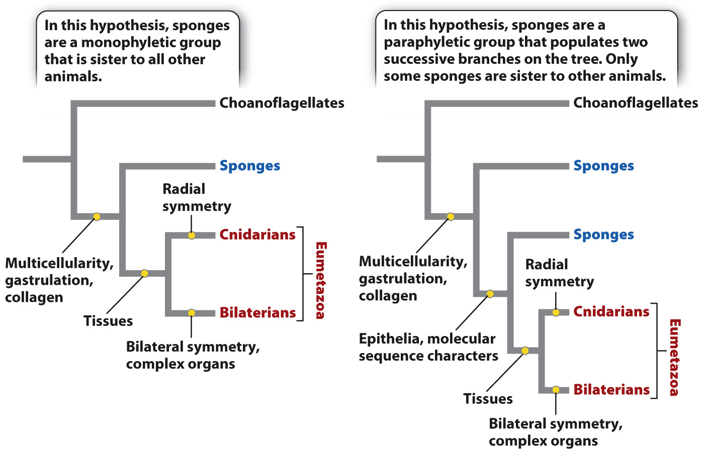
Until recently, most biologists regarded sponges as a monophyletic group, one that includes all the descendants of a common ancestor. Some molecular data, however, now suggest that sponges are paraphyletic—