Signals are often amplified in the cytosol.
Because different G protein-coupled receptors have different effects in different cells, we will follow the steps in cell signaling by following a specific example. Adrenaline, discussed earlier, binds to a G protein-coupled receptor. When adrenaline binds to its G protein-coupled receptor on cardiac muscle cells, GDP in the G protein is replaced by GTP and the G protein is activated.
The GTP-bound α subunit then binds to and activates an enzyme in the cell membrane called adenylyl cyclase (Fig. 9.10). Adenylyl cyclase converts the nucleotide ATP into cyclic AMP (cAMP). Cyclic AMP is known as a second messenger. The term “second messenger” was coined to differentiate it from signaling molecules like adrenaline and growth factors, which are considered first messengers. Second messengers are signaling molecules found inside cells that relay information to the next target in the signal transduction pathway. In the case of cAMP, it binds to and activates another molecule, a kinase called protein kinase A (PKA).
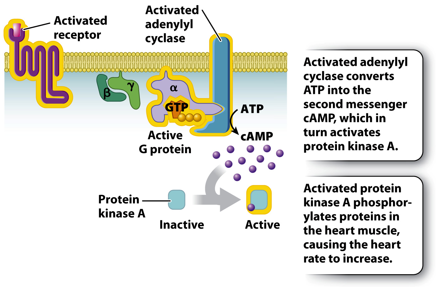
FIG. 9.10 Adrenaline signaling in heart muscle. Adrenaline binds to a G protein-coupled receptor, leading to production of the second messenger cAMP and activation of protein kinase A. The cell’s response is increased heart rate.
A little adrenaline goes a long way as a result of signal amplification (Fig. 9.11). First, a single receptor bound to adrenaline can activate several G protein molecules. Second, each molecule of adenylyl cyclase catalyzes the production of large amounts of the second messenger cAMP. And third, each PKA molecule activates multiple protein targets by phosphorylation. These sequential molecular changes in the cytosol amplify the signal so that a very small amount of signaling molecule has a large effect on a responding cell.
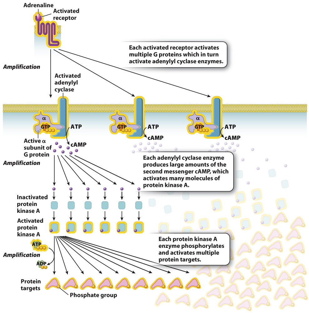
FIG. 9.11 Amplification of G protein-coupled signaling. Signaling through G protein-coupled receptors is amplified at several places, so a small amount of signal can produce a large response in the cell.