19.1 Light Energy and Eye Structures
19-
Our eyes receive light energy and transduce (transform) it into neural messages that our brain then processes into what we consciously see. How does such a taken-
The Stimulus Input: Light Energy
When you look at a bright red tulip, the stimuli striking your eyes are not particles of the color red but pulses of electromagnetic energy that your visual system perceives as red. What we see as visible light is but a thin slice of the whole spectrum of electromagnetic energy, ranging from imperceptibly short gamma waves to the long waves of radio transmission (FIGURE 19.1). Other organisms are sensitive to differing portions of the spectrum. Bees, for instance, cannot see what we perceive as red but can see ultraviolet light.
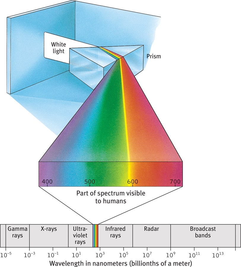
The wavelengths we see What we see as light is only a tiny slice of a wide spectrum of electromagnetic energy, which ranges from gamma rays as short as the diameter of an atom to radio waves over a mile long. The wavelengths visible to the human eye (shown enlarged) extend from the shorter waves of blue-
Two physical characteristics of light help determine our sensory experience. Light’s wavelength—the distance from one wave peak to the next (FIGURE 19.2a)—determines its hue (the color we experience, such as the tulip’s red petals or green leaves). Intensity—the amount of energy in light waves (determined by a wave’s amplitude, or height)—influences brightness (FIGURE 19.2b). To understand how we transform physical energy into color and meaning, consider the eye.
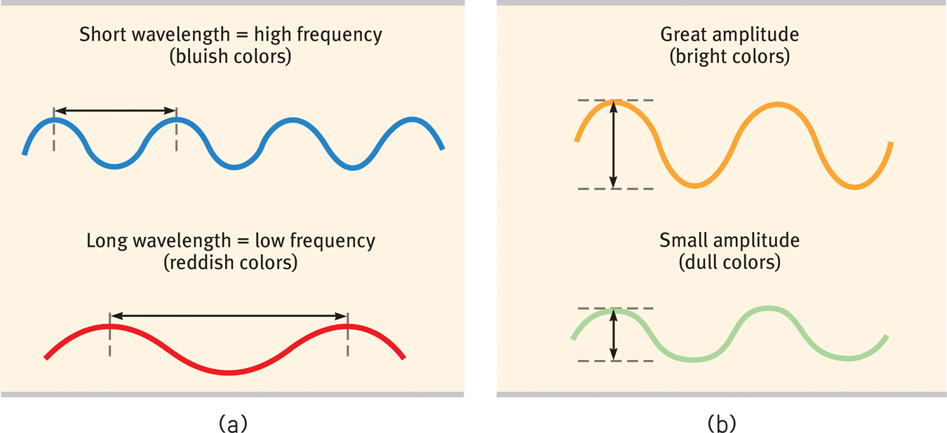
The physical properties of waves (a) Waves vary in wavelength (the distance between successive peaks). Frequency, the number of complete wavelengths that can pass a point in a given time, depends on the wavelength. The shorter the wavelength, the higher the frequency. Wavelength determines the perceived color of light. (b) Waves also vary in amplitude (the height from peak to trough). Wave amplitude influences the perceived brightness of colors.
240
The Eye
Light enters the eye through the cornea, which bends light to help provide focus (FIGURE 19.3). The light then passes through the pupil, a small adjustable opening. Surrounding the pupil and controlling its size is the iris, a colored muscle that dilates or constricts in response to light intensity—
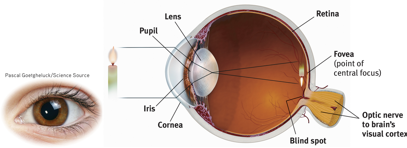
The eye Light rays reflected from a candle pass through the cornea, pupil, and lens. The curvature and thickness of the lens change to bring nearby or distant objects into focus on the retina. Rays from the top of the candle strike the bottom of the retina, and those from the left side of the candle strike the right side of the retina. The candle’s image on the retina thus appears upside down and reversed.
Behind the pupil is a transparent lens that focuses incoming light rays into an image on the retina, a multilayered tissue on the eyeball’s sensitive inner surface. The lens focuses the rays by changing its curvature and thickness in a process called accommodation.
For centuries, scientists knew that when an image of a candle passes through a small opening, it casts an inverted mirror image on a dark wall behind. If the image passing through the pupil casts this sort of upside-
Today’s answer: The retina doesn’t “see” a whole image. Rather, its millions of receptor cells convert particles of light energy into neural impulses and forward those to the brain. There, the impulses are reassembled into a perceived, upright-
241