5.2 Neural Communication
For scientists, it is a happy fact of nature that the information systems of humans and other animals operate similarly—
Neurons
5-
Our body’s neural information system is complexity built from simplicity. Its building blocks are neurons, or nerve cells. To fathom our thoughts and actions, our memories and moods, we must first understand how neurons work and communicate.
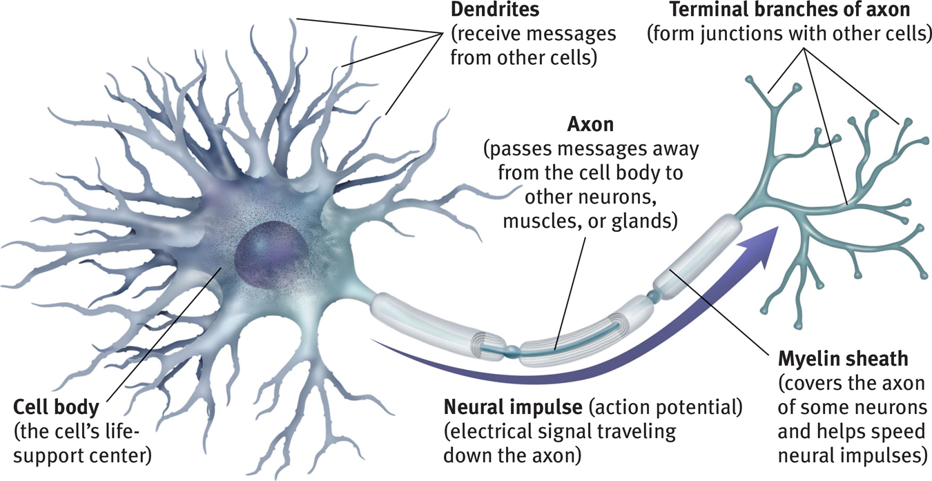
Neurons differ, but all are variations on the same theme (FIGURE 5.2). Each consists of a cell body and its branching fibers. The bushy dendrite fibers receive information and conduct it toward the cell body. From there, the cell’s single lengthy axon fiber passes the message through its terminal branches to other neurons or to muscles or glands. Dendrites listen. Axons speak.
A motor neuron

Unlike the short dendrites, axons may be very long, projecting several feet through the body. A human neuron carrying orders to a leg muscle, for example, has a cell body and axon roughly on the scale of a basketball attached to a 4-
54
 To review and assess your understanding of neurons, visit LaunchPad’s Concept Practice: Structure of a Motor Neuron.
Supporting these billions of nerve cells are spidery glial cells (“glue cells”). Neurons are like queen bees; on their own they cannot feed or sheathe themselves. Glial cells are worker bees. They provide nutrients and insulating myelin, guide neural connections, and mop up ions and neurotransmitters. Glia also play a role in learning and thinking. By “chatting” with neurons they participate in information transmission and memory (Fields, 2011, 2013; Miller, 2005).
In more complex animal brains, the proportion of glia to neurons increases. A postmortem analysis of Einstein’s brain did not find more or larger-
The Neural Impulse
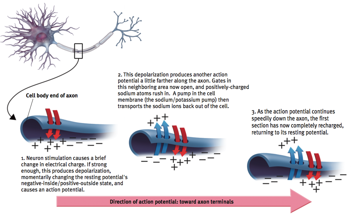
Neurons transmit messages when stimulated by signals from our senses or when triggered by chemical signals from neighboring neurons. In response, a neuron fires an impulse, called the action potential—a brief electrical charge that travels down its axon.
Depending on the type of fiber, a neural impulse travels at speeds ranging from a sluggish 2 miles per hour to more than 200 miles per hour. But even its top speed is 3 million times slower than that of electricity through a wire. We measure brain activity in milliseconds (thousandths of a second) and computer activity in nanoseconds (billionths of a second). Thus, unlike the nearly instantaneous reactions of a computer, your reaction to a sudden event, such as a child darting in front of your car, may take a quarter-
“I sing the body electric.”
Walt Whitman, “Children of Adam” (1855)
Like batteries, neurons generate electricity from chemical events. In the neuron’s chemistry-
55
When a neuron fires, however, the security parameters change: The first section of the axon opens its gates, rather like sewer covers flipping open, and positively charged sodium ions flood in (FIGURE 5.3). The loss of the inside/outside charge difference, called depolarization, causes the next axon channel to open, and then the next, like falling dominos, each tripping the next. This temporary inflow of positive ions is the neural impulse—
Action potential
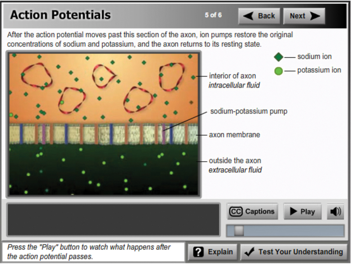
For an animated explanation of this process, visit LaunchPad’s Concept Practice: Action Potentials.
During a resting pause called the refractory period, the neuron pumps the positively charged sodium ions back outside. Then it can fire again. (In myelinated neurons, as in Figure 5.2, the action potential speeds up by hopping from the end of one myelin “sausage” to the next.) The mind boggles when imagining this electrochemical process repeating up to 100 or even 1000 times a second. But this is just the first of many astonishments.
Each neuron is itself a miniature decision-
Increasing the level of stimulation above the threshold will not increase the neural impulse’s intensity. The neuron’s reaction is an all-or-none response: Like guns, neurons either fire or they don’t. How, then, do we detect the intensity of a stimulus? How do we distinguish a gentle touch from a big hug? A strong stimulus can trigger more neurons to fire, and to fire more often. But it does not affect the action potential’s strength or speed. Squeezing a trigger harder won’t make a bullet go faster.
“What one neuron tells another neuron is simply how much it is excited.”
Francis Crick, The Astonishing Hypothesis, 1994
56
RETRIEVAL PRACTICE
- When a neuron fires an action potential, the information travels through the axon, the dendrites, and the cell body, but not in that order. Place these three structures in the correct order.
dendrites, cell body, axon
- How does our nervous system allow us to experience the difference between a slap and a tap on the back?
Stronger stimuli (the slap) cause more neurons to fire and to fire more frequently than happens with weaker stimuli (the tap).
How Neurons Communicate
5-
Neurons interweave so intricately that even with a microscope you would have trouble seeing where one neuron ends and another begins. Scientists once believed that the axon of one cell fused with the dendrites of another in an uninterrupted fabric. Then British physiologist Sir Charles Sherrington (1857–
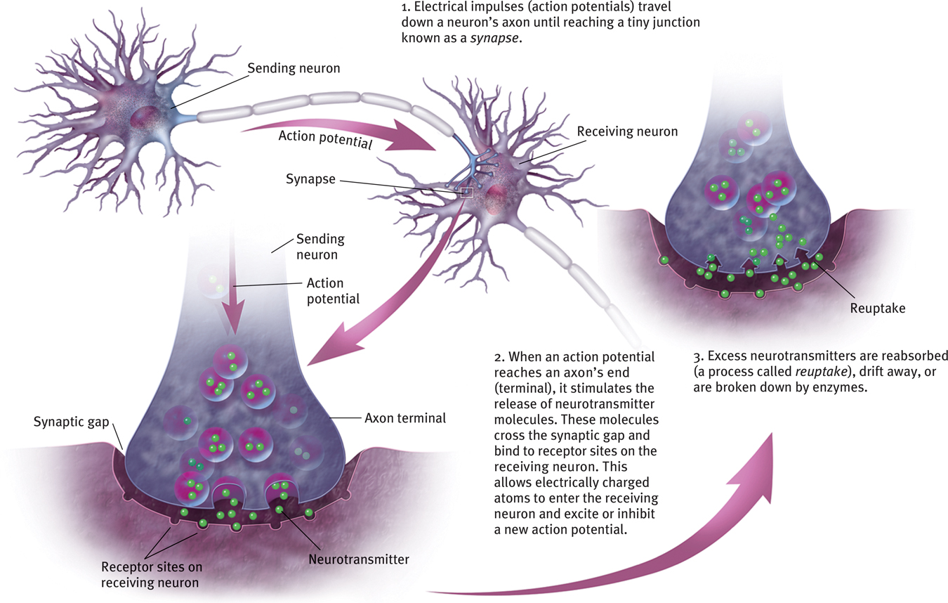
We now know that the axon terminal of one neuron is in fact separated from the receiving neuron by a synaptic gap (or synaptic cleft) less than a millionth of an inch wide. Spanish anatomist Santiago Ramón y Cajal (1852–
“All information processing in the brain involves neurons ‘talking to’ each other at synapses.”
Neuroscientist Solomon H. Snyder (1984)
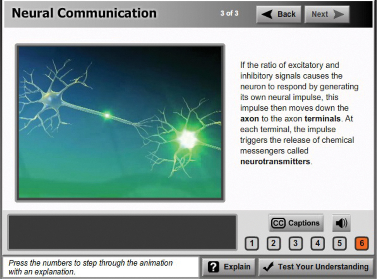
When an action potential reaches the knob-
How neurons communicate
RETRIEVAL PRACTICE
- What happens in the synaptic gap?
Neurons send neurotransmitters (chemical messengers) across this tiny space between one neuron’s terminal branch and the next neuron’s dendrite or cell body.
- What is reuptake? What two other things can happen to excess neurotransmitters after a neuron reacts?
Reuptake occurs when excess neurotransmitters are reabsorbed by the sending neuron. (They can also drift away or be broken down by enzymes.)
How Neurotransmitters Influence Us
“When it comes to the brain, if you want to see the action, follow the neurotransmitters.”
Neuroscientist Floyd Bloom (1993)
5-
In their quest to understand neural communication, researchers have discovered several dozen neurotransmitters and as many new questions: Are certain neurotransmitters found only in specific places? How do they affect our moods, memories, and mental abilities? Can we boost or diminish these effects through drugs or diet?
For an animated explanation, visit LaunchPad’s Concept Practice activities: Structure of a Synapse and Neural Communication.
57
Other modules explore neurotransmitter influences on hunger and thinking, depression and euphoria, addictions and therapy. For now, let’s glimpse how neurotransmitters influence our motions and our emotions. A particular brain pathway may use only one or two neurotransmitters (FIGURE 5.5), and particular neurotransmitters may affect specific behaviors and emotions (TABLE 5.1). But neurotransmitter systems don’t operate in isolation; they interact, and their effects vary with the receptors they stimulate. Acetylcholine (ACh), which is one of the best-
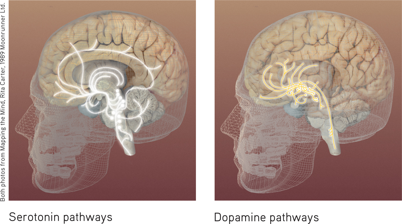
Neurotransmitter pathways Each of the brain’s differing chemical messengers has designated pathways where it operates, as shown here for serotonin and dopamine (Carter, 1998).
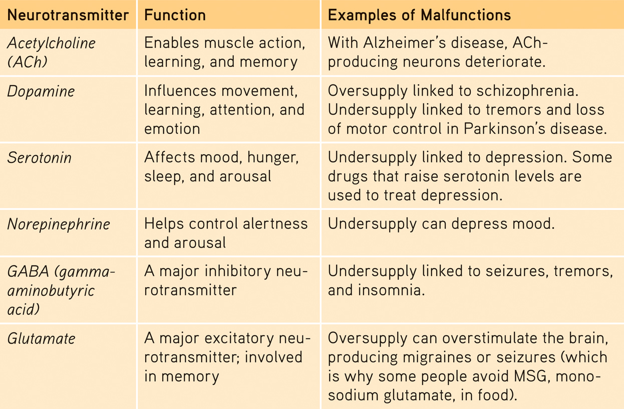
Some Neurotransmitters and Their Functions
58
Candace Pert and Solomon Snyder (1973) made an exciting discovery about neurotransmitters when they attached a radioactive tracer to morphine, showing where it was taken up in an animal’s brain. The morphine, an opiate drug that elevates mood and eases pain, bound to receptors in areas linked with mood and pain sensations. But why would the brain have these “opiate receptors”? Why would it have a chemical lock, unless it also had a natural key to open it?
Physician Lewis Thomas, on the endorphins: “There it is, a biologically universal act of mercy. I cannot explain it, except to say that I would have put it in had I been around at the very beginning, sitting as a member of a planning committee.”
The Youngest Science, 1983
Researchers soon confirmed that the brain does indeed produce its own naturally occurring opiates. Our body releases several types of neurotransmitter molecules similar to morphine in response to pain and vigorous exercise. These endorphins (short for endogenous [produced within] morphine) help explain good feelings such as the “runner’s high,” the painkilling effects of acupuncture, and the indifference to pain in some severely injured people. But once again, new knowledge led to new questions.
RETRIEVAL PRACTICE
- Serotonin, dopamine, and endorphins are all chemical messengers called _________.
neurotransmitters
How Drugs and Other Chemicals Alter NeurotransmissionIf indeed the endorphins lessen pain and boost mood, why not flood the brain with artificial opiates, thereby intensifying the brain’s own “feel-
59
Drugs and other chemicals affect brain chemistry, often by either exciting or inhibiting neurons’ firing. Agonist molecules increase a neurotransmitter’s action. Agonists may increase the production or release of neurotransmitters, or block reuptake in the synapse. Other agonists may be similar enough to a neurotransmitter to bind to its receptor and mimic its excitatory or inhibitory effects. Some opiate drugs are agonists and produce a temporary “high” by amplifying normal sensations of arousal or pleasure.
Antagonists decrease a neurotransmitter’s action by blocking production or release. Botulin, a poison that can form in improperly canned food, causes paralysis by blocking ACh release. (Small injections of botulin—
RETRIEVAL PRACTICE
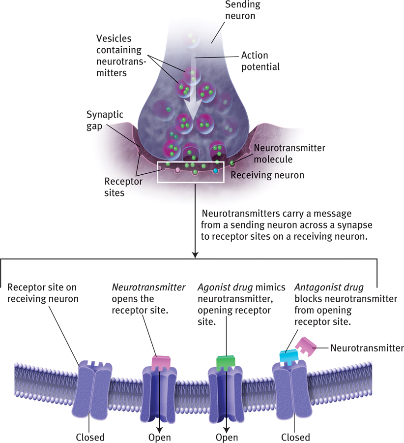
Agonists and antagonists
- Curare poisoning paralyzes its victims by blocking ACh receptors involved in muscle movements. Morphine mimics endorphin actions. Which is an agonist, and which is an antagonist?
Morphine is an agonist; curare is an antagonist.
For an illustrated review of neural communication, visit LaunchPad’s PsychSim 6: Neural Messages.
60