Chapter 1. Receptive Fields and Visual Perception
1.0.1 Receptive Fields and Visual Perception
Receptive Fields and Visual Perception
By: Dr. Diana Lim, Concordia University
1.1 Neuroscience in Action
Look around. Perhaps you are outside on a sunny day or sitting at a desk in a dimly lit room. Notice the objects around you and how their shapes are defined by the light shining on them. Your eyes capture a wide variety of visual information, but how is that information converted to a form that your brain can process? This activity introduces you to the receptive fields of the cells within the visual system.
After completing this activity, you should be able to:
- Explain how the off-center and on-center ganglion cells influence the perception of shapes.
- Distinguish between simple, complex, and hypercomplex cells in the primary visual cortex.
- Describe two kinds of functional columns in the primary visual cortex.
This activity relates to the following principles of nervous system function:
- Principle 6: Brain Systems Are Organized Hierarchically and in Parallel
- Principle 8: The Brain Divides Sensory Input for Object Recognition and Movement
- Principle 10: The Nervous System Works by Juxtaposing Excitation and Inhibition
1.2 The Pathway from Eye to Brain
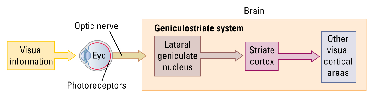
Visual information enters the eye and activates specialized sensory receptors, known as photoreceptors, on the retina. Photoreceptors transduce (convert) light energy, in the form of photons, into a neural signal and then pass that information on to retinal ganglion cells (RGCs). The axons of the RGCs make up the optic nerve, which travels from the eye to the optic chiasm and finally synapses in the lateral geniculate nucleus (LGN) of the thalamus. Projections from the thalamus go to the primary visual cortex (V1), sometimes referred to as striate cortex, and then on to other visual cortical areas, such as the secondary visual cortex (V2) and beyond.
1.3 Receptive Fields
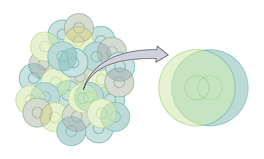
Every neuron within a sensory receptor organ (for example, the skin or the eye) has a receptive field, a specific part of the world to which it responds. The receptive field of a neuron within a visual cortical area corresponds to the part of the retina to which that neuron is connected. As a rule of thumb, cells in the cortex have much larger receptive fields than RGCs do. This large field size means that the receptive field of a cortical neuron must be composed of the receptive fields of many RGCs, as shown in the video below.
1.4 Processing in the Retinal Ganglion Cells: On-Center Cells
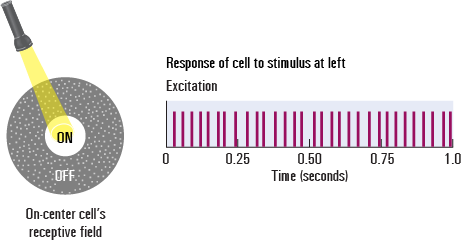
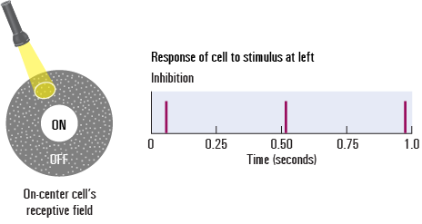
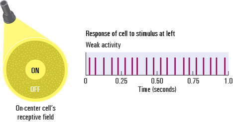
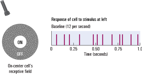
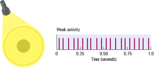
Neurons in the retina respond to the presence or absence of light in their receptive fields. The receptive field of a ganglion cell has a concentric circle arrangement, as illustrated below. For a type of ganglion cell called an on-center cell, a spot of light falling directly in the center of the receptive field excites the cell. A spot of light falling on the edge (periphery) of the receptive field inhibits the cell. A spot of light falling across the entire receptive field weakly increases the cell’s firing rate.
Microelectrodes can be used to record the excitation and inhibition of a ganglion cell, relative to their baseline firing rate. Below, you will be able to see (and hear!) the cell’s activity as you change the light stimulus.
Using the buttons below, watch what happens to the activity of the cell when you change the location and size of the light stimulus on the receptive field of this cell.
Question
LwWVyzOYW0F6fg8uz1nmqbDwgMcZWM2H0PUERyaiFuTV6BVNPMULAzSsAnx6qz8XiHABvbdzS9RUBfnFE9oWczNVS1HXlur5rfitliCj0/608ZbyRtc8C9Svojm2dhjNJ2pf8tXjIzqYYucGUaBR7qrmsz1IgkwPozrOgvtJ1IHuhxM/h58+1567vzroG+lPNQYU7JSIbPbbLL2T2clNFjFWj2Rl/l1ev1c17lCpOewNI6g+1.5 Processing in the Retinal Ganglion Cells: Off-Center Cells
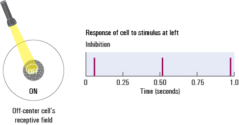
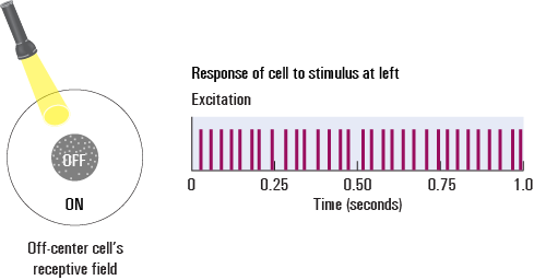
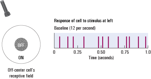
Another type of retinal ganglion cell, called an off-center cell, has the opposite arrangement of the on-center cell. When light strikes the center of an off-center cell, activity is reduced. When light strikes the periphery, activity is increased.
Watch what happens to the activity of the cell when you change the location and size of the light stimulus on the receptive field of this cell. Notice how this cell responds differently than the on-center cell.
Question
LwWVyzOYW0F6fg8uz1nmqbDwgMcZWM2H0PUERyaiFuTV6BVNPMULAzSsAnx6qz8XiHABvbdzS9RUBfnFE9oWczNVS1HXlur5rfitliCj0/608ZbyRtc8C9Svojm2dhjNJ2pf8tXjIzqYYucGUaBR7qrmsz1IgkwPozrOgvtJ1IHuhxM/h58+1567vzroG+lPNQYU7JSIbPbbLL2T2clNFjFWj2Rl/l1ev1c17lCpOewNI6g+Retinal ganglion cells (RGCs) are arranged in an overlapping pattern of on- and off-center cells to cover the visual field. This overlap and the on/off arrangement of the RGC receptive fields together make these cells especially responsive to tiny spots of light in the visual field.
1.6 Processing Shape in Retinal Ganglion Cells
How do overlapping RGCs help the brain understand shape? Imagine that a spotlight is striking an object, creating a deep shadow behind it. The receptive fields of the ganglion cells cover the entire visual field. Some of the ganglion cells’ receptive fields will be in the dark area, others will be in the light area, and still others will straddle the edge between the light and dark areas.
Visual information is transmitted from the to the visual areas of the brain, but the brain does not give equal weight to all visual field regions. Because they receive greater stimulation, greater emphasis is placed on information coming from RGCs in regions containing differences in luminance—areas along the edges. In this way, RGCs are really sending signals about edges, and edges are critical to helping us determine the shape of objects.
[Insert animation]
1.7 Check Your Understanding: RGCs
Before we proceed, review what you know about the retinal ganglion cells. For each question, choose the correct response.
Question
TslAOyjs5U56vchY2WtR1sWJgZjF15WVcdBWa5PWK33oY9WL2Uto5HVFbzBd1bVHRZ7seWB3ecWnW9/TVeqjXWdODybuZpYy6YPXutH+5dsD3BiaJCjLDzvO0+T5SQTkCoUQQ71jYVLfOTR6Y1x2eJ4RGa91qnm/2mu743OhSOraD5ulqaXYLpTgQlpDPNzYrI/SXFnLJ7PrudSvyYKTcV4qVnqEdfvN/6PU9ti37FvGmzs36n5SFCEHlZWTkwU36sOHboTzfIgucIuSFvh3v3ozm/nrIVszoA4l2N4UPJvQIXpKKIBN23FfXByyXYPt3FwGhrgGb8nrRwn88LRo6lQ/5gSoeR/RuPC8AF5Spfv6/lLXhdNHskSfpV/0XzphmLwHdaw8zFYj06kPlSTlHweTHYrXJl8PiwHNcuYM0JaSETSlV/dUNPnI3XNuwEUUdFW8ylQCkpPi11TPW1V0eRXwWI22AZ2TNNQh3R3mCAEeXfe5zpfUU0gFaanHQ8n/AZ0CxOwQIKfLUxdsndvmEihv05MQe5NK Wh3Tsvu+EpA4D9gCZPFbnVI20Pu9eJNvHxbsmdln7CJ8dkYPnkfA1H1zkByiM5XLtGcW9PYS2ep7GdaRwidi3nTORnSVLAtDEgrXnKLXNADg9FWfnCtE9PwKGx59CvD9EZLVEfH/EZwWXXYMB147N3xb4OCR901gvwCUp87Ium0f/zl/TebRUBg0x4R762IgbXMO+63o2bXU9cvSJ23rC9t8/bRo1qdHWANEceMPV/JE7ExXvVaYAqwEyQSnUS7+BX9G6IrLw/Nt+Pf+YQ+Xn61BMb2PyTx/wcxo930e+ifqongvOLNq/sMWHx1nDAklS+ABn7KcYJ6GtEwBu29IBK1hF+VxvQrbqvSYzojr25WV/TNcCXow9WwMkyWnrv2W0fi+U57cVSQwkCZA4etavhao6Hzsj/vRAVwU0GClpxiIbwe7Oac3hn/DifNmoJ2kJRv2XkN7iXSDYr8ZaQj9fo35WB+GTCAFWY0FnlnKWe43Rh4ULG8PI6Dccq8nBg+TWP3XwVu3ihPfkz2lij6nbpQNuY0= fCcAeuGn5J6Qq5po+MbXaAjLquWTEkZTejfLzu1iheDALR14imKuhEfcw35XOIdzn0tpnBLtd7ATrPMJUu0AJEPBAfJ8Uu3Uip5WFyVYDUoLRuiOXFuz7yQNvFpfEgD2aw8Dc6kZL2YmGZQUkjh24a+O62MisMm2Zqq5v7ynfYsWN+XjDUXye9VH/ry9RI27kizOX+1ZsRxR/eoAOGJ/y+tCi0mXH4ytH6ODvLvNY5eM7sX7Nl+Tg6jYMVXqA6/wwAgUgLfxhLk9RtwuNGBEbusj4iPLsYhKqDWrNar+KpCsKIitavYttg==1.8 Processing in Primary Visual Cortex (V1) Cells: Simple Cells
Now let’s see what happens when visual information reaches the brain. Multiple RGC inputs converge on a single primary visual cortex (V1) cell, which means the receptive fields of the V1 neurons are much larger than those of retinal neurons. Consequently, V1 cells respond to more complex light compared to RGCs. V1 cells are maximally excited by bars of light oriented in a particular direction.
Let’s see how one type of V1 cells, called simple cells, respond to different bars of light.
[Insert animation]
1.9 Processing in Primary Visual Cortex (V1) Cells: Complex Cells
In contrast to simple cells, complex cells are maximally excited by bars of light moving in a particular direction through the visual field. Let’s see how complex cells respond to different types of light stimuli.
[Insert animation]
1.10 Processing in Primary Visual Cortex (V1) Cells: Hypercomplex Cells
Some V1 cells are maximally excited by bars of light that are moving in a particular direction through the visual field and have an inhibitory area at one end of their receptive field. These types of cells are known as hypercomplex cells. Let’s see how this third type of V1 cell responds to different types of light stimuli.
[Insert animation]
1.11 Check Your Understanding: V1 Cells
Before we go deeper into the primary visual cortex, let’s review the different types of V1 cells.
Question
jNBi3cRYQGJcSRCjgDT1xbvwHJY2/qvwzBCR9h6D6z8XZzQhlRGc75IpN6kHUKzddCQBAOIXNRsy8nTw83J3OsqXTTNs1q5Hcp9YOe80hK72Fx3BdMwKQbbfyvXEcI4A5IFCXzFlKzNH4QCyXiPUWLhC3XPf9gpHbkr+KGFh29PmtaaIhPN9G5AOhWCwkGW6UnzFv9dt2eEqJVlZdiUTc7VmHTllrJxhwosrX8Yj5n6PU69aalttFLZwnhThbIBHm9sqTndDla+9pPhOQ85B/qDJefnC/SymIjAbwDNhlzDouJeg43nXv71e+5qIcgYwRjjBJX0oINlSebm9cKbggg== KZIx8MnGEjh7tR/sBZ2WV0tmzzXGPix14oHryClTx0KwtNbDXg6/or7L+ZRT9mwM07UUWr22SpbhbWz7fICSYAE+aEB1mPL4VghCACBVI4FhneCDdjpnqewFvrSdclUZidOJNR59v58vUo4ztKkOu8RQY28UsHAJPZmWGWOVv35RHhu0bajuw7RcRCQZdwLH+8oG96IziVVhOcMZj1i9BseWhFpVgmuQzfzU0Lyrfv7QVFv5f3j8vR7vbMyfIhBJmqXnDMFI1bmRbG9WXWtejLco9DkGSWoiVAIrkK13KR4FvdhGlzkZzdcFZ2N4YKuLwNd7lmh8Lg53JL5ufm/2Kg== vpkq6CjiAah8VvvhE9Us+U7umC+fGahSKDLZ8B4vqwe+PUrQoWCM+cnxZQ8cR+vAROJB+R1HtgCG2sETld7nEcPozTgBtdZZ2bc151Uu7qsuwm4WUmhweyLjOwqlbN9QDr0s8HBpn4ph9I1HQEM7ZEkserC4q9+LHgtz1L1jAJCAqYsvU87LeaIZxDq0OcOKMOUMNB6MXMY/yVS/g9TicS5bhF62I/KusOt3l51bBezzRXQ0FYdk7ma5bPQLDlYuj9D3UktQ6lWnPqC/viqTR93ukGLEvEfWj+GvtOcNMWrcSM+7ioVD18AULCfU0jBxW0lnefdBYtdZWreI08nMZxwD2zaltvfYltPimghBC8Cs4GMKXfIa3PmOzzA1rsghH76EPIugaKjPpEizmKlBEt9xbFx0yB2/1.12 Cortical Columns in the Primary Visual Cortex
Finally, let’s explore the two types of functional columns that comprise the primary visual cortex (V1): orientation columns and ocular dominance columns.
[Insert animation]
1.13 Check Your Understanding: Functional Columns
What are the two kinds of columns in the primary visual cortex, and how are they arranged?
Question 1.1
Y7XxM02sErG18w4JIYir5oGri4DGa9VRfqhB0RPKtW3j4keF1k0iDK100M45h19L2FTGHARNNLttKIDtj9ppoPWEpYk=An ocular dominance column is a functional column in the visual cortex that is maximally responsive to information coming from one eye (left or right). The ocular dominance columns are arranged in alternating fashion so that there is one column that responds maximally to the right eye next to a column that responds maximally to the left eye, and so on.
Your answer has been provisionally accepted. You'll get full credit for now, but your instructor may update your grade later after evaluating it.
1.14 Summary
Congratulations! You have successfully completed the activity. You learned about the receptive fields of the cells within the visual system and how the off-center and on-center ganglion cells influence the perception of shape. You also saw how the cells of the primary visual system can be classified as simple, complex, or hypercomplex cells and how these cells are arranged in functional columns within the visual cortex.
The visual system is complex, and this activity only begins to describe the intricate organization of the visual system. By understanding how the receptive fields of the cells of the visual system are stimulated, you can begin to understand how the visual system processes more complex features in our environment, such as edges, shapes, and objects.
Your instructor may now have you take a short quiz about this activity. Good luck!