5.2 Vision: Sensory and Perceptual Processing
Your eyes receive light energy and transform it into neural messages that your brain then processes into what you consciously see. How does such a taken-
Light Energy and Eye Structures
LOQ 5-
The Stimulus Input: Light Energy
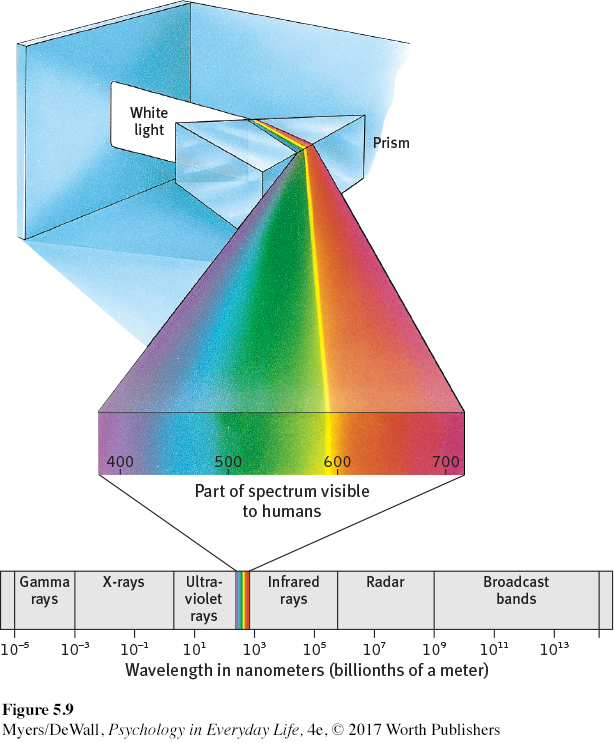
When you look at a bright red tulip, what strikes your eyes are not bits of the color red but pulses of energy that your visual system perceives as red. What we see as visible light is but a thin slice of the wide spectrum of electromagnetic energy shown in FIGURE 5.9. On one end of this spectrum are the short gamma waves, no longer than the diameter of an atom. On the other end are the mile-
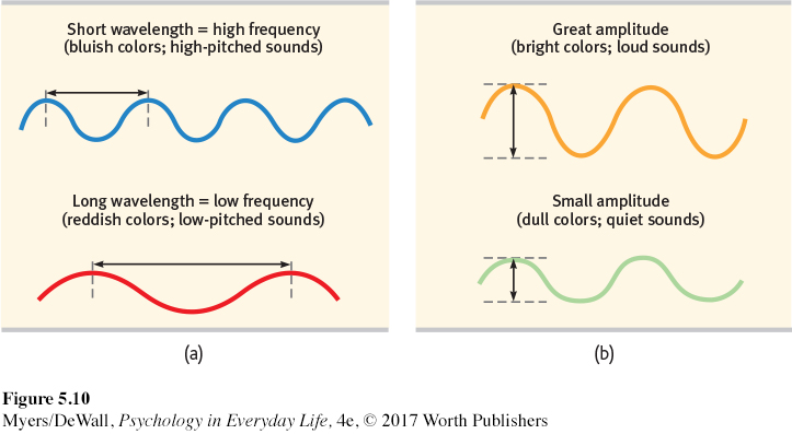
wavelength the distance from the peak of one light wave or sound wave to the peak of the next.
hue the dimension of color that is determined by the wavelength of light; what we know as the color names blue, green, and so forth.
intensity the amount of energy in a light wave or sound wave, which influences what we perceive as brightness or loudness. Intensity is determined by the wave’s amplitude (height).
Light travels in waves, and the shape of those waves influences what we see. Light’s wavelength is the distance from one wave peak to the next (FIGURE 5.10a). Wavelength determines hue—the color we experience, such as a tulip’s red petals. A light wave’s amplitude, or height, determines its intensity—the amount of energy the wave contains. Intensity influences brightness (FIGURE 5.10b).
Understanding the characteristics of the physical energy we see as light is one part of understanding vision. But to appreciate how we transform that energy into color and meaning, we need to know more about vision’s window—
The Eye
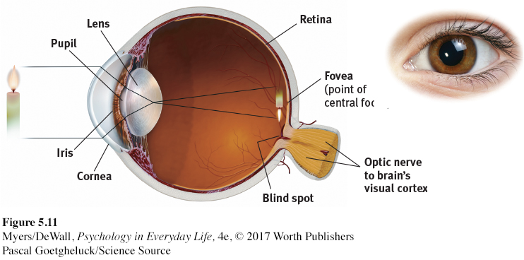
What color are your eyes? Asked this question, most people describe their iris, the doughnut-
141
retina the light-
After passing through your pupil, light hits the lens in your eye. The lens then focuses the light rays into an image on your eyeball’s inner surface, the retina. For centuries, scientists knew that an image of a candle passing through a small opening will cast an upside-
Eventually the answer became clear: The retina doesn’t “see” a whole image. Rather, its millions of receptor cells behave like the prankster engineering students who make news by taking a car apart and rebuilding it in a friend’s third-
Information Processing in the Eye and Brain
142
LOQ 5-
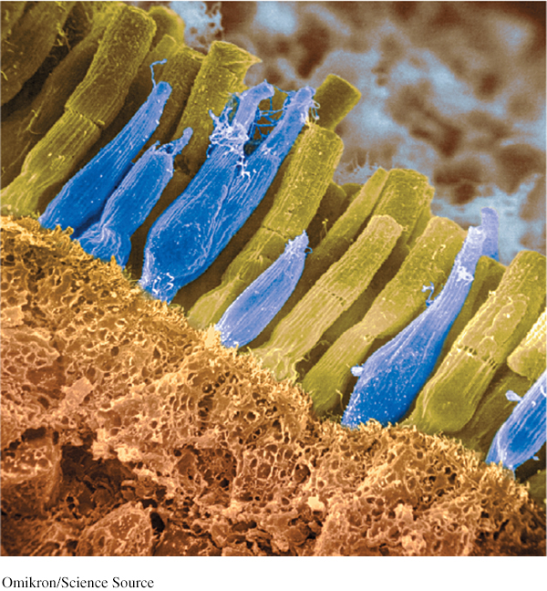
rods retinal receptors that detect black, white, and gray, and are sensitive to movement; necessary for peripheral and twilight vision, when cones don’t respond.
cones retinal receptors that are concentrated near the center of the retina; in daylight or well-
optic nerve the nerve that carries neural impulses from the eye to the brain.
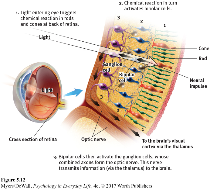
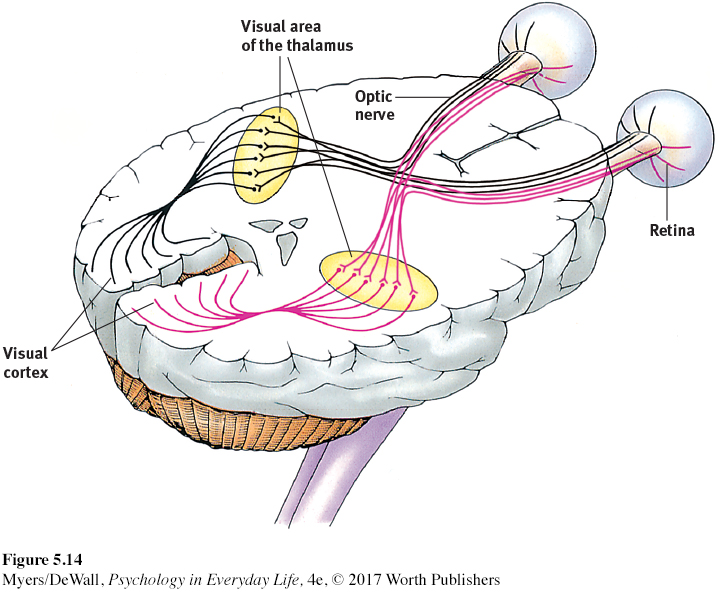
Imagine that you could follow a single light-
blind spot the point at which the optic nerve leaves the eye; this part of the retina is “blind” because it has no receptor cells.
The optic nerve is an information highway from the eye to the brain. This nerve can send nearly 1 million messages at once through its nearly 1 million ganglion fibers. We pay a small price for this high-
Retrieve + Remember

Question 5.5
ANSWER: Your blind spot is on the nose side of each retina, which means that objects to your right may fall onto the right eye’s blind spot. Objects to your left may fall on the left eye’s blind spot. The blind spot does not normally impair your vision, because your eyes are moving and because one eye catches what the other misses.
The retina’s two types of receptor cells, rods and cones, differ in where they’re found and in what they do (TABLE 5.1). Cones cluster in and around the fovea, the retina’s area of central focus. Many cones have their own hotline to the brain. One cone transmits a precise message to a single bipolar cell, which relays it to the visual cortex. Thanks to cones, you can see fine details and perceive color—
| Cones | Rods | |
|---|---|---|
| Number | 6 million | 120 million |
| Location in retina | Center | Periphery |
| Sensitivity in dim light | Low | High |
| Color sensitivity | High | Low |
| Detail sensitivity | High | Low |
143
Rods, which are located around the outer regions (the periphery) of your retina, remain sensitive in dim light. If cones are soloists, rods perform as a chorus. They enable black-
Stop for a minute and experience the rod-
So, cones and rods each provide a special sensitivity: Cones are sensitive to detail and color. Rods are sensitive to faint light and peripheral motion. But these receptor cells do more than simply pass along electrical impulses. They begin processing sensory information by coding and analyzing it. (In a frog’s eye, for example, the “bug detector” cells that fire when they respond to moving fly-
144
Retrieve + Remember
Question 5.6
•Some night-
ANSWERS: rods; cones; color
Question 5.7
•Cats are able to open their ____ much wider than we can, which allows more light into their eyes so they can see better at night.
ANSWER: pupils
Color Processing
LOQ 5-
We talk as though objects possess color: “A tomato is red.” Recall the old question, “If a tree falls in the forest and no one hears it, does it make a sound?” We can ask the same of color: If no one sees the tomato, is it red?
The answer is No. First, the tomato is everything but red, because it rejects (reflects) the long wavelengths of red. Second, the tomato’s color is our mental construction. As the famous physicist Sir Isaac Newton (1704) observed more than three centuries ago, “The [light] rays are not colored.” Color, like all aspects of vision, lives not in the object but in the theater of our brain. Even while dreaming, we may perceive things in color.
One of vision’s most basic and intriguing mysteries is how we see the world in color. How, from the light energy striking your retina, does your brain construct your experience of color—
Modern detective work on the mystery of color vision began in the nineteenth century, when Hermann von Helmholtz built on the insights of an English physicist, Thomas Young. The clue that led to their breakthrough was the knowledge that any color can be created by combining the light waves of three primary colors—
Young-
Years later, researchers confirmed the Young-
By one estimate, we can see differences among more than 1 million color variations (Neitz et al., 2001). At least most of us can. About 1 person in 50 is “colorblind.” That person is usually male, because the defect is genetically sex linked. Most people with color-
But why do people blind to red and green often still see yellow? And why does yellow appear to be a pure color, not a mixture of red and green, the way purple is of red and blue? As Ewald Hering soon noted, trichromatic theory leaves some parts of the color vision mystery unsolved.
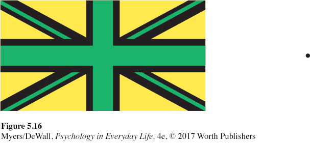
Hering, a physiologist, found a clue in afterimages. Stare at a green square for a while and then look at a white sheet of paper, and you will see red, green’s opponent color. Stare at a yellow square and its opponent color, blue, will appear on the white paper. (To experience this, try the flag demonstration in FIGURE 5.16.) Hering proposed that color vision must involve two additional processes: one responsible for red-
opponent-
A century later, researchers confirmed Hering’s proposal, now called the opponent-
How does opponent-
145
First, you stared at green bars, which tired the green part of the green-
red pairing in your eyes. Then you stared at a white area. White contains all colors, including red.
Because you had tired your green response, only the red part of the green-
red pairing fired normally.
 For an interactive review and demonstration of these color vision principles, visit LaunchPad’s PsychSim 6: Colorful World.
The present solution to the mystery of color vision is therefore roughly this: Color processing occurs in two stages.
The retina’s red, green, and blue cones respond in varying degrees to different color stimuli, as the Young-
Helmholtz trichromatic theory suggested. The cones’ responses are then processed by opponent-
process cells, as Hering’s opponent- process theory proposed.
Retrieve + Remember
Question 5.8
•What are two key theories of color vision? Do they contradict each other, or do they make sense together? Explain.
ANSWER: The Young-
Feature Detection
LOQ 5-
feature detectors nerve cells in the brain that respond to specific features of a stimulus, such as shape, angles, or movement.
Scientists once compared the brain to a movie screen, on which the eye projected images. But along came David Hubel and Torsten Wiesel (1979), who showed that at an early stage, our visual processing system takes images apart and later reassembles them. Hubel and Wiesel received a Nobel Prize for their work on feature detectors, nerve cells in the brain that respond to a scene’s specific features—
One temporal lobe area by your right ear enables you to perceive faces and, thanks to a specialized neural network, to recognize them from many viewpoints (Connor, 2010). If this region is damaged, people still may recognize other forms and objects, but, like Heather Sellers, they cannot recognize familiar faces. How do we know this? In part because in laboratory experiments, researchers have used magnetic pulses to disrupt that brain area, producing a temporary loss of face recognition. The interaction between feature detectors and supercells provides instant analyses of objects in the world around us.
Parallel Processing
146
LOQ 5-
parallel processing processing many aspects of a problem or scene at the same time; the brain’s natural mode of information processing for many functions, including vision.
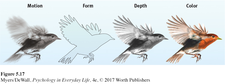
One of the most amazing aspects of visual information processing is the brain’s ability to divide a scene into its parts. Using parallel processing, your brain assigns different teams of nerve cells the separate tasks of simultaneously processing a scene’s movement, form, depth, and color (FIGURE 5.17). You then construct your perceptions by integrating the work of these different visual teams (Livingstone & Hubel, 1988).
Destroy or disable the neural workstation for a visual subtask, and something peculiar results, as happened to “Mrs. M.” (Hoffman, 1998). After a stroke damaged areas near the rear of both sides of her brain, she could not perceive movement. People in a room seemed “suddenly here or there but I [had] not seen them moving.” Pouring tea into a cup was a challenge because the fluid appeared frozen—
* * *
“I am fearfully and wonderfully made.”
King David, Psalm 139:14
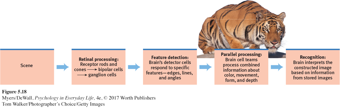
Think about the wonders of visual processing. As you read this page, the letters reflect light rays onto your retina, which then sends formless nerve impulses to several areas of your brain, which integrate the information and decode its meaning. The amazing result: we have transferred information across time and space, from our minds to your mind (FIGURE 5.18). That all of this happens instantly, effortlessly, and continuously is awe-
Retrieve + Remember
Question 5.9
•What is the rapid sequence of events that occurs when you see and recognize the crouching animal in FIGURE 5.18?
ANSWER: Light waves reflect off the image and travel into your eyes. Receptor cells in your retina convert the light waves’ energy into millions of neural impulses sent to your brain. Your brain’s detector cells and work teams process the different parts of this visual input—
Perceptual Organization
LOQ 5-
It’s one thing to understand how we see colors and shapes. But how do we organize and interpret those sights (or sounds or tastes or smells) so that they become meaningful perceptions—
gestalt an organized whole. Gestalt psychologists emphasized our tendency to integrate pieces of information into meaningful wholes.
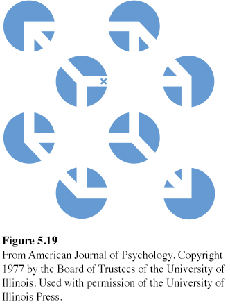
Early in the twentieth century, a group of German psychologists noticed that people who are given a cluster of sensations tend to organize them into a gestalt, a German word meaning a “form” or a “whole.” As we look straight ahead, we cannot separate the perceived scene into our left and right fields of view. Our conscious perception is, at every moment, one whole, seamless scene. Consider FIGURE 5.19. The individual elements of this figure, called a Necker cube, are really nothing but eight blue circles, with three white lines meeting near the center. When we view all these elements together, however, we see a cube that sometimes reverses direction. The Necker cube nicely illustrates a famous saying of Gestalt psychologists: In perception, the whole may exceed the sum of its parts.
147
Over the years, the Gestalt psychologists demonstrated many principles we use to organize our sensations into perceptions. Underlying all of them is a basic truth: Our brain does more than register information about the world. Perception is not just opening a shutter and letting a picture print itself on the brain. We filter incoming information and we construct perceptions. Mind matters.
How Do We Perceive Form?
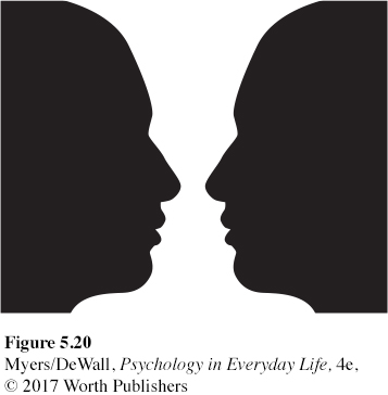
figure-
Imagine designing a video-
grouping the perceptual tendency to organize stimuli into meaningful groups.
While telling figure from ground, we (and our video-
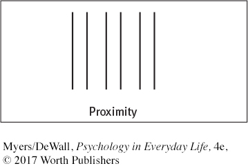
Proximity We group nearby figures together. We see not six separate lines, but three sets of two lines:
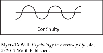
Continuity We perceive smooth, continuous patterns rather than discontinuous ones. This pattern could be a series of alternating semicircles, but we perceive it as two continuous lines—
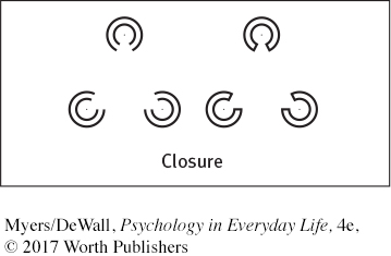
Closure We fill in gaps to create a complete, whole object. Thus, we assume that the circles on the left are complete but partially blocked by the (illusory) triangle. Add nothing more than little lines to close off the circles, and your brain stops constructing a triangle:
148
Retrieve + Remember
Question 5.10
•In terms of perception, a band’s lead singer would be considered _______________ (figure/ground), and the other musicians would be considered ________________ (figure/ground).
ANSWERS:
figure
ground
Question 5.11
•What do we mean when we say that, in perception, “the whole may exceed the sum of its parts”?
ANSWER: Gestalt psychologists used this saying to describe our perceptual tendency to organize clusters of stimuli into meaningful forms or groups.
How Do We Perceive Depth?
LOQ 5-
depth perception the ability to see objects in three dimensions, although the images that strike the retina are two-
Our brain performs many amazing tricks, but one of its best is depth perception. From the two-
visual cliff a laboratory device for testing depth perception in infants and young animals.
As Eleanor Gibson picnicked on the rim of the Grand Canyon, her scientific curiosity kicked in. She wondered, Would a toddler peering over the rim perceive the dangerous drop-
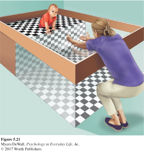
Had they learned to perceive depth? Crawling, no matter when it begins, seems to increase an infant’s fear of heights (Adolph et al., 2014; Campos et al., 1992). But depth perception is also partly innate. Mobile newborn animals—
If we were to build this ability into our video-
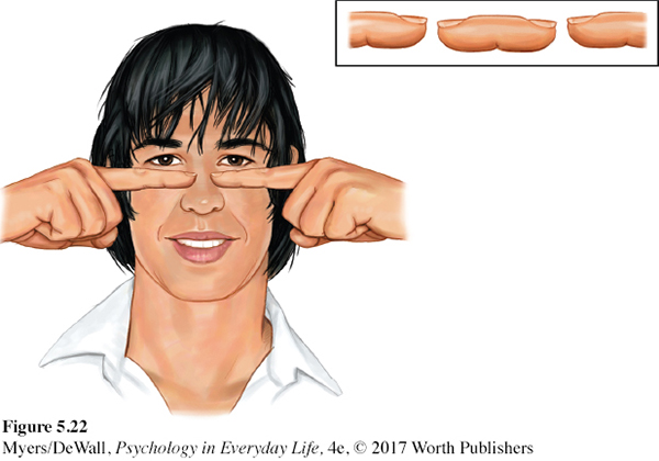
binocular cue a depth cue, such as retinal disparity, that depends on the use of two eyes.
BINOCULAR CUES People who see with two eyes perceive depth thanks partly to binocular cues. Here’s an example. With both eyes open, hold two pens or pencils in front of you and touch their tips together. Now do so with one eye closed. A more difficult task, yes?
retinal disparity a binocular cue for perceiving depth. By comparing images from the two eyes, the brain computes distance—
We use binocular cues to judge the distance of nearby objects. One such cue is retinal disparity. Because your eyes are about 2½ inches apart, your retinas receive slightly different images of the world. By comparing these two images, your brain can judge how close an object is to you. The greater the disparity (the difference) between the two retinal images, the closer the object. Try it. Hold your two index fingers, with the tips about half an inch apart, directly in front of your nose, and your retinas will receive quite different views. If you close one eye and then the other, you can see the difference. (You can also create a finger sausage, as in FIGURE 5.22.) At a greater distance—
We could easily include retinal disparity in our video-
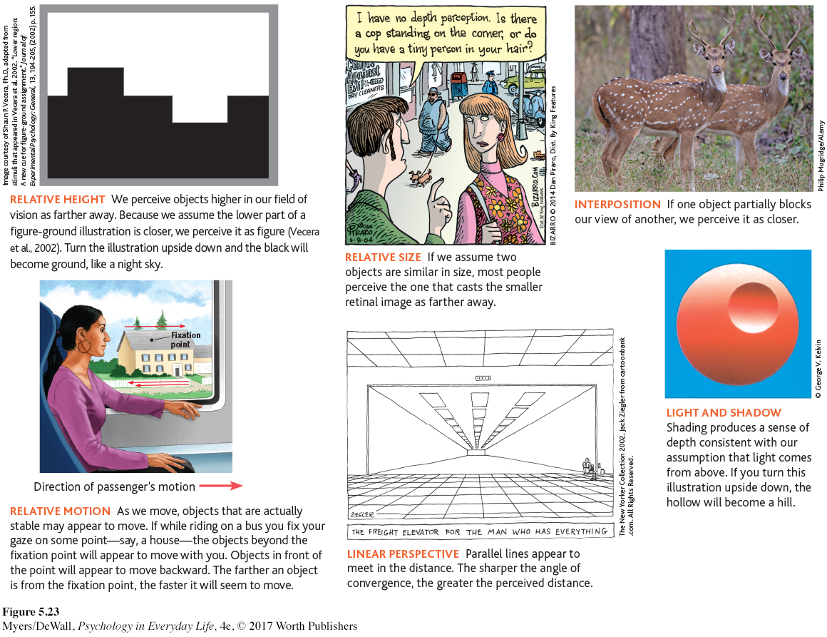
monocular cue a depth cue, such as interposition or linear perspective, available to either eye alone.
MONOCULAR CUES How do we judge whether a person is 10 or 100 yards away? Retinal disparity won’t help us here, because there won’t be much difference between the images cast on our right and left retinas. At such distances, we depend on monocular cues (depth cues available to each eye separately). See FIGURE 5.23 for some examples.
149
150
For animated demonstrations and explanations of these cues, visit LaunchPad’s Concept Practice: Depth Cues.
Retrieve + Remember
Question 5.12
•How do we normally perceive depth?
ANSWER: We are normally able to perceive depth thanks to (1) binocular cues (which are based on our retinal disparity), and (2) monocular cues (which include relative height, relative size, interposition, linear perspective, light and shadow, and relative motion).
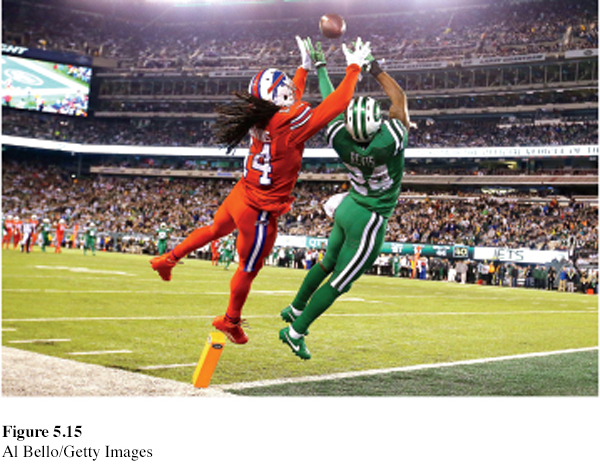
How Do We Perceive Motion?
Imagine that you could perceive the world as having color, form, and depth but that you could not see motion. You would be unable to bike or drive, and writing, eating, and walking would be a challenge.
Normally your brain computes motion based partly on its assumption that shrinking objects are moving away (not getting smaller) and enlarging objects are approaching. But you are imperfect at motion perception. In young children, this ability to correctly perceive approaching (and enlarging) vehicles is not yet fully developed, which puts them at risk for pedestrian accidents (Wann et al., 2011).
It’s not just children who have occasional difficulties with motion perception. Our adult brain is sometimes tricked into believing what it is not seeing. When large and small objects move at the same speed, the large objects appear to move more slowly. Thus, trains seem to move slower than cars, and jumbo jets seem to land more slowly than smaller jets.
Perceptual Constancy
LOQ 5-
perceptual constancy perceiving objects as unchanging (having consistent color, brightness, shape, and size) even as illumination and retinal images change.
So far, we have noted that our video-
color constancy perceiving familiar objects as having consistent color, even if changing illumination alters the wavelengths reflected by the object.
COLOR CONSTANCY Our experience of color depends on an object’s context. This would be clear if you viewed an isolated tomato through a paper tube over the course of the day. As the light—
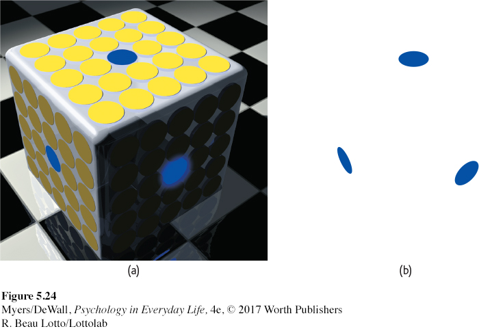
We see color thanks to our brain’s ability to decode the meaning of the light reflected by an object relative to the objects surrounding it. FIGURE 5.24 dramatically illustrates the ability of a blue object to appear very different in three different contexts. Yet we have no trouble seeing these disks as blue. Paint manufacturers have learned this lesson. Knowing that your perception of a paint color will be determined by other colors in your home, many now offer trial samples you can test in that context. The take-
“From there to here, from here to there, funny things are everywhere.”
Dr. Seuss, One Fish, Two Fish, Red Fish, Blue Fish, 1960
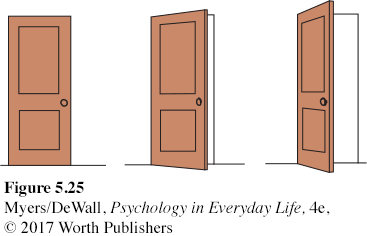
SHAPE AND SIZE CONSTANCIES Thanks to shape constancy, we usually perceive the form of familiar objects, such as the door in FIGURE 5.25, as constant even while our retinas receive changing images of them. Thanks to size constancy, we perceive objects as having a constant size even while our distance from them varies. We assume a car is large enough to carry people, even when we see its tiny image from two blocks away. This assumption also shows the close connection between perceived distance and perceived size. Perceiving an object’s distance gives us cues to its size. Likewise, knowing its general size—
Even in size-
151
Mistaken judgments like these reveal the workings of our normally effective perceptual processes. The perceived relationship between distance and size is usually valid. But under special circumstances it can lead us astray.
To experience more visual illusions, and to understand what they reveal about how you perceive the world, visit LaunchPad’s PsychSim 6: Visual Illusions.
Form perception, depth perception, and perceptual constancies illuminate how we organize our visual experiences. Perceptual organization applies to our other senses, too. Listening to an unfamiliar language, we have trouble hearing where one word stops and the next one begins. Listening to our own language, we automatically hear distinct words. We even organize a string of letters—
Perceptual Interpretation
The debate over whether our perceptual abilities spring from our nature or our nurture has a long history. To what extent do we learn to perceive? German philosopher Immanuel Kant (1724–
Experience and Visual Perception
LOQ 5-
RESTORED VISION AND SENSORY RESTRICTION Writing to John Locke, a friend wondered what would happen if “a man born blind, and now adult, [was] taught by his touch to distinguish between a cube and a sphere.” Could he, if made to see, visually distinguish the two? Locke’s answer was No, because the man would never have learned to see the difference.
This question has since been put to the test with a few dozen adults who, though blind from birth, later gained sight (Gregory, 1978; Huber et al., 2015; von Senden, 1932). Most were born with cataracts—
Seeking to gain more control than is provided by clinical cases, researchers have outfitted infant kittens and monkeys with goggles through which they could see only diffuse, unpatterned light (Wiesel, 1982). After infancy, when the goggles were removed, the animals’ reactions were much like those of humans born with cataracts. They could distinguish color and brightness but not form. Their eyes were healthy. Their retinas still sent signals to their visual cortex. But without early stimulation, the brain’s cortical cells had not developed normal connections. Thus, the animals remained functionally blind to shape.
152
Surgery on blind children in India reveals that children blind from birth can benefit from cataract surgery. Although their vision may never be as sharp as normal, the younger they are, the more they will benefit (Sinha, 2013). There is a critical period for normal sensory and perceptual development—a limited time when exposure to certain stimuli or experiences is required.
In humans and other animals, similar sensory restrictions later in life do no permanent harm. When researchers cover an adult animal’s eye for several months, its vision will be unaffected after the eye patch is removed. When surgeons remove cataracts that developed during late adulthood, most people are thrilled at the return to normal vision.
perceptual adaptation the ability to adjust to changed sensory input, including an artificially displaced or even inverted visual field.
PERCEPTUAL ADAPTATION Given a new pair of glasses, we may feel a little strange, even dizzy. Within a day or two, we adjust. Our perceptual adaptation to changed visual input makes the world seem normal again. But imagine wearing a far more dramatic pair of new glasses—
Could you adapt to this distorted world? Not if you were a baby chicken. When fitted with such lenses, baby chicks continue to peck where food grains seem to be (Hess, 1956; Rossi, 1968). But we humans adapt to distorting lenses quickly. Within a few minutes, your throws would again be accurate, your stride on target. Remove the lenses and you would experience an aftereffect. At first your throws would err in the opposite direction, sailing off to the right. But again, within minutes you would adjust.
Indeed, given an even more radical pair of glasses—
At first, when Stratton wanted to walk, he found himself searching for his feet, which were now “up.” Eating was nearly impossible. He became nauseated and depressed. But he persisted, and by the eighth day he could comfortably reach for an object and walk without bumping into things. When Stratton finally removed the headgear, he readapted quickly. So did research participants who also wore such optical gear—
So do we learn to perceive the world? In part we do, as we constantly adjust to changed sensory input. Research on critical periods teaches us that early nurture sculpts what nature has provided. In less dramatic ways, nurture continues to do this throughout our lives. Experience guides, sustains, and maintains the pathways in our brain that enable our perceptions.