23.17–23.19: The brain is organized into distinct structures dedicated to specific functions.

An adult human brain weighs only a bit over 3 pounds, or 1.4 kilograms. It’s fairly uniform in color, a drab whitish-
949
From a cellular perspective, all animal brains function in the same general way. A neuron is a neuron is a neuron. But this isn’t the whole picture. Each region of the brain is characterized by neurons that are shaped slightly differently, have different axon and dendrite branching patterns and complexities, and use different neurotransmitters at their synapses. Each of these regions of the brain is the control center for various activities in the body.
Among different species, evolutionary changes have resulted in differences in the relative amount of the brain dedicated to various body structures. These differences have, in turn, led to corresponding differences in abilities and behaviors. In the cerebral cortex, for example, humans have significantly more neurons than do other species. The extra brain matter in this particular part of the brain (which we examine in more detail later in the chapter) is associated with many abilities that are particularly well developed in humans, including abstract thought, perception, and language. Similarly, a rat’s brain has an unusually large number of smell-
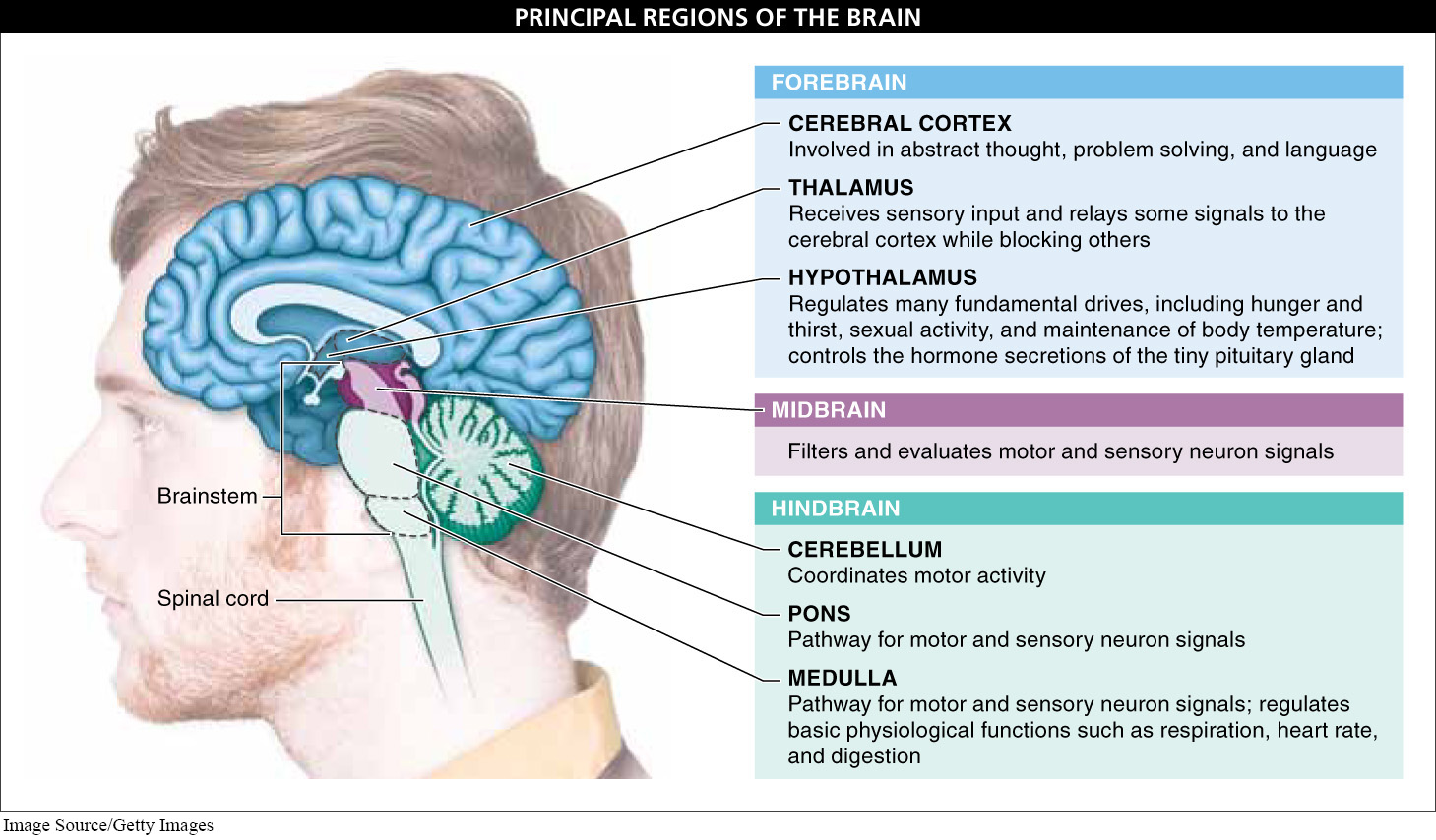
In this section and the next, we explore the three principal regions of the vertebrate brain—
Hindbrain The central nervous system is made up of the brain and the spinal cord. The spinal cord extends from the brain down through the backbone (though not down the entire length of the backbone), protected by the vertebral column. Branching off from the spinal cord at regular intervals are bundles of axons, some carrying motor information to muscles and glands, others carrying in sensory information from the senses to the brain. The top of the spinal cord is the beginning of the brain. Here, the spinal cord expands into the hindbrain. Three important structures are located in the hindbrain: the medulla, the pons, and the cerebellum. Because these structures are not involved in higher thought, hindbrain damage can lead to paralysis without harming consciousness.
Medulla and pons. Running through these structures are the many motor neurons traveling from the brain to the spinal cord and the many sensory neurons traveling from all parts of the body to the brain. Additionally, within the medulla are lots of collections of neurons that regulate basic physiological functions such as respiration, heart rate, and digestion. Consequently, injury to these structures—
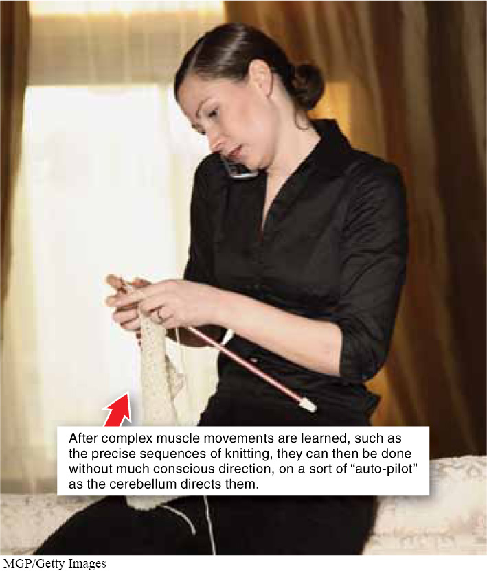
Cerebellum. This structure coordinates numerous types of motor activity. When it works properly, graceful motion is possible. The coordination of many complex movements is overseen by the cerebellum. Knitting or typing, for example, involves a wide range of complex muscle movements that must be executed in precise sequences. Once learned, these movements run on “auto-
Midbrain The midbrain, along with the medulla and pons of the hindbrain, makes up the brainstem. Most of the sensory information and motor neuron connections coming into and out of the brain pass through the midbrain. These receptors and effectors frequently have synapses with each other, as the midbrain helps filter and evaluate the importance of each signal. The midbrain is greatly enlarged and plays a more significant role in cold-
Forebrain In humans, the forebrain is the largest region of the brain. It includes two control and relay structures, the thalamus and hypothalamus, as well as the cerebrum. The cerebral cortex, which makes up the bulk of the cerebrum, is responsible for most of what we consider “higher” thought, including perception, memory, language, intelligence, and personality.
950
Thalamus and hypothalamus. The thalamus is one of the primary switchboards in the brain. It receives most of the visual, auditory, and touch input and, like a secretary deciding which calls to let through, relays some signals to the cerebral cortex while blocking others. The hypothalamus (part of the limbic system, discussed later in this section) is one of the chief regulatory centers of the brain. It regulates many fundamental drives, including hunger and thirst, sexual activity, and maintenance of body temperature. It also controls the hormone secretions of the tiny pituitary gland, located right next to it, which play important roles in reproduction and development.
“The mind is its own place, and in itself Can make a heaven of hell, a hell of heaven.”
— JOHN MILTON, Paradise Lost, 1667
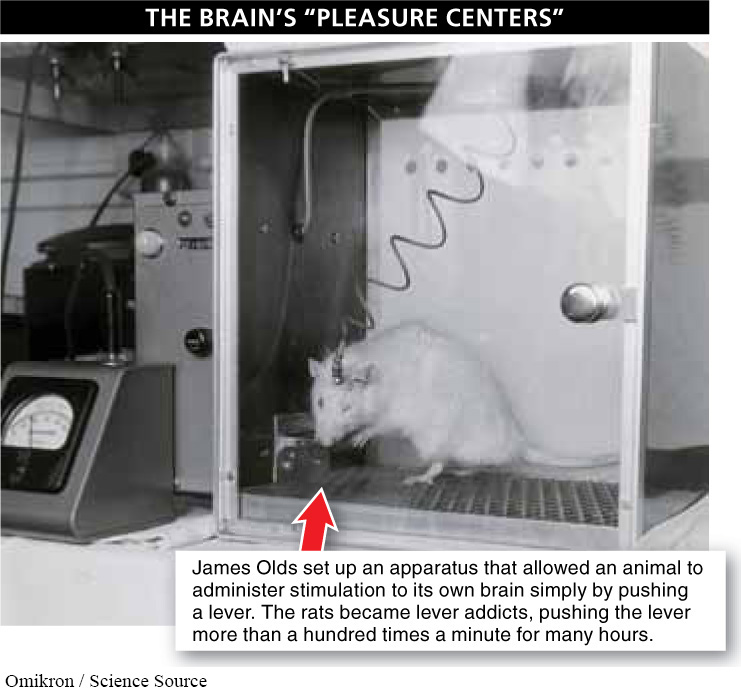
In the 1950s, James Olds discovered something very peculiar about the hypothalamus. When he inserted a tiny electrode into the hypothalamus of a rat’s brain, the animal seemed to experience great pleasure. Of course, Olds couldn’t be certain that it was pleasure the rats were experiencing, but he hypothesized that this was so by noting how the rats would work very hard (for instance, by learning to run very complex mazes) to get the stimulation. When he set up the apparatus so that an animal could administer the stimulation to its own brain simply by pushing a lever, the rats became lever addicts, pushing the lever more than a hundred times a minute for many hours (FIGURE 23-41). They would even choose to push the lever over eating food or drinking water, with some rats starving to death while pushing the lever rather than taking even a short break to eat.
This discovery that the brain has “pleasure centers” or “do-

Cerebral cortex. For humans, the cerebral cortex is the most sophisticated part of the brain. Involved in abstract thought, problem solving, language, and more, this is the part of the brain most responsible for the traits that set us apart from other animal species.
One of the most dramatic demonstrations of the complexity of the cerebral cortex resulted from a tragic accident and apparently miraculous recovery. In 1848, a railroad worker named Phineas Gage was packing explosives into a hole when they exploded unexpectedly, blasting a three-
Gage died about 13 years after the accident, and his skull was preserved. Recent analyses of the skull indicate that Gage suffered from severe damage to part of his frontal cortex. In other case studies of patients with damage to the same part of their frontal cortex, such as that caused by certain tumors, similar personality changes have been noted, with patients frequently exhibiting radically changed emotional responses.
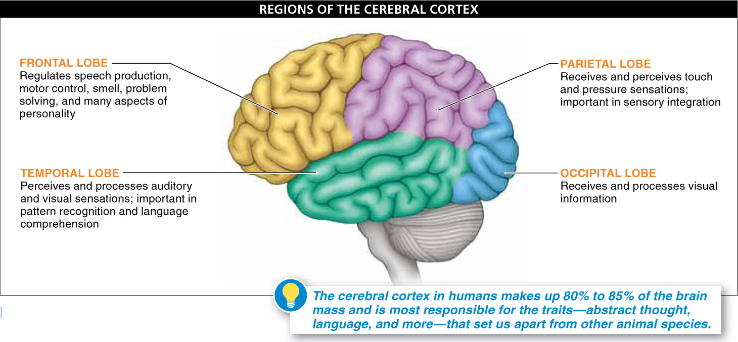
The highly folded cerebral cortex is by far the largest brain structure, covering all of the brain except for the cerebellum (FIGURE 23-43). The cerebral cortex makes up 80% to 85% of the brain mass in humans, with about 10 billion neurons and hundreds of billions of synapses. Although the brain (including the cerebral cortex) looks fairly symmetrical, functions are not divided in a symmetrical way. The brain is divided into a left and a right hemisphere, connected by a broad, thick band of neurons, called the corpus callosum. The left hemisphere in most, but not all, individuals is the site of areas specializing in language, logic, and mathematical skills, while the right hemisphere is more commonly home to larger areas dedicated to emotions, intuitive thinking, and artistic expression.
951
Each hemisphere of the cerebral cortex has four different lobes: frontal, temporal, parietal, and occipital. Each lobe is associated with specific functions (but each also has many other subareas that control a variety of additional functions; see Figure 23-43).
- 1. Frontal lobe: important in speech production, motor control, smell detection, problem solving, and many aspects of personality.
- 2. Temporal lobe: perceives and processes auditory and visual sensations; also important in pattern recognition and language comprehension. Damage to part of the right temporal lobe—
usually due to a stroke or tumor— has been implicated in a condition known as prosopagnosia, or face blindness. Individuals with this condition find it difficult or impossible to recognize faces. - 3. Parietal lobe: receives and perceives touch and pressure sensations; also important in sensory integration, the process of incorporating information from all of the senses to form a single perception of something.
- 4. Occipital lobe: receives and processes visual information.
952
Limbic system. Within the forebrain, near the center of the brain, is a set of structures that together make up the limbic system. It includes parts of the cerebral cortex and two structures deeper within the center of the brain, the hippocampus and the amygdala, and is responsible for many of our physiological drives and instincts. Together, these structures also play an important role in emotions, learning, and memory, which we discuss in the next section.
TAKE-HOME MESSAGE 23.17
There are several distinct regions in the brain, each of which is the control center for various activities in the body. The hindbrain regulates basic physiological functions such as respiration, heart rate, and digestion, and coordinates numerous types of motor activity. The midbrain, part of the brainstem, helps filter and evaluate the importance of sensory and motor information. In humans, the forebrain is the largest region of the brain, responsible for most of what we consider higher thought, including perception, memory, language, intelligence, and personality.
What are the three principle regions of the forebrain, and which region controls hormone secretion by the pituitary?