A Royal Disease
383
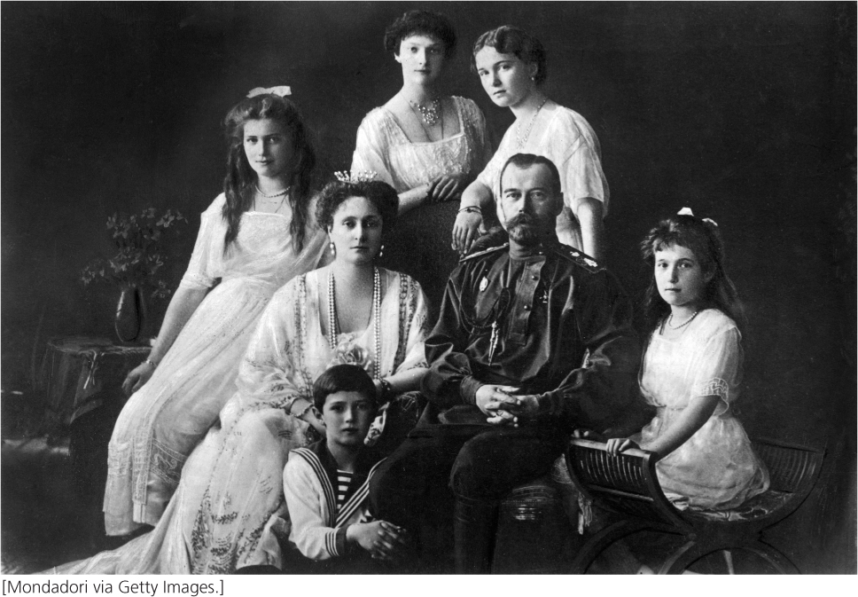
On August 12, 1904, Tsar Nicholas Romanov II of Russia wrote in his diary: “A great never-to-be forgotten day when the mercy of God has visited us so clearly.” That day Alexei, Nicholas’s first son and heir to the Russian throne, had been born.
At birth, Alexei was a large and vigorous baby with yellow curls and blue eyes, but, at 6 weeks of age, he began spontaneously hemorrhaging from the navel. The bleeding persisted for several days and caused great alarm. As he grew and began to walk, Alexei often stumbled and fell, as all children do. Even his small scrapes bled profusely, and minor bruises led to significant internal bleeding. It soon became clear that Alexei had hemophilia.
Marked by slow clotting and excessive bleeding, hemophilia is caused by a mutation in one of several genes that encode proteins involved in the process of blood clotting. In those with hemophilia, minor injuries can result in life-threatening blood loss, and spontaneous bleeding into joints erodes the bone with crippling consequences.
Alexei suffered from classic hemophilia, which is an X-linked genetic disorder. He inherited the hemophilia gene from his mother, Alexandra, who was a carrier. The gene appears to have originated with Queen Victoria of England (see Figure 6.8). In all, ten of Queen Victoria’s male descendants suffered from hemophilia. Six female descendants, including her granddaughter Alexandra (Alexei’s mother), were carriers.
During his childhood, Alexei experienced a number of severe bleeding episodes. The royal physicians were often helpless during these crises—they had no treatment that would stop the bleeding. At this time the Russian Revolution broke out. Bolsheviks captured the tsar and his family and held them captive in the city of Yekaterinburg. On the night of July 16, 1918, a firing squad executed the royal family and their attendants, including Alexei and his four sisters. For many years, the bodies of the Tsar’s family were lost, but their skeletons were eventually recovered from two graves outside Yekaterinburg. Comparisons of mitochondrial DNA and nuclear DNA from the bones and from descendants of relatives of the family verified that the bones were indeed those of the royal family. Although this analysis determined the identity of the remains, the molecular nature of the royal hemophilia long remained unknown.
In 2009, geneticists analyzed DNA from the bones of the Tsar’s family to determine the genetic nature of the royal hemophilia. In people with X-linked hemophilia, a mutation commonly occurs in one of two genes on the X chromosome, either the gene for blood coagulation factor VIII or the gene for factor IX.
384
In eukaryotic cells, genes are often interrupted by noncoding sequences. Those parts of a gene that encode the amino acids of a protein are called exons, while the noncoding sequences are called introns. Initially both exons and introns are transcribed into pre-mRNA but, in a process called RNA splicing, the introns are later cut out and the exons pasted together. Examination of the DNA sequences for the two clotting-factor genes in Alexandra revealed no mutations in the nucleotides that code for amino acids, but a mutation did occur in one of the introns of her gene for factor IX. This mutation altered the splicing of the exons, creating a new stop signal in the coding sequence of the mRNA and producing a truncated, defective clotting factor IX. Both normal and mutant alleles were detected in DNA from Alexandra, as is expected of a heterozygous carrier. DNA from Alexei carried only the mutant allele, which caused his hemophilia.
As illustrated by Alexei’s hemophilia, RNA processing is critically important for the proper synthesis of proteins. In eukaryotic cells, RNA molecules are often extensively modified after transcription: for genes that encode proteins, a special nucleotide called the cap is added to the 5′ end, a tail of adenine nucleotides is added to the 3′ end, and introns are cut out of the middle. In both prokaryotes and eukaryotes, rRNAs and tRNAs are also modified after transcription. In Chapter 13, we focused on transcription—the process by which RNA molecules are synthesized. In this chapter, we examine the function and processing of RNA.
We begin by taking a careful look at the nature of the gene. We then examine messenger RNA (mRNA), its structure, and how it is modified in eukaryotes after transcription. Then we turn to transfer RNA (tRNA), the adapter molecule that forms the interface between amino acids and mRNA in protein synthesis. We examine ribosomal RNA (rRNA), the structure and organization of rRNA genes, and how rRNAs are processed. Finally, we consider a newly discovered group of very small RNAs that play important roles in numerous biological functions.
As we explore the world of RNA and its role in gene function, we will see evidence of two important characteristics of this nucleic acid. First, RNA is extremely versatile, both structurally and biochemically. It can assume a number of different secondary structures, which provide the basis for its functional diversity. Second, RNA processing and function frequently include interactions between two or more RNA molecules.