2.3 Sexual Reproduction Produces Genetic Variation Through the Process of Meiosis
If all reproduction were accomplished through mitosis, life would be quite dull because mitosis produces only genetically identical progeny. With only mitosis, you, your children, your parents, your brothers and sisters, your cousins, and many people whom you don’t even know would be clones—copies of one another. Only the occasional mutation would introduce any genetic variability. All organisms reproduced in this way for the first 2 billion years of Earth’s existence (and it is the way in which some organisms still reproduce today). Then, about 1.5 billion to 2 billion years ago, something remarkable evolved: cells that produce genetically variable offspring through sexual reproduction.
The evolution of sexual reproduction is among the most-significant events in the history of life. As will be discussed in Chapters 24-25, the pace of evolution depends on the amount of genetic variation present. By shuffling the genetic information from two parents, sexual reproduction greatly increases the amount of genetic variation and allows for accelerated evolution. Most of the tremendous diversity of life on Earth is a direct result of sexual reproduction.
28
Sexual reproduction consists of two processes. The first is meiosis, which leads to gametes in which the number of chromosomes is reduced by half. The second process is fertilization, in which two haploid gametes fuse and restore the number of chromosomes to its original diploid value.
Meiosis
The words mitosis and meiosis are sometimes confused. They sound a bit alike, and both refer to chromosome division and cytokinesis. But don’t be deceived. The outcomes of mitosis and meiosis are radically different, and several unique events that have important genetic consequences take place only in meiosis.
How does meiosis differ from mitosis? Mitosis consists of a single nuclear division and is usually accompanied by a single cell division. Meiosis, on the other hand, consists of two divisions. After mitosis, chromosome number in newly formed cells is the same as that in the original cell, whereas meiosis causes chromosome number in the newly formed cells to be reduced by half. Finally, mitosis produces genetically identical cells, whereas meiosis produces genetically variable cells. Let’s see how these differences arise.
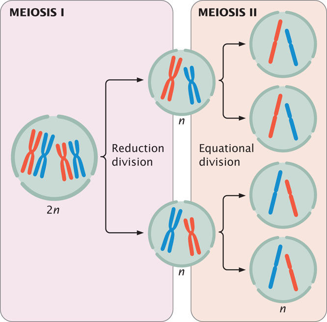
Like mitosis, meiosis is preceded by an interphase stage that includes G1, S, and G2 phases. Meiosis consists of two distinct processes: meiosis I and meiosis II, each of which includes a cell division. The first division, which comes at the end of meiosis I, is termed the reduction division because the number of chromosomes per cell is reduced by half (Figure 2.13). The second division, which comes at the end of meiosis II, is sometimes termed the equational division. The events of meiosis II are similar to those of mitosis. However, meiosis II differs from mitosis in that chromosome number has already been halved in meiosis I, and the cell does not begin with the same number of chromosomes as it does in mitosis (see Figure 2.13).
Meiosis I
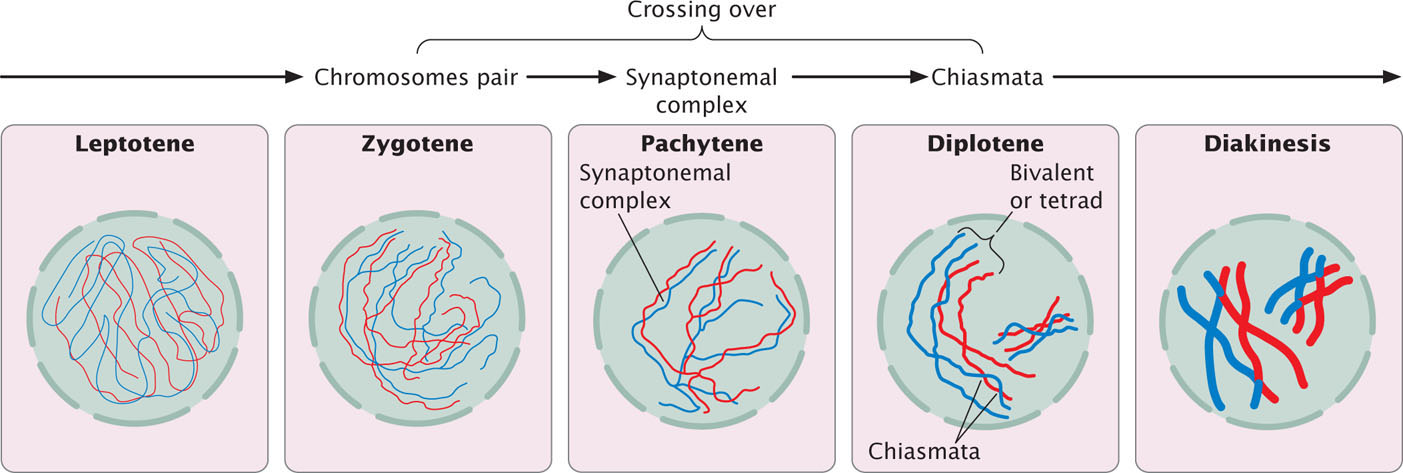
During interphase, the chromosomes are relaxed and visible as diffuse chromatin. Prophase I is a lengthy stage, divided into five substages (Figure 2.14). In leptotene, the chromosomes contract and become visible. In zygotene, the chromosomes continue to condense; homologous chromosomes pair up and begin synapsis, a very close pairing association. Each homologous pair of synapsed chromosomes consists of four chromatids called a bivalent or tetrad. In pachytene, the chromosomes become shorter and thicker, and a three-part synaptonemal complex develops between homologous chromosomes. The function of the synaptonemal complex is unclear, but the chromosomes of many cells deficient in this complex do not separate properly.
29
Crossing over takes place in prophase I, in which homologous chromosomes exchange genetic information. Crossing over generates genetic variation (see Sources of Genetic Variation in Meiosis later in this chapter) and is essential for the proper alignment and separation of homologous chromosomes. The centromeres of the paired chromosomes move apart in diplotene; the two homologs remain attached at each chiasma (plural, chiasmata), which is the result of crossing over. Near the end of prophase I, the nuclear membrane breaks down and the spindle forms, setting the stage for metaphase I. The stages of meiosis are outlined in Figure 2.15.
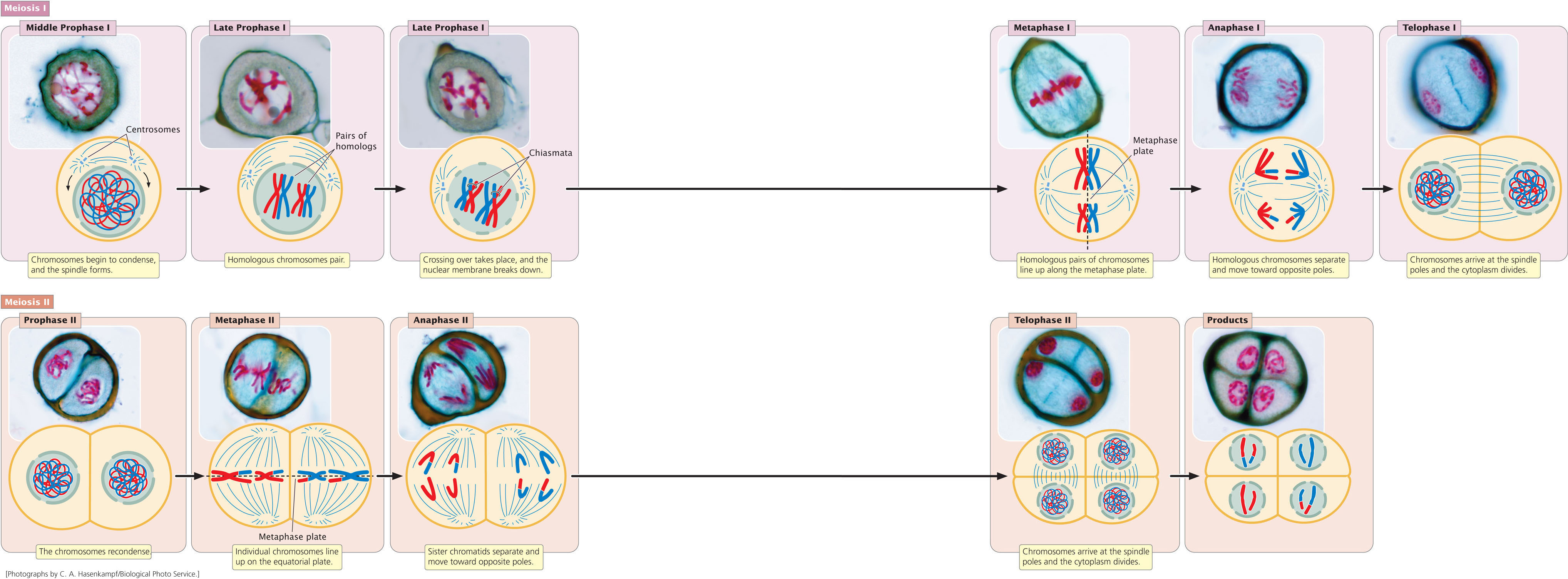
Metaphase I is initiated when homologous pairs of chromosomes align along the metaphase plate (see Figure 2.15). A microtubule from one pole attaches to one chromosome of a homologous pair, and a microtubule from the other pole attaches to the other member of the pair. Anaphase I is marked by the separation of homologous chromosomes. The two chromosomes of a homologous pair are pulled toward opposite poles. Although the homologous chromosomes separate, the sister chromatids remain attached and travel together. In telophase I, the chromosomes arrive at the spindle poles and the cytoplasm divides.
Meiosis II
The period between meiosis I and meiosis II is interkinesis, in which the nuclear membrane re-forms around the chromosomes clustered at each pole, the spindle breaks down, and the chromosomes relax. These cells then pass through prophase II, in which the events of interkinesis are reversed: the chromosomes recondense, the spindle reforms, and the nuclear envelope once again breaks down. In interkinesis in some types of cells, the chromosomes remain condensed, and the spindle does not break down. These cells move directly from cytokinesis into metaphase II, which is similar to metaphase of mitosis: the individual chromosomes line up on the metaphase plate, with the sister chromatids facing opposite poles.
In anaphase II, the kinetochores of the sister chromatids separate and the chromatids are pulled to opposite poles. Each chromatid is now a distinct chromosome. In telophase II, the chromosomes arrive at the spindle poles, a nuclear envelope re-forms around the chromosomes, and the cytoplasm divides. The chromosomes relax and are no longer visible.
The major events of meiosis are summarized in Table 2.2. To examine the details of meiosis and the consequences of its failure, take a look at  Animation 2.2.
| Stage | Major Features |
|---|---|
| Meiosis I | |
| Prophase I | Chromosomes condense, homologous chromosomes synapse, crossing over takes place, the nuclear envelope breaks down, and the mitotic spindle forms. |
| Metaphase I | Homologous pairs of chromosomes line up on the metaphase plate. |
| Anaphase I | The two chromosomes (each with two chromatids) of each homologous pair separate and move toward opposite poles. |
| Telophase I | Chromosomes arrive at the spindle poles. |
| Cytokinesis | The cytoplasm divides to produce two cells, each having half the original number of chromosomes. |
| Interkinesis | In some types of cells, the spindle breaks down, chromosomes relax, and a nuclear envelope re-forms, but no DNA synthesis takes place. |
| Meiosis II | |
| Prophase II* | Chromosomes condense, the spindle forms, and the nuclear envelope disintegrates. |
| Metaphase II | Individual chromosomes line up on the metaphase plate. |
| Anaphase II | Sister chromatids separate and move as individual chromosomes toward the spindle poles. |
| Telophase II | Chromosomes arrive at the spindle poles; the spindle breaks down and a nuclear envelope re-forms. |
| Cytokinesis | The cytoplasm divides. |
| *Only in cells in which the spindle has broken down, chromosomes have relaxed, and the nuclear envelope has re-formed in telophase I. Other types of cells proceed directly to metaphase II after cytokinesis. | |
30
CONCEPTS
Meiosis consists of two distinct processes: meiosis I and meiosis II. Meiosis I includes the reduction division, in which homologous chromosomes separate and chromosome number is reduced by half. In meiosis II (the equational division) chromatids separate.
 CONCEPT CHECK 5
Which of the following events takes place in metaphase I?
- Crossing over
- Chromosomes contract.
- Homologous pairs of chromosomes line up on the metaphase plate.
- Individual chromosomes line up on the metaphase plate.
31
Sources of Genetic Variation in Meiosis
What are the overall consequences of meiosis? First, meiosis comprises two divisions; so each original cell produces four cells (there are exceptions to this generalization, as, for example, in many female animals; see Figure 2.20b). Second, chromosome number is reduced by half; so cells produced by meiosis are haploid. Third, cells produced by meiosis are genetically different from one another and from the parental cell. Genetic differences among cells result from two processes that are unique to meiosis: crossing over and the random separation of homologous chromosomes.
Crossing Over
Crossing over, which takes place in prophase I, refers to the exchange of genetic material between nonsister chromatids (chromatids from different homologous chromosomes). Evidence from yeast suggests that crossing over is initiated in zygotene, before the synaptonemal complex develops, and is not completed until near the end of prophase I (see Figure 2.14). In other organisms, crossing over is initiated after the formation of the synaptonemal complex and, in yet others, there is no synaptonemal complex.
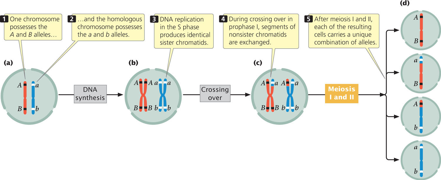
After crossing over has taken place, the sister chromatids are no longer identical. Crossing over is the basis for intrachromosomal recombination, creating new combinations of alleles on a chromatid. To see how crossing over produces genetic variation, consider two pairs of alleles, which we will abbreviate Aa and Bb. Assume that one chromosome possesses the A and B alleles and its homolog possesses the a and b alleles (Figure 2.16a). When DNA is replicated in the S phase, each chromosome duplicates, and so the resulting sister chromatids are identical (Figure 2.16b).
32
In the process of crossing over, there are breaks in the DNA strands and the breaks are repaired in such a way that segments of nonsister chromatids are exchanged (Figure 2.16c). The molecular basis of this process will be described in more detail in Chapter 12. The important thing here is that, after crossing over has taken place, the two sister chromatids are no longer identical: one chromatid has alleles A and B, whereas its sister chromatid (the chromatid that underwent crossing over) has alleles a and B. Likewise, one chromatid of the other chromosome has alleles a and b, and the other chromatid has alleles A and b. Each of the four chromatids now carries a unique combination of alleles: A B, a B, A b, and a b. Eventually, the two homologous chromosomes separate, each going into a different cell. In meiosis II, the two chromatids of each chromosome separate, and thus each of the four cells resulting from meiosis carries a different combination of alleles (Figure 2.16d). You can see how crossing over affects genetic variation by viewing Animation 2.3.
Random Separation of Homologous Chromosomes
The second process of meiosis that contributes to genetic variation is the random distribution of chromosomes in anaphase I after their random alignment in metaphase I. To illustrate this process, consider a cell with three pairs of chromosomes, I, II, and III (Figure 2.17a). One chromosome of each pair is maternal in origin (Im, IIm, and IIIm); the other is paternal in origin (Ip, IIp, and IIIp). The chromosome pairs line up in the center of the cell in metaphase I and, in anaphase I the chromosomes of each homologous pair separate.
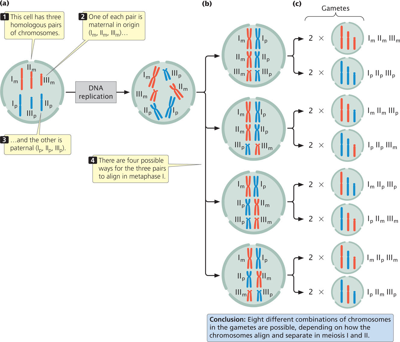
How each pair of homologs aligns and separates is random and independent of how other pairs of chromosomes align and separate (Figure 2.17b). By chance, all the maternal chromosomes might migrate to one side, with all the paternal chromosomes migrating to the other. After division, one cell would contain chromosomes Im, IIm, and IIIm, and the other, Ip, IIp, and IIIp. Alternatively, the Im, IIm, and IIIp chromosomes might move to one side, and the Ip, IIp, and IIIm chromosomes to the other. The different migrations would produce different combinations of chromosomes in the resulting cells (Figure 2.17c). There are four ways in which a diploid cell with three pairs of chromosomes can divide, producing a total of eight different combinations of chromosomes in the gametes. In general, the number of possible combinations is 2n, where n equals the number of homologous pairs. As the number of chromosome pairs increases, the number of combinations quickly becomes very large. In humans, who have 23 pairs of chromosomes, there are 223, or 8,388,608, different combinations of chromosomes possible from the random separation of homologous chromosomes. You can explore the random distribution of chromosomes by viewing Animation 2.3. The genetic consequences of this process, termed independent assortment, will be explored in more detail in Chapter 3.
In summary, crossing over shuffles alleles on the same chromosome into new combinations, whereas the random distribution of maternal and paternal chromosomes shuffles alleles on different chromosomes into new combinations. Together, these two processes are capable of producing tremendous amounts of genetic variation among the cells resulting from meiosis. ![]() TRY PROBLEMS 33 AND 34
TRY PROBLEMS 33 AND 34
CONCEPTS
The two mechanisms that produce genetic variation in meiosis are crossing over and the random distribution of maternal and paternal chromosomes.
33
CONNECTING CONCEPTS: Mitosis and Meiosis Compared
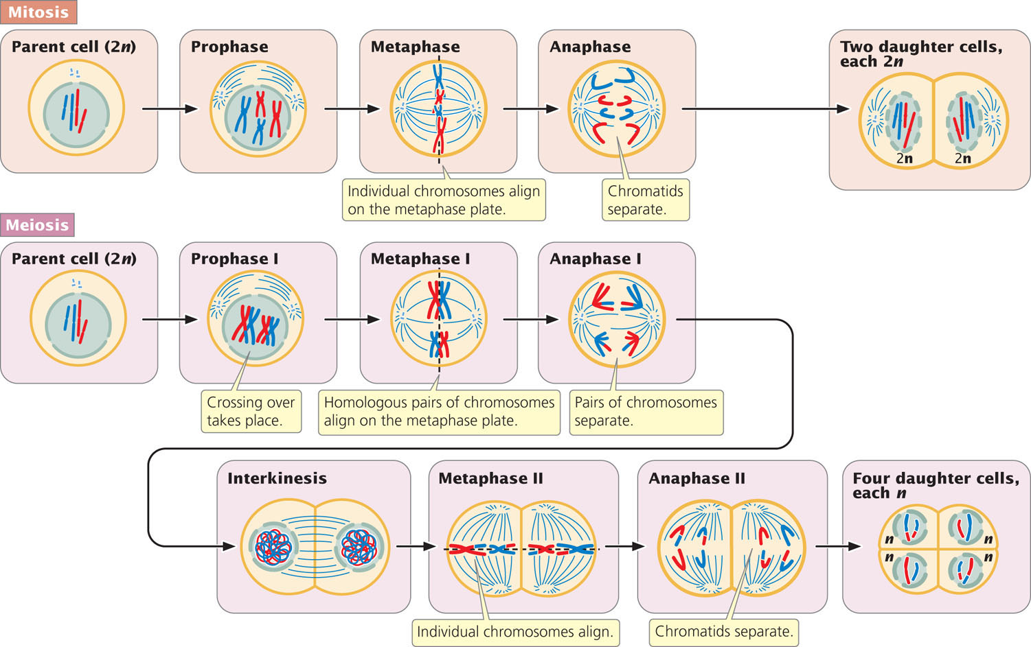
Now that we have examined the details of mitosis and meiosis, let’s compare the two processes (Figure 2.18 and Table 2.3). In both mitosis and meiosis, the chromosomes contract and become visible; both processes include the movement of chromosomes toward the spindle poles, and both are accompanied by cell division. Beyond these similarities, the processes are quite different.
| Event | Mitosis | Meiosis I | Meiosis II |
|---|---|---|---|
| Cell division | Yes | Yes | Yes |
| Chromosome reduction | No | Yes | No |
| Genetic variation produced | No | Yes | No |
| Crossing over | No | Yes | No |
| Random distribution of maternal and paternal chromosomes | No | Yes | No |
| Metaphase | Individual chromosomes line up | Homologous pairs line up | Individual chromosomes line up |
| Anaphase | Chromatids separate | Homologous chromosomes separate | Chromatids separate |
Mitosis results in a single cell division and usually produces two daughter cells. Meiosis, in contrast, comprises two cell divisions and usually produces four cells. In diploid cells, homologous chromosomes are present before both meiosis and mitosis, but the pairing of homologs takes place only in meiosis.
Another difference is that, in meiosis, chromosome number is reduced by half as a consequence of the separation of homologous pairs of chromosomes in anaphase I, but no chromosome reduction takes place in mitosis. Furthermore, meiosis is characterized by two processes that produce genetic variation: crossing over (in prophase I), and the random distribution of maternal and paternal chromosomes (in anaphase I). There are normally no equivalent processes in mitosis.
Mitosis and meiosis also differ in the behavior of chromosomes in metaphase and anaphase. In metaphase I of meiosis, homologous pairs of chromosomes line up on the metaphase plate, whereas individual chromosomes line up on the metaphase plate in metaphase of mitosis (and in metaphase II of meiosis). In anaphase I of meiosis, paired chromosomes separate and migrate toward opposite spindle poles, each chromosome possessing two chromatids attached at the centromere. In contrast, in anaphase of mitosis (and in anaphase II of meiosis), sister chromatids separate, and each chromosome that moves toward a spindle pole is unreplicated. ![]() TRY PROBLEMS 28 AND 29
TRY PROBLEMS 28 AND 29
The Separation of Sister Chromatids and Homologous Chromosomes
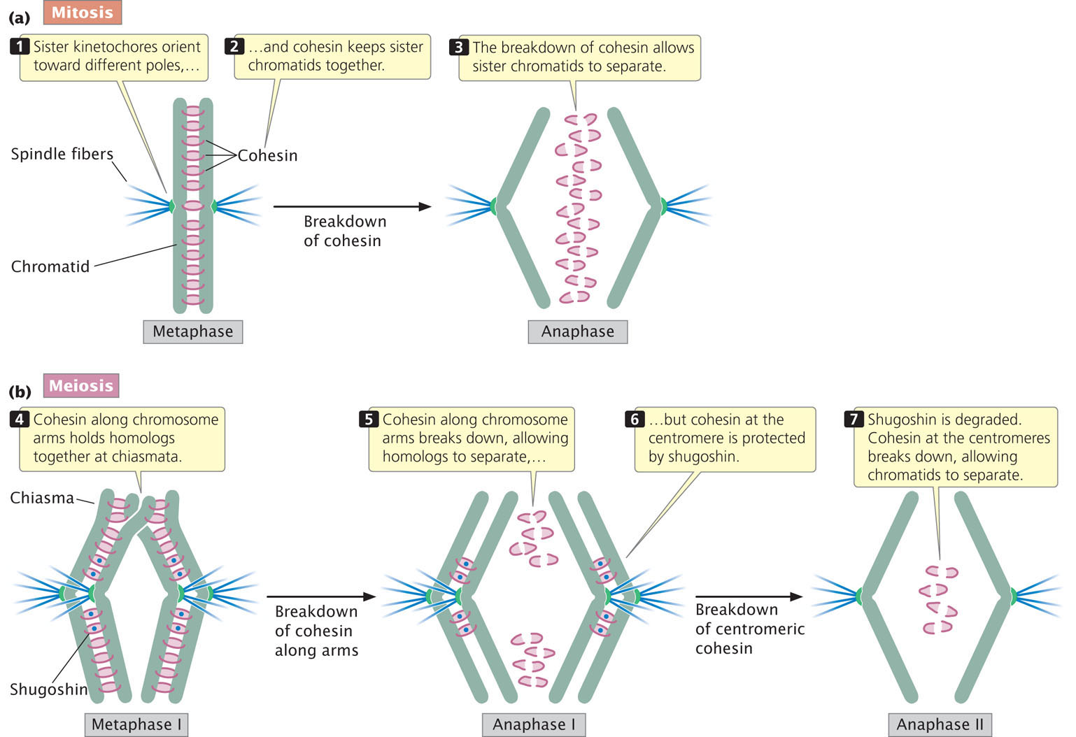
In recent years, some of the molecules required for the joining and separation of chromatids and homologous chromosomes have been identified. Cohesin, a protein that holds the chromatids together, is key to the behavior of chromosomes in mitosis and meiosis (Figure 2.19a). The sister chromatids are held together by cohesin, which is established in the S phase and persists through G2 and early mitosis. In anaphase of mitosis, cohesin along the entire length of the chromosome is broken down by an enzyme called separase, allowing the sister chromatids to separate.
34
As we have seen, mitosis and meiosis differ fundamentally in the behavior of chromosomes in anaphase (see Figure 2.18). Why do homologs separate in anaphase I of meiosis, whereas chromatids separate in anaphase of mitosis and anaphase II of meiosis? It is important to note that the forms of cohesin used in mitosis and meiosis differ. At the beginning of meiosis, the meiosis-specific cohesin is found along the entire length of a chromosome’s arms (Figure 2.19b). The cohesin also acts on the chromosome arms of homologs at the chiasmata, tethering two homologs together at their ends.
In anaphase I, cohesin along the chromosome arms is broken, allowing the two homologs to separate. However, cohesin at the centromere is protected by a protein called shugoshin, which means “guardian spirit” in Japanese. Because of this protective action by shugoshin, the centromeric cohesin remains intact and prevents the separation of the two sister chromatids during anaphase I of meiosis. Shugoshin is subsequently degraded. At the end of metaphase II, the centromeric cohesin—no longer protected by shugoshin—breaks down, allowing the sister chromatids to separate in anaphase II, just as they do in mitosis (Figure 2.19b). ![]() TRY PROBLEM 30
TRY PROBLEM 30
35
CONCEPTS
Cohesin holds sister chromatids together during the early part of mitosis. In anaphase, cohesin breaks down, allowing sister chromatids to separate. In meiosis, cohesin is protected at the centromeres during anaphase I, and so homologous chromosomes, but not sister chromatids, separate in meiosis I. The breakdown of centromeric cohesin allows sister chromatids to separate in anaphase II of meiosis.
CONCEPT CHECK 6
How does shugoshin affect sister chromatids in meiosis I and meiosis II?
Meiosis in the Life Cycles of Animals and Plants
The overall result of meiosis is four haploid cells that are genetically variable. Let’s now see where meiosis fits into the life cycles of a multicellular animal and a multicellular plant.
Meiosis in Animals
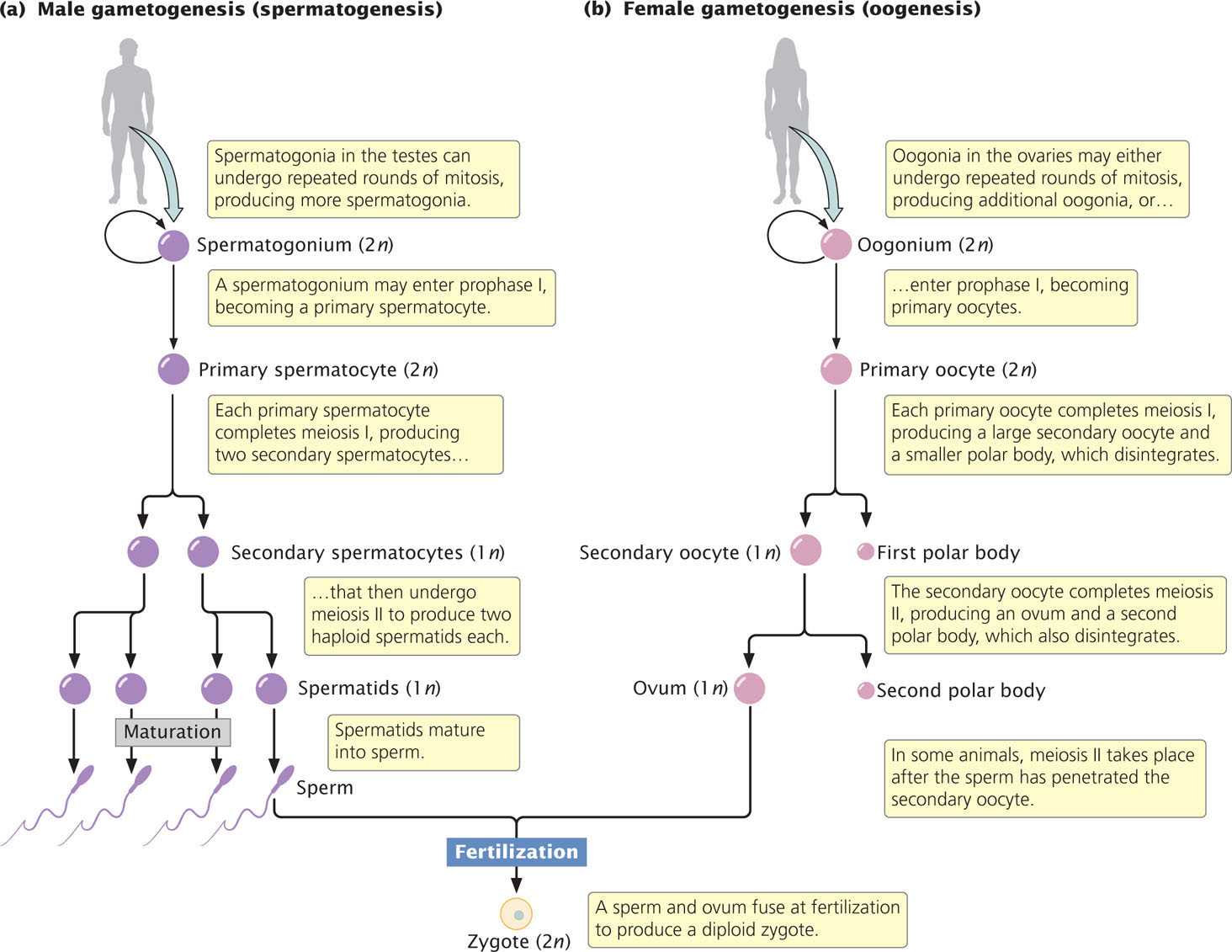
The production of gametes in a male animal, a process called spermatogenesis, takes place in the testes. There, diploid primordial germ cells divide mitotically to produce diploid cells called spermatogonia (Figure 2.20a). Each spermatogonium can undergo repeated rounds of mitosis, giving rise to numerous additional spermatogonia. Alternatively, a spermatogonium can initiate meiosis and enter into prophase I. Now called a primary spermatocyte, the cell is still diploid because the homologous chromosomes have not yet separated. Each primary spermatocyte completes meiosis I, giving rise to two haploid secondary spermatocytes that then undergo meiosis II, with each producing two haploid spermatids. Thus, each primary spermatocyte produces a total of four haploid spermatids, which mature and develop into sperm.
36
The production of gametes in a female animal, a process called oogenesis, begins much as spermatogenesis does. Within the ovaries, diploid primordial germ cells divide mitotically to produce oogonia (Figure 2.20b). Like spermatogonia, oogonia can undergo repeated rounds of mitosis or they can enter into meiosis. When they enter prophase I, these still-diploid cells are called primary oocytes. Each primary oocyte completes meiosis I and divides.
At this point, the process of oogenesis begins to differ from that of spermatogenesis. In oogenesis, cytokinesis is unequal: most of the cytoplasm is allocated to one of the two haploid cells, the secondary oocyte. The smaller cell, which contains half of the chromosomes but only a small part of the cytoplasm, is called the first polar body; it may or may not divide further. The secondary oocyte completes meiosis II, and, again, cytokinesis is unequal—most of the cytoplasm passes into one of the cells. The larger cell, which acquires most of the cytoplasm, is the ovum, the mature female gamete. The smaller cell is the second polar body. Only the ovum is capable of being fertilized, and the polar bodies usually disintegrate. Oogenesis, then, produces a single mature gamete from each primary oocyte.
In mammals, oogenesis differs from spermatogenesis in another way. The formation of sperm takes place continuously in a male throughout his adult reproductive life. The formation of female gametes, however, is often a discontinuous process, and may take place over a period of years. Oogenesis begins before birth; at this time, oogonia initiate meiosis and give rise to primary oocytes. Meiosis is then arrested, stalled in prophase I. A female is born with primary oocytes arrested in prophase I. In humans, this period of suspended animation may last 30 or 40 years. Before ovulation, rising hormone levels stimulate one or more of the primary oocytes to recommence meiosis. The first division of meiosis is completed and a secondary oocyte is ovulated from the ovary. In humans and many other species, the second division of meiosis is then delayed until contact with the sperm. When the sperm penetrates the outer layer of the secondary oocyte, the second meiotic division takes place, the second polar body is extruded from the egg, and the nuclei of the sperm and newly formed ovum fuse, giving rise to the zygote.
37
CONCEPTS
In the testes, a diploid spermatogonium undergoes meiosis, producing a total of four haploid sperm cells. In the ovary, a diploid oogonium undergoes meiosis to produce a single large ovum and smaller polar bodies that normally disintegrate.
CONCEPT CHECK 7
A secondary spermatocyte has 12 chromosomes. How many chromosomes will be found in the primary spermatocyte that gave rise to it?
- 6
- 12
- 18
- 24
Meiosis in Plants
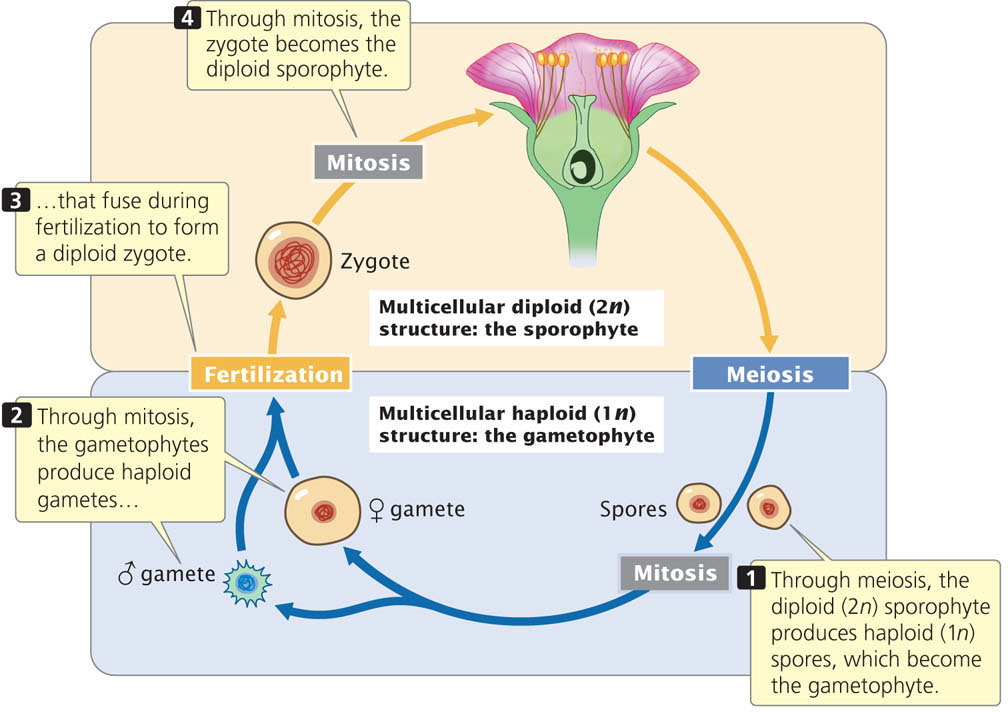
Most plants have a complex life cycle that includes two distinct structures (generations): a multicellular diploid sporophyte and a multicellular haploid gametophyte. These two generations alternate; the sporophyte produces haploid spores through meiosis, and the gametophyte produces haploid gametes through mitosis (Figure 2.21). This type of life cycle is sometimes called alternation of generations. In this cycle, the immediate products of meiosis are called spores, not gametes; the spores undergo one or more mitotic divisions to produce gametes. Although the terms used for this process are somewhat different from those commonly used in regard to animals (and from some of those employed so far in this chapter), the processes in plants and animals are basically the same: in both, meiosis leads to a reduction in chromosome number, producing haploid cells.
 ; male,
  ).
).
In flowering plants, the sporophyte is the obvious, vegetative part of the plant; the gametophyte consists of only a few haploid cells within the sporophyte. The flower, which is part of the sporophyte, contains the reproductive structures. In some plants, both male and female reproductive structures are found in the same flower; in other plants, they exist in different flowers. In either case, the male part of the flower, the stamen, contains diploid reproductive cells called microsporocytes, each of which undergoes meiosis to produce four haploid microspores (Figure 2.22a). Each microspore divides mitotically, producing an immature pollen grain consisting of two haploid nuclei. One of these nuclei, called the tube nucleus, directs the growth of a pollen tube. The other, termed the generative nucleus, divides mitotically to produce two sperm cells. The pollen grain, with its two haploid nuclei, is the male gametophyte.
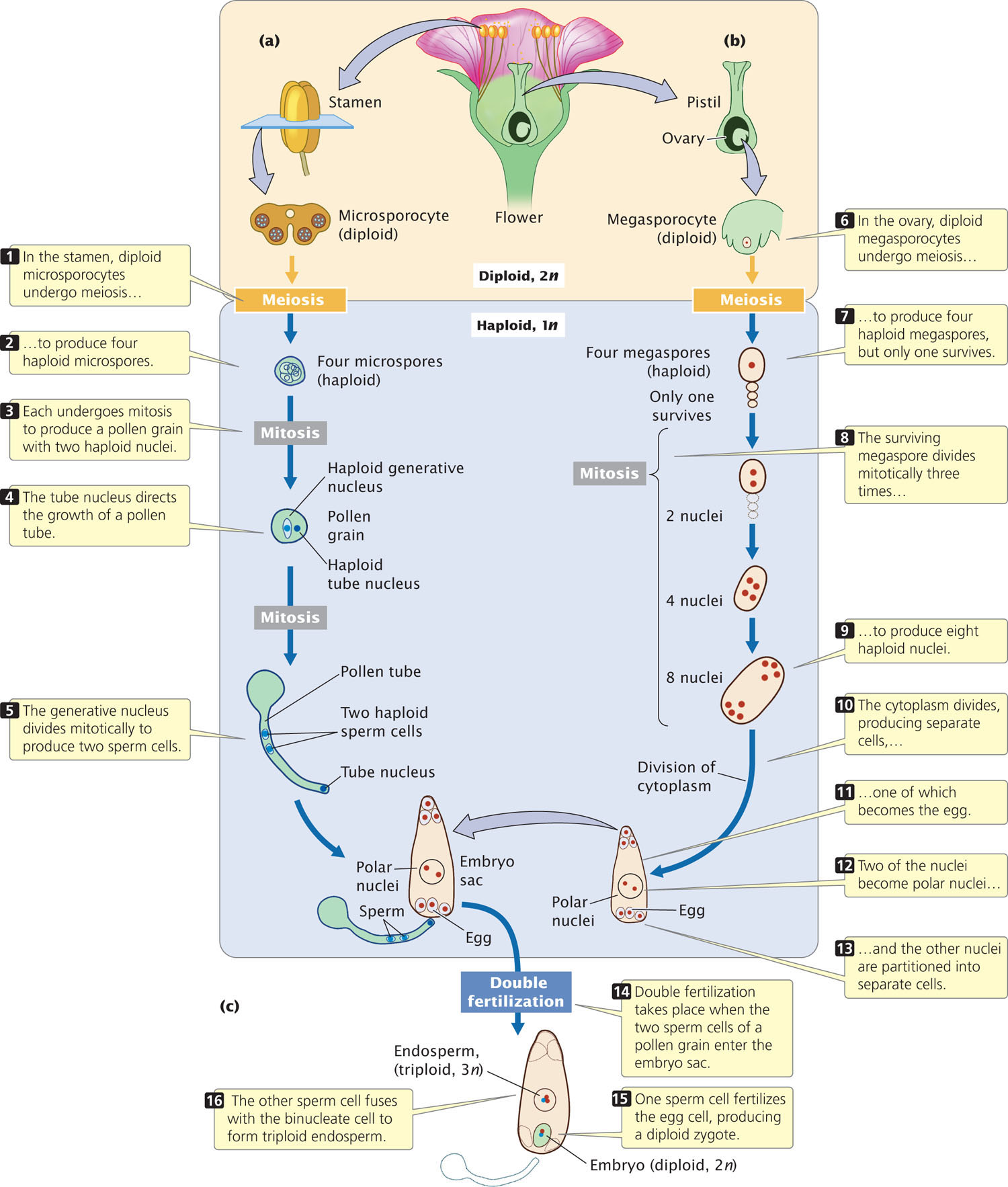
The female part of the flower, the ovary, contains diploid cells called megasporocytes, each of which undergoes meiosis to produce four haploid megaspores (Figure 2.22b), only one of which survives. The nucleus of the surviving megaspore divides mitotically three times, producing a total of eight haploid nuclei that make up the female gametophyte, otherwise known as the embryo sac. Division of the cytoplasm then produces separate cells, one of which becomes the egg.
When the plant flowers, the stamens open and release pollen grains. Pollen lands on a flower’s stigma—a sticky platform that sits on top of a long stalk called the style. At the base of the style is the ovary. If a pollen grain germinates, it grows a tube down the style into the ovary. The two sperm cells pass down this tube and enter the embryo sac (Figure 2.22c). One of the sperm cells fertilizes the egg cell, producing a diploid zygote, which develops into an embryo. The other sperm cell fuses with two nuclei enclosed in a single cell, giving rise to a 3n (triploid) endosperm, which stores food that will be used later by the embryonic plant. These two fertilization events are termed double fertilization.
38
39
CONCEPTS
In the stamen of a flowering plant, meiosis produces haploid microspores that divide mitotically to produce haploid sperm in a pollen grain. Within the ovary, meiosis produces four haploid megaspores, only one of which divides mitotically three times to produce eight haploid nuclei. After pollination, one sperm fertilizes the egg cell, producing a diploid zygote; the other fuses with two nuclei to form the endosperm.
CONCEPT CHECK 8
Which structure is diploid?
- Microspore
- Megaspore
- Egg
- Microsporocyte
We have now examined the place of meiosis in the sexual cycle of two organisms, a typical multicellular animal and a flowering plant. These cycles are just two of the many variations found among eukaryotic organisms. Although the cellular events that produce reproductive cells in plants and animals differ in the number of cell divisions, the number of haploid gametes produced, and the relative size of the final products, the overall result is the same: meiosis gives rise to haploid, genetically variable cells that then fuse during fertilization to produce diploid progeny. ![]() TRY PROBLEMS 36 AND 38
TRY PROBLEMS 36 AND 38