8.1 Chromosome Mutations Include Rearrangements, Aneuploids, and Polyploids
Before we consider the different types of chromosome mutations, their effects, and how they arise, we will review the basics of chromosome structure.
Chromosome Morphology
Each functional chromosome has a centromere, to which spindle fibers attach, and two telomeres, which stabilize the chromosome (see Figure 2.7). Chromosomes are classified into four basic types:
- 1. Metacentric. The centromere is located approximately in the middle, and so the chromosome has two arms of equal length.
- 2. Submetacentric. The centromere is displaced toward one end, creating a long arm and a short arm. (On human chromosomes, the short arm is designated by the letter p and the long arm by the letter q.)
- 3. Acrocentric. The centromere is near one end, producing a long arm and a knob, or satellite, at the other end.
- 4. Telocentric. The centromere is at or very near the end of the chromosome (see Figure 2.8).
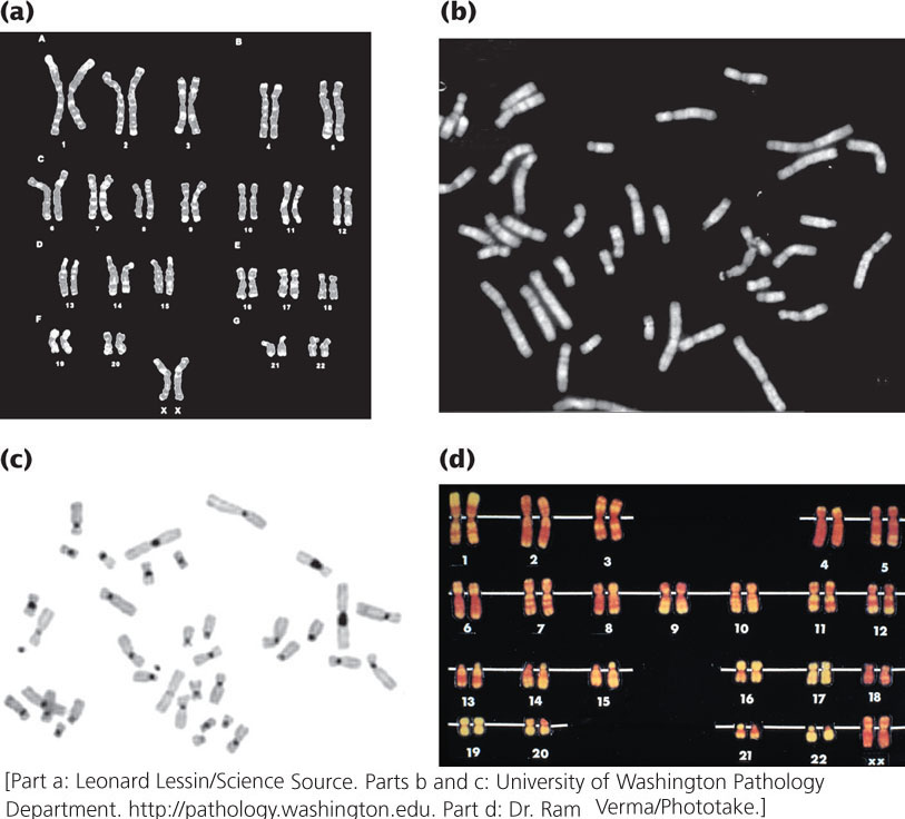
The complete set of chromosomes possessed by an organism is called its karyotype and is usually presented as a picture of metaphase chromosomes lined up in descending order of their size (Figure 8.1). Karyotypes are prepared from actively dividing cells, such as white blood cells, bone-marrow cells, or cells from meristematic tissues of plants. After treatment with a chemical (such as colchicine) that prevents them from entering anaphase, the cells are chemically preserved, spread on a microscope slide, stained, and photographed. The photograph is then enlarged, and the individual chromosomes are cut out and arranged in a karyotype. For human chromosomes, karyotypes are often routinely prepared by automated machines, which scan a slide using a video camera attached to a microscope, looking for chromosome spreads. When a spread has been located, the camera takes a picture of the chromosomes, the image is digitized, and the chromosomes are sorted and arranged electronically by a computer.
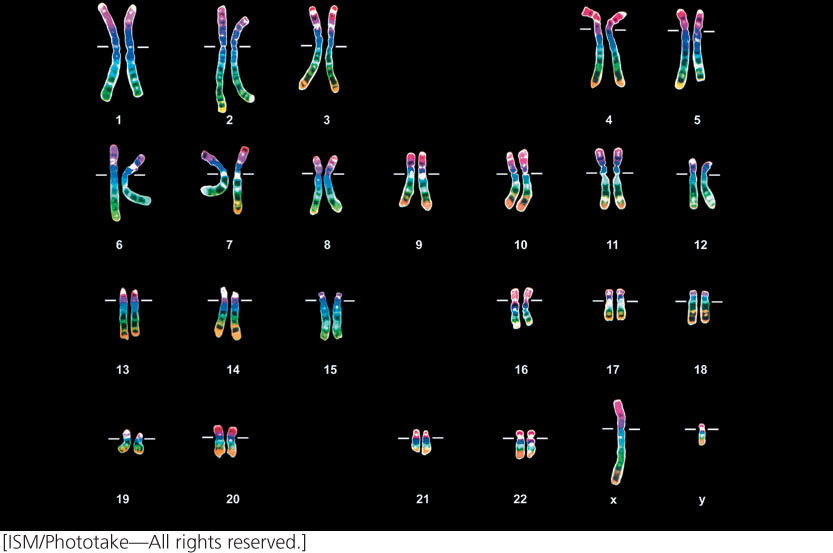
Preparation and staining techniques help to distinguish among chromosomes of similar size and shape. For instance, special preparation and staining of chromosomes with a special dye called Giemsa reveals G bands, which distinguish areas of DNA that are rich in adenine–thymine (A–T) base pairs (Figure 8.2a) see Chapter 10). Q bands (Figure 8.2b) are revealed by staining chromosomes with quinacrine mustard and viewing the chromosomes under ultraviolet light; variation in the brightness of Q bands results from differences in the relative amounts of cytosine–guanine (C–G) and adenine–thymine base pairs. Other techniques reveal C bands (Figure 8.2c), which are regions of DNA occupied by centromeric heterochromatin, and R bands (Figure 8.2d), which are rich in cytosine–guanine base pairs.
211
Types of Chromosome Mutations
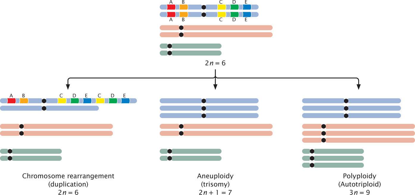
Chromosome mutations can be grouped into three basic categories: chromosome rearrangements, aneuploids, and polyploids (Figure 8.3). Chromosome rearrangements alter the structure of chromosomes; for example, a piece of a chromosome might be duplicated, deleted, or inverted. In aneuploidy, the number of chromosomes is altered: one or more individual chromosomes are added or deleted. In polyploidy, one or more complete sets of chromosomes are added. A polyploid is any organism that has more than two sets of chromosomes (3n, 4n, 5n, or more).
212