Viewing DNA Fragments
After the completion of a restriction reaction, a number of questions arise. Did the restriction enzyme cut the DNA? Into how many fragments was the DNA cut? What are the sizes of the resulting fragments? Gel electrophoresis provides us with a means of answering these questions.
Electrophoresis is a standard technique for separating molecules on the basis of their size and electrical charge. There are a number of different types of electrophoresis; to separate DNA molecules, gel electrophoresis is used. A porous gel is often made from agarose (a polysaccharide isolated from seaweed), which is melted in a buffer solution and poured into a plastic mold. As it cools, the agarose solidifies, making a gel that looks something like stiff gelatin.
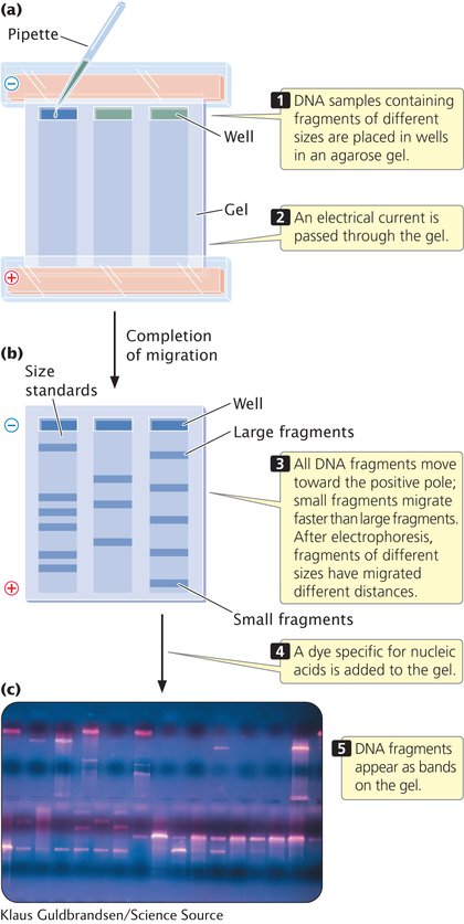
Small wells are made at one end of the gel to hold solutions of DNA fragments (Figure 14.2a), and an electrical current is passed through the gel. Because the phosphate group on each DNA nucleotide carries a negative charge, the DNA fragments migrate toward the positive end of the gel. During this migration, the porous gel acts as a sieve, separating the DNA fragments by size. Small DNA fragments migrate more rapidly than large ones do, so over time, the fragments separate on the basis of their size. Typically, DNA fragments of known length (a marker sample) are placed in one of the wells. By comparing the migration distance of the unknown fragments with the distance traveled by the marker sample, one can determine the approximate size of the unknown fragments (Figure 14.2b).
376
The DNA fragments are still too small to see, so the problem of visualizing the DNA needs to be addressed. Visualization can be accomplished in several ways. The simplest procedure is to stain the gel with a dye specific for nucleic acids, such as ethidium bromide, which wedges itself tightly (intercalates) between the bases of DNA and fluoresces orange when exposed to UV light, producing brilliant orange bands on the gel (Figure 14.2c).
Alternatively, DNA fragments can be visualized by adding a label to the DNA before it is placed in the gel. For example, chemical labels can be detected by adding antibodies or other substances that carry a dye and will attach to the relevant DNA, which can then be visualized directly.  TRY PROBLEM 20
CONCEPTS
DNA fragments can be separated, and their sizes determined, with the use of gel electrophoresis. The fragments can be viewed by using a dye that is specific for nucleic acids or by labeling the fragments with a chemical tag.
 CONCEPT CHECK 2
DNA fragments that are 500 bp, 1000 bp, and 2000 bp in length are separated by gel electrophoresis. Which fragment will migrate farthest in the gel?
The 2000-
bp fragment. The 1000-
bp fragment. The 500-
bp fragment. All will migrate equal distances.
c