DNA as the Source of Genetic Information
While chemists were working out the structure of DNA, biologists were attempting to identify the source of genetic information. Mendel identified the basic rules of heredity in 1866, but he had no idea about the physical nature of hereditary information. By the early 1900s, biologists had concluded that genes reside on chromosomes, which were known to contain both DNA and protein. Two sets of experiments, one conducted on bacteria and the other on viruses, provided pivotal evidence that DNA, rather than protein, was the genetic material.
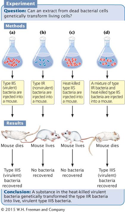
THE DISCOVERY OF THE TRANSFORMING PRINCIPLE The first clue that DNA was the carrier of hereditary information came with the demonstration that DNA was responsible for a phenomenon called transformation (see Chapter 7). This phenomenon was first observed in 1928 by Fred Griffith, an English physician whose special interest was the bacterium that causes pneumonia, Streptococcus pneumoniae. Griffith had succeeded in isolating several different strains of S. pneumoniae (type I, II, III, and so forth). In the virulent (disease-
Griffith observed that small amounts of living type IIIS bacteria injected into mice caused the mice to develop pneumonia and die; when he examined the dead mice, he found large amounts of type IIIS bacteria in their blood (Figure 8.1a). When Griffith injected type IIR bacteria into mice, the mice lived, and no bacteria were recovered from their blood (Figure 8.1b). Griffith knew that boiling killed all bacteria and destroyed their virulence; when he injected large amounts of heat-
The results of these experiments were not unusual. However, Griffith got a surprise when he injected his mice with a small amount of living type IIR bacteria along with a large amount of heat-
210
Griffith concluded that the type IIR bacteria had somehow been transformed, acquiring the genetic virulence of the dead type IIIS bacteria, and that this transformation had produced a permanent genetic change in the bacteria. Although Griffith didn’t understand the nature of transformation, he theorized that some substance in the polysaccharide coat of the dead bacteria might be responsible. He called this substance the transforming principle.  TRY PROBLEM 19
IDENTIFICATION OF THE TRANSFORMING PRINCIPLE At the time of Griffith’s report, Oswald Avery was a microbiologist at the Rockefeller Institute. At first, Avery was skeptical of Griffith’s results, but after other microbiologists successfully repeated Griffith’s experiments with other bacteria, Avery set out to understand the nature of the transforming principle.
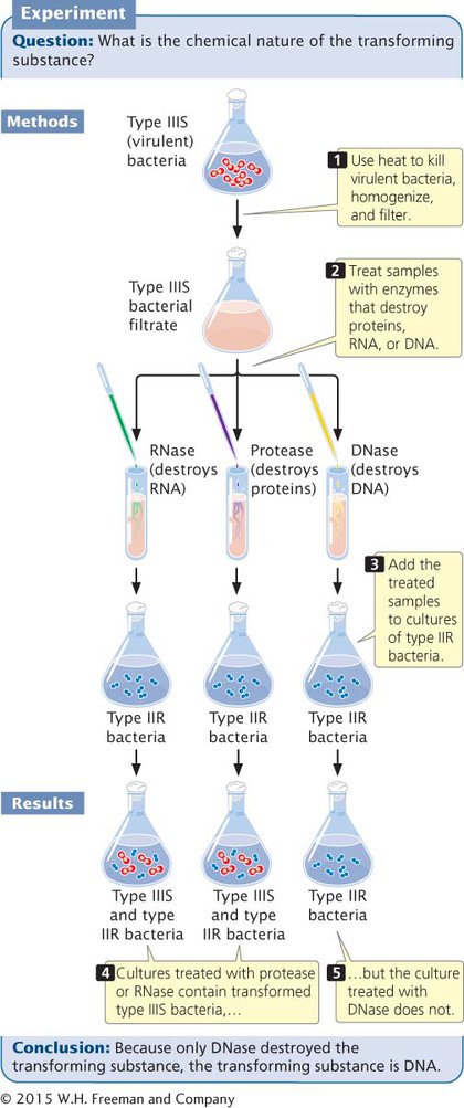
After 10 years of research, Avery, Colin MacLeod, and Maclyn McCarty succeeded in isolating and partially purifying the transforming substance. They showed that it had a chemical composition closely matching that of DNA and quite different from that of proteins. Enzymes such as trypsin and chymotrypsin, known to break down proteins, had no effect on the transforming substance. Ribonuclease, an enzyme that destroys RNA, also had no effect. Enzymes capable of destroying DNA, however, eliminated the biological activity of the transforming substance (Figure 8.2). Avery, MacLeod, and McCarty showed that the transforming substance precipitated at about the same rate as purified DNA and that it absorbed ultraviolet light at the same wavelengths as DNA. These results, published in 1944, provided compelling evidence that the transforming principle—
211
CONCEPTS
The process of transformation indicates that some substance—
 CONCEPT CHECK 2
If Avery, MacLeod, and McCarty had found that samples of heat-
Protease carries out transformation.
RNA and DNA are the genetic materials.
Protein is the genetic material.
RNase and DNase are necessary for transformation.
c
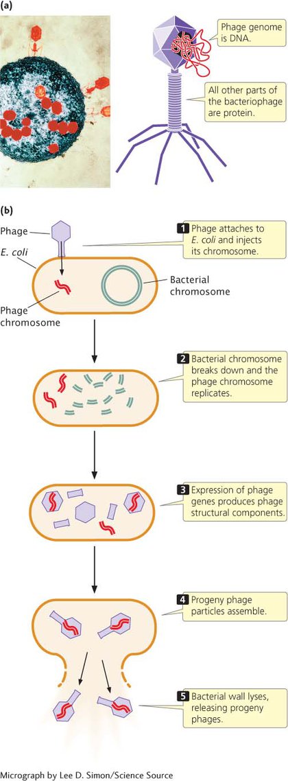
THE HERSHEY–CHASE EXPERIMENT A second piece of evidence that indicated DNA was the genetic material resulted from a study of the T2 bacteriophage conducted by Alfred Hershey and Martha Chase. The T2 bacteriophage infects the bacterium Escherichia coli (Figure 8.3a). As we saw in Chapter 7, a phage reproduces by attaching to the outer wall of a bacterial cell and injecting its DNA into the cell, where it replicates and directs the cell to synthesize phage protein. The phage DNA becomes encapsulated within the proteins, producing progeny phages that lyse (break open) the cell and escape (Figure 8.3b).
At the time of the Hershey–Chase study (their paper was published in 1952), biologists did not understand exactly how phages reproduce. What they did know was that the T2 phage is approximately 50% protein and 50% DNA, that a phage infects a cell by first attaching to the cell wall, and that progeny phages are ultimately produced within the cell. Because the progeny carry the same traits as the infecting phage, they knew that genetic material from the infecting phage must be transmitted to the progeny, but how this transmission takes place was unknown.
212
Hershey and Chase designed a series of experiments to determine whether the phage protein or the phage DNA is transmitted in phage reproduction. To follow the fates of protein and DNA, they used radioactive forms, or isotopes, of phosphorus and sulfur. A radioactive isotope can be used as a tracer to identify the location of a specific molecule because any molecule containing the isotope will be radioactive and therefore easily detected. DNA contains phosphorus, but not sulfur. Thus, Hershey and Chase used a radioactive isotope of phosphorus (32P) to follow phage DNA during reproduction. Protein contains sulfur, but not phosphorus, so they used a radioactive isotope of sulfur (35S) to follow the protein.
Hershey and Chase grew one batch of E. coli in a medium containing 32P and infected the bacteria with T2 phage so that all the new phages would have DNA labeled with 32P (Figure 8.4). They grew a second batch of E. coli in a medium containing 35S and infected these bacteria with T2 phage so that all these new phages would have proteins labeled with 35S. Hershey and Chase then infected separate batches of unlabeled E. coli with the 35S-
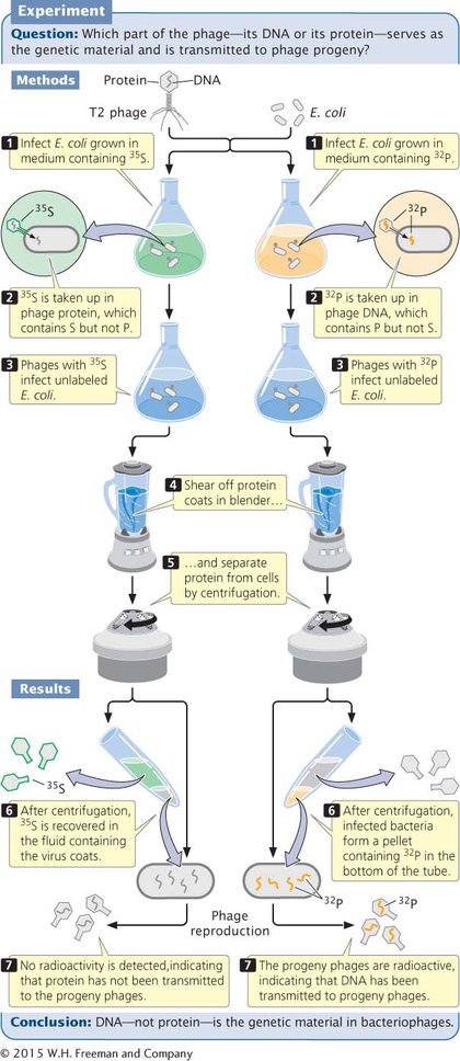
When phages labeled with 35S infected the bacteria, most of the radioactivity was detected in the protein coats and little was detected in the cells. Furthermore, when new phages emerged from the cells, they contained almost no 35S (see Figure 8.4). This result indicated that the protein component of a phage does not enter the cell and is not transmitted to progeny phages.
In contrast, when Hershey and Chase infected bacteria with 32P- TRY PROBLEM 36
CONCEPTS
Using radioactive isotopes, Hershey and Chase traced the movement of DNA and protein during phage infection of bacteria. They demonstrated that DNA, not protein, enters the bacterial cell during phage reproduction and that only DNA is passed on to progeny phages.
CONCEPT CHECK 3
Could Hershey and Chase have used a radioactive isotope of carbon instead of 32P? Why or why not?
No: carbon is found in both protein and nucleic acid.