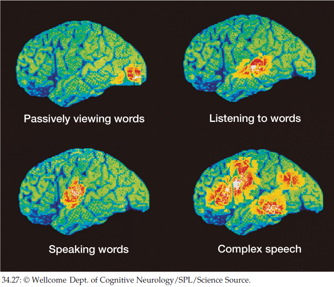
Figure 34.27: Imaging Techniques Reveal Active Parts of the Brain Positron emission tomography (PET) scanning reveals the brain regions activated by various aspects of language use. Radioactively labeled glucose is given to the subject. Brain areas take up radioactivity in proportion to their metabolic use of glucose. The PET scan visualizes levels of radioactivity in specific brain regions when a particular activity is performed. The red and yellow areas are the most active, green and blue the least active.