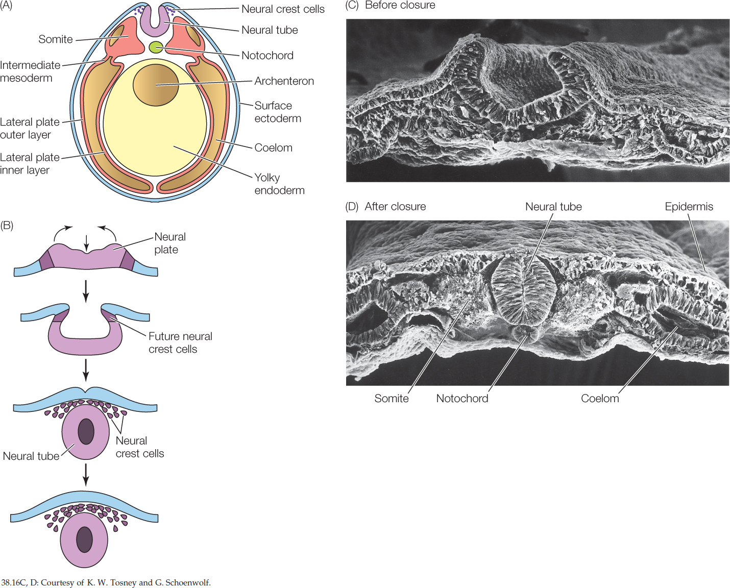
Figure 38.16: Neurulation and Differentiation of Mesoderm in Vertebrates (A) This illustration of a frog egg shows how the neural folds draw together and close, releasing neural crest cells. The mesoderm extends between the ectoderm and endoderm. The three divisions of trunk mesoderm—somite, intermediate mesoderm, and lateral plate mesoderm—are apparent. (B) At the start of vertebrate neurulation, the ectoderm of the neural plate is flat. The neural plate invaginates and folds, forming a tube. Neural crest cells are released. (C,D) Scanning electron micrograph of the neural tube in a chick, before and after closure.