Concept 7.2: Both Binary Fission and Mitosis Produce Genetically Identical Cells
Cell division by either binary fission or mitosis produces two genetically identical cells. This is the basis of asexual reproduction in single-celled organisms: prokaryotes reproduce by binary fission, and single-celled eukaryotes reproduce by mitosis. In multicellular organisms, mitosis is a way to build tissues and organs during development and to repair damaged tissues once development is complete.
In order for any cell to divide, the following events must occur:
- There must be one or more reproductive signals. These signals initiate cell division and may originate from either inside or outside the cell.
- DNA replication (i.e., replication of the genetic material) must occur so that each of the two new cells will have a full complement of genes to complete cell functions.
- The cell must distribute the replicated DNA to each of the two new cells. This process is called DNA segregation.
- The cytoplasm must divide to form the two new cells, each surrounded by a cell membrane and a cell wall in organisms that have one. This process is called cytokinesis.
Let’s see how these events occur during the processes of binary fission in prokaryotes and mitosis in eukaryotes.
Prokaryotes divide by binary fission
In prokaryotes, cell division results in the reproduction of the entire single-celled organism. The cell grows in size, replicates its DNA, and then separates the cytoplasm and DNA into two new cells by a process called binary fission.
Reproductive Signals
External factors such as environmental conditions and nutrient concentrations are common reproductive signals for prokaryotes. For example, the bacterium Bacillus subtilis can divide every 30 minutes under ideal conditions. But when nutrients in its environment are low, it stops dividing. It then resumes dividing when conditions improve.
Dna Replication
In most prokaryotic cells, almost all of the genetic information is carried on one single chromosome. In many cases the ends of the single DNA molecule are covalently joined, making the chromosome circular. Two regions of the prokaryotic chromosome play functional roles in cell reproduction:
- ori: the site where replication of the circular chromosome starts (the origin of replication)
- ter: the site where replication ends (the terminus of replication)
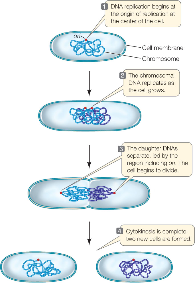
Chromosome replication takes place as the DNA is threaded through a “replication complex” of proteins near the center of the cell. Replication begins at the ori site and moves toward the ter site. When replication is complete, the two daughter DNA molecules separate and segregate from one another at opposite ends of the cell. In rapidly dividing prokaryotes, DNA replication occupies the entire time between cell divisions.
Dna Segregation
Replication begins near the center of the cell, and as it proceeds, the ori regions move toward opposite ends of the cell (FIGURE 7.4). DNA sequences adjacent to the ori region bind proteins that are essential for this segregation. This is an active process, since the binding proteins hydrolyze ATP. Components of the prokaryotic cytoskeleton are involved in the segregation process. In particular, a bacterial protein that is structurally related to actin but functionally related to tubulin provides a filament along which the ori regions and their associated proteins move.
Cytokinesis
The actual division of a single cell and its contents into two cells begins immediately after chromosome segregation. Initially, there is a pinching in of the cell membrane caused by the contraction of a ring of fibers on the inside surface of the membrane (similar to a drawstring on shorts being tightened). In this case, the major component of these fibers is structurally similar to eukaryotic tubulin (which makes up microtubules), but its function is analogous to that of actin in the contractile ring of an animal cell (see below). As the membrane pinches in, new cell wall materials are deposited, which finally separate the two cells.
133
Eukaryotic cells divide by mitosis followed by cytokinesis
As in prokaryotes, cell division in eukaryotes entails reproductive signals, DNA replication, DNA segregation, and cytokinesis. Some of the details, however, are quite different:
- Reproductive signals. Unlike prokaryotes, eukaryotic cells do not constantly divide whenever environmental conditions are adequate. In fact, most cells in a multicellular organism are specialized and do not divide. In a eukaryotic organism, the signals for cell division are usually not related to the environment of a single cell, but to the function of the entire organism. We will discuss the signals that control eukaryotic cell division in Concept 7.3.
- DNA replication. Unlike prokaryotes, eukaryotes have more than one chromosome. But the replication of each eukaryotic DNA molecule is similar to replication in prokaryotes, in that it is achieved by threading the long strands through replication complexes (see Concept 9.2). DNA replication occurs only during a specific stage of the cell cycle.
- DNA segregation. This is much more complicated than in prokaryotes, because first, there is a nuclear envelope, and second, there are multiple chromosomes. When a cell divides, one copy of each chromosome must end up in each of the two new cells—for example, each new somatic cell in a human will have all 46 chromosomes. In eukaryotes, the pairs of newly replicated chromosomes are initially attached to one another. They become highly condensed, and then the pairs are pulled apart before segregating into two new nuclei. The cytoskeleton is involved in this process.
- Cytokinesis. The process of cytokinesis in plant cells (which have cell walls) is different than in animal cells (which do not have cell walls). We describe both processes below.
LINK
The cytoskeleton is crucial for cell division. Review the description of the cytoskeleton and its molecular components in Concept 4.4
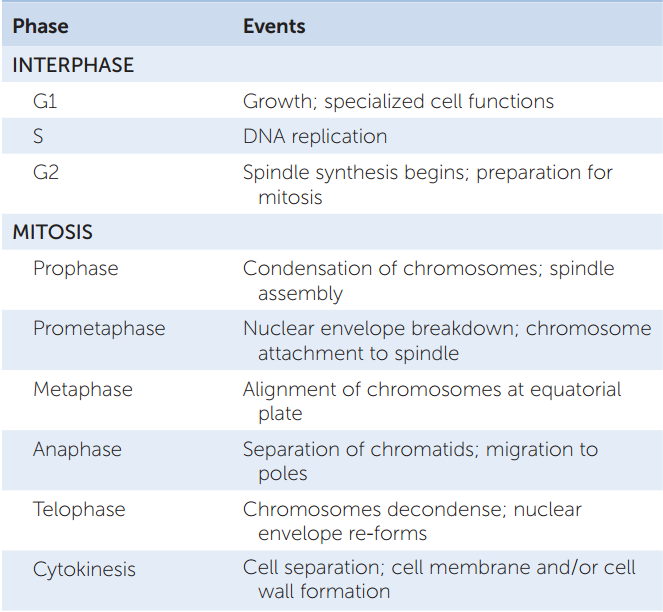
These events occur within the context of the cell cycle: the period from one cell division to the next. In eukaryotes, the cell cycle can be divided into several stages (FIGURE 7.5):
- Mitosis is the set of processes in which the chromosomes become condensed and then segregate into two new nuclei.
- Cytokinesis usually follows immediately after mitosis, and these two stages—mitosis and cytokinesis—are referred to as M phase.
- M phase is followed by a much longer period called interphase, when the cell nucleus is visible and typical cell functions occur—including DNA replication in cells that are preparing to divide.
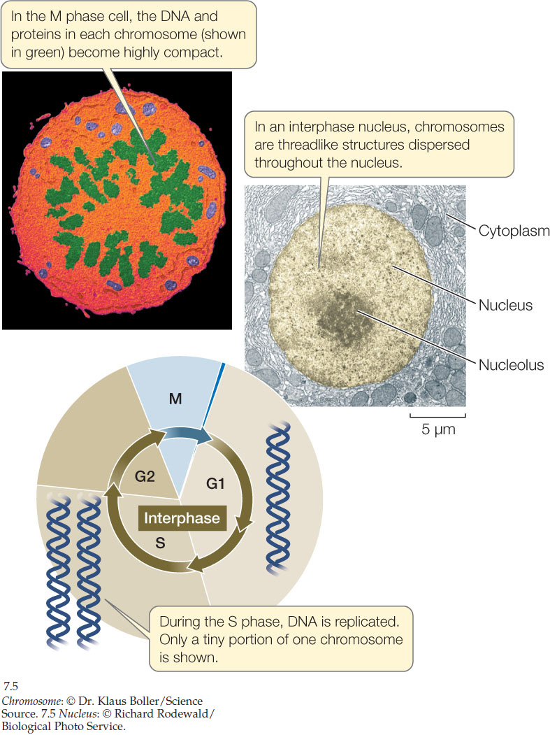
Interphase has three subphases called G1, S, and G2 (the G stands for gap). G1 is quite variable, and a cell may spend a long time in this phase carrying out its specialized functions. The cell’s DNA is replicated during S phase (S for synthesis). During G2, the cell makes preparations for mitosis—for example, by synthesizing components of the microtubules that will move the segregating chromosomes to opposite ends of the dividing cell.
In mitosis, a single nucleus gives rise to two daughter nuclei that each contain the same number of chromosomes as the parent nucleus. Although mitosis is a continuous process in which each event flows smoothly into the next, it is convenient to subdivide it into a series of stages: prophase, prometaphase, metaphase, anaphase, and telophase. Next we will look at these stages in more detail.

Go to ANIMATED TUTORIAL 7.1 Mitosis
PoL2e.com/at7.1
Prophase sets the stage for DNA segregation
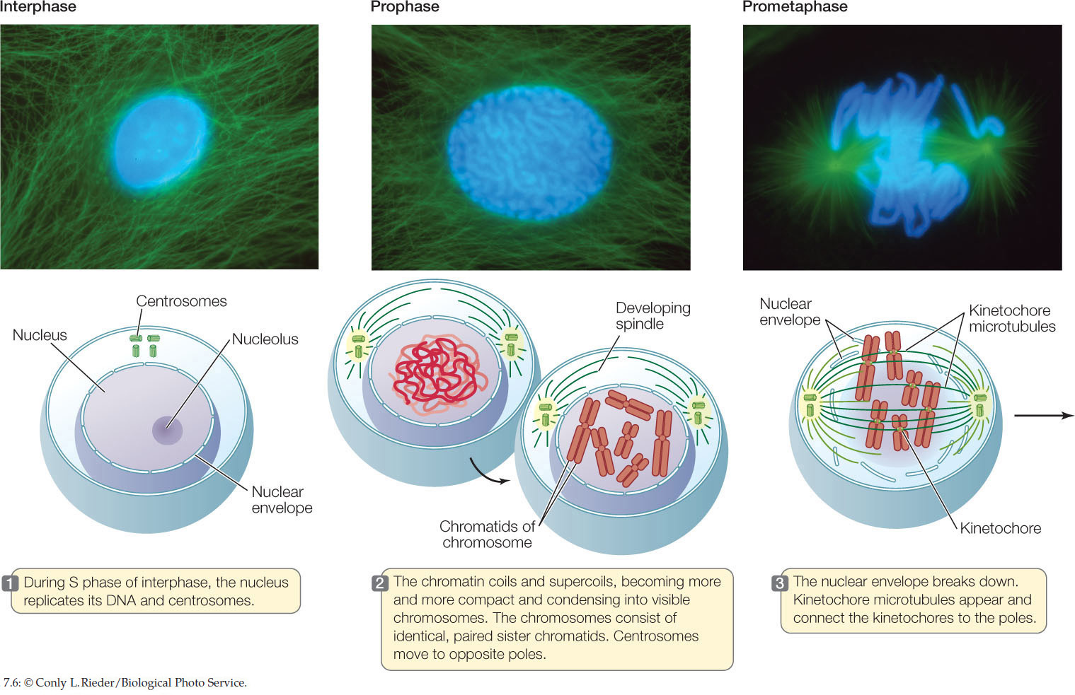
During interphase, only the nuclear envelope and the nucleolus (the region of the nucleus where ribosomes are formed; see Concept 4.3) are visible under the light microscope. The chromatin (the DNA with its associated proteins) is not yet condensed, and individual chromosomes cannot be discerned. The appearance of the nucleus changes as the cell enters prophase—the beginning of mitosis. Here we describe three structures that appear during prophase and contribute to the orderly segregation of the replicated DNA: the condensed chromosomes, the reoriented centrosomes, and the spindle.
Condensed Chromosomes
Before S phase of interphase, each chromosome contains one very long double-stranded DNA molecule. If all of the DNA in a typical human cell were put end to end, it would be nearly 2 meters long. Yet the nucleus is only 5 μm (0.000005 m) in diameter. So even during interphase, eukaryotic DNA is packaged in a highly organized way. The DNA is wound around specific proteins, and other proteins coat the DNA coils. During prophase the chromosomes become much more tightly coiled and condensed.
LINK
DNA “packaging” and the specialized proteins that accomplish it are described in detail in Concept 11.3
After DNA replication, each chromosome has two DNA molecules, known as sister chromatids. Until they are separated during anaphase (see below), the chromatids are held together at a region called the centromere. During prophase the chromosomes become so compact that they can be seen clearly with a light microscope after staining with special dyes. They are even more clearly visualized with an electron microscope:
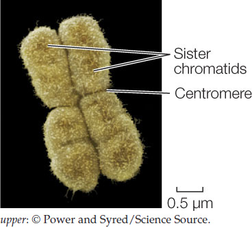
Specialized protein structures called kinetochores assemble on the centromeres, one on each chromatid. These structures are important for chromosome movement.
For a given organism, the number and sizes of the condensed chromosomes constitute the karyotype. Each chromosome has a particular length, and the centromere is located at a particular position along its length. For example, humans have 46 chromosomes (23 homologous pairs) that can be distinguished from one another by their sizes and centromere positions. (In the image below, each chromosome is composed of two chromatids, but the individual chromatids cannot be distinguished. The colors are from dyes that were used to help identify the different chromosomes.)
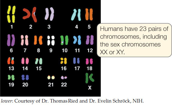
In the past, karyotype analysis was used as a way to identify organisms, and this method is still used to detect chromosomal abnormalities in humans. However, DNA sequence analysis is now much more commonly used to identify individuals and to classify related organisms (see Concept 16.2).
Reoriented Centrosomes
Before the spindle apparatus forms (see below), its orientation is determined. In many cells this is accomplished by the centrosome (“central body”), an organelle in the cytoplasm near the nucleus. The centrosome consists of a pair of centrioles, each one a hollow tube formed by nine triplets of microtubules. During S phase the centrosome becomes duplicated, and at the G2–M transition, the two centrosomes separate from one another, moving to opposite sides of the nucleus. Eventually these identify “poles” toward which chromosomes move during segregation.
The positions of the centrosomes determine the plane at which the cell divides; therefore they determine the spatial relationship between the two new cells. This relationship may be of little consequence to single free-living cells such as yeasts, but it is important for development in a multicellular organism. For example, during the development of an embryo, the daughter cells from some divisions must be positioned correctly to receive signals to form new tissues. Plant cells lack centrosomes, but distinct microtubule organizing centers at each end of the cell play the same role.
135
Spindle
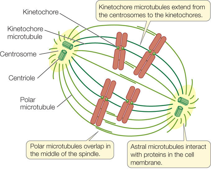
Each of the two centrosomes, when positioned on opposite sides of the nucleus, serves as a pole toward which the chromosomes move. Tubulin dimers from around the centrosomes aggregate into microtubules that extend from the poles into the middle region of the cell. Together these microtubules make up a spindle. The spindle forms during prophase and prometaphase, when the nuclear envelope breaks down. The microtubules are initially unstable, constantly forming and falling apart, until they contact kinetochores or microtubules from the other half-spindle and become more stable.
There are three types of microtubules in the spindle:
- Polar microtubules overlap in the middle region of the cell and keep the two poles apart.
- Astral microtubules interact with proteins attached to the cell membrane, and also assist in keeping the poles apart.
- Kinetochore microtubules attach to the kinetochores on the chromosomes. The two sister chromatids in each chromosome become attached to kinetochore microtubules from opposite sides of the cell. This ensures that the two chromatids will move to opposite poles.
Separation of the chromatids and movement of the daughter chromosomes (which the sister chromatids become after separation) is the central feature of mitosis. It accomplishes the DNA segregation that is needed for cell division and completion of the cell cycle. Note the difference between chromatids and chromosomes:
- Chromatids share a centromere.
- Chromosomes have their own centromere.
Go to ACTIVITY 7.2 The Mitotic Spindle
PoL2e.com/ac7.2
Chromosome separation and movement are highly organized
During the next three phases of mitosis—prometaphase, metaphase, and anaphase—dramatic changes take place in the cell and the chromosomes (FIGURE 7.6):
- In prometaphase the nuclear envelope breaks down and the compacted chromosomes, each consisting of two chromatids, attach to the kinetochore microtubules.
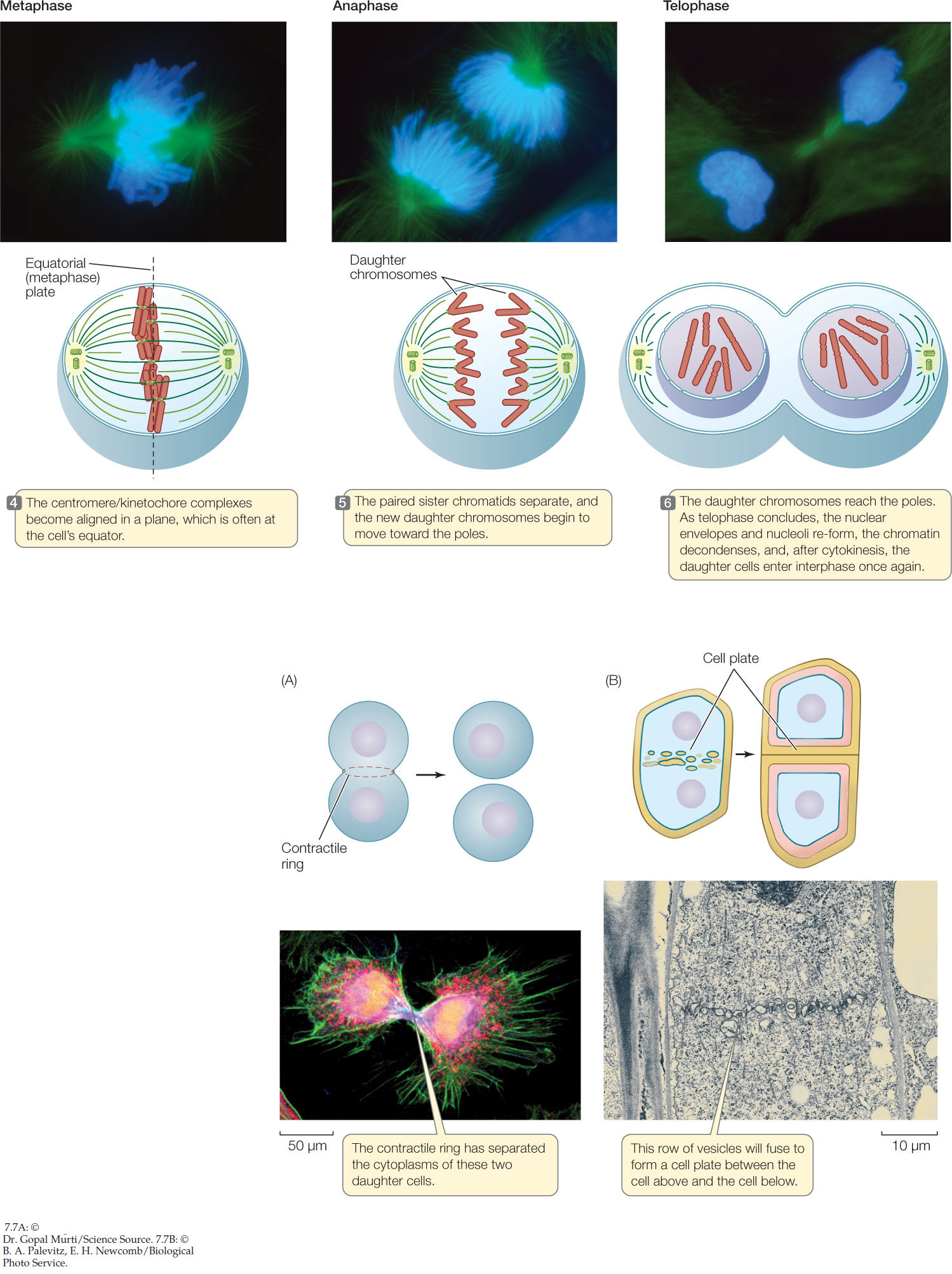
- In metaphase the chromosomes line up at the midline of the cell (the equatorial position).
- In anaphase the chromatids separate, and the daughter chromosomes move away from each other toward the poles.
Go to ACTIVITY 7.3 Images of Mitosis
PoL2e.com/ac7.3

Go to MEDIA CLIP 7.1 Mitosis: Live and Up Close
PoL2e.com/mc7.1
The separation of chromatids into daughter chromosomes occurs at the beginning of anaphase. The migration of the daughter chromosomes to the poles of the cell is a highly organized, active process. Two mechanisms operate to move the chromosomes along. First, the kinetochores contain molecular motor proteins, including kinesin and dynein (see Concept 4.4), which use energy from ATP hydrolysis to move the chromosomes along the microtubules. Second, the kinetochore microtubules shorten from the poles, drawing the chromosomes toward the poles.
Telophase occurs after the chromosomes have separated and is the last phase of mitosis. During this period, a nuclear envelope forms around each set of new chromosomes, nucleoli appear, and the chromosomes become less compact. The spindle also disappears at this stage. As a result, there are two new nuclei in a single cell.
Cytokinesis is the division of the cytoplasm
Mitosis refers only to the division of the nucleus. Cytokinesis, the division of the cell’s cytoplasm, is the final stage of cell reproduction. This process occurs differently in plants and animals.
Animal Cells
Cytokinesis usually begins with a furrowing of the cell membrane, as if an invisible thread were cinching the cytoplasm between the two nuclei (FIGURE 7.7A). This contractile ring is composed of microfilaments of actin and myosin, which form a ring on the cytoplasmic surface of the cell membrane. These two proteins interact to produce a contraction (just as they do in muscles; described in Concept 33.1), pinching the cell in two. The microfilaments assemble rapidly from actin monomers that are present in the interphase cytoskeleton. Their assembly is controlled by calcium ions (commonly used in cellular signaling) that are released from storage sites in the center of the cell.
Plant Cells
In plant cells, the cytoplasm divides differently because plants have cell walls. As the spindle breaks down after mitosis, vesicles derived from the Golgi apparatus appear along the plane of cell division, roughly midway between the two daughter nuclei. The vesicles are propelled along microtubules by the motor protein kinesin and fuse to form a new cell membrane. At the same time they contribute their contents to a cell plate, which is the beginning of a new cell wall between the two daughter cells (FIGURE 7.7B).
136
Following cytokinesis, each daughter cell contains all the components of a complete cell. A precise distribution of chromosomes is ensured by mitosis. In contrast, organelles such as mitochondria and chloroplasts are not necessarily distributed equally, although at least one of each must be present in each daughter cell. The orientation of cell division is important in development (see above), but there does not appear to be a precise mechanism for the distribution of the cytoplasmic contents.
TABLE 7.1 summarizes the major events of the eukaryotic cell cycle.
137
138
CHECKpoint CONCEPT 7.2
- How does the mitotic spindle ensure that each daughter cell receives a full complement of the genetic material in the cell nucleus?
- Compare the cell cycles of prokaryotes and eukaryotes with regard to reproductive signals for initiation, how many chromosomes are present, and how the replicated DNA segregates.
- Sketch the five stages of mitosis for a diploid organism with four chromosomes (two pairs). Clearly label chromosomes and chromatids and note the number of double-stranded DNA molecules in each structure at each stage.
- The drug cytochalasin B blocks the assembly and function of microfilaments. What would you expect to happen in dividing animal cells treated with this drug after telophase but before cytokinesis?
Light microscopists have studied the dramatic events of mitosis and cell division since the 1880s, and we now have detailed descriptions of these processes. More recently, biologists have focused on the mechanisms controlling cell reproduction, which will be our next topic.