Many different types of parasites affect the abundance of host species
Parasites typically have specific habitat needs and, as a result, often live in particular places on a host organism. In humans, for instance, parasites find certain parts of the body to be highly suitable habitats. As illustrated in Figure 15.1, head lice live in the hair, liver flukes reside in the liver, the fungus that causes athlete’s foot resides in the feet, and so on.

Ectoparasite A parasite that lives on the outside of an organism.
Endoparasite A parasite that lives inside an organism.
We can categorize the wide variety of parasites as either ectoparasites or endoparasites. Both are common in plants and animals. Ectoparasites are parasites that live on the outside of organisms whereas endoparasites are parasites that live inside organisms. Each lifestyle has advantages and disadvantages, as summarized in Table 15.1. For example, because ectoparasites live on the outside of their hosts, they do not have to combat the immune system of a host and they can easily move on and off a host. In addition, they do not directly cause diseases in their host. In contrast, because endoparasites live inside their hosts, they must contend with the immune system of their host, and they may have a difficult time getting in and out of their host’s body. Endoparasites have the advantage of being protected from the external environment and are therefore not exposed to most of their enemies. In addition, living inside the host gives endoparasites easy access to the host’s body fluids on which they feed. In contrast, ectoparasites are exposed to the variable conditions of the external environment, including natural enemies, and they must find a way to pierce the flesh of their host to feed. In this section, we will discuss some of the most common types of ectoparasites and endoparasites and the effects they have on their hosts.
346
COMPARING THE CONSEQUENCES OF ENDOPARASITE AND ECTOPARASITE LIFESTYLES
|
FACTOR |
ECTOPARASITES |
ENDOPARASITES |
|
Exposure to natural enemies |
High |
Low |
|
Exposure to external environment |
High |
Low |
|
Difficulty of movement to and from host, for parasite or its offspring |
Low |
High |
|
Exposure to host’s immune system |
Low |
High |
|
Ease of feeding on host |
Low |
High |
Ectoparasites
A variety of organisms live as ectoparasites, as shown in Figure 15.2. Most ectoparasites that attack animals are arthropods. Among the best known ectoparasites are two groups of arachnids—ticks and mites—and two groups of insects—lice and fleas. There are many other groups of ectoparasites, including leeches and some species of lamprey fish. Each of these animals attaches to a host and consumes the host’s blood and other body fluids.

347
Plants also have ectoparasites, but they are most commonly either nematodes—also known as roundworms—or other species of plants. Nematodes that act as plant ectoparasites live in the soil and feed on plant roots. The 1-mm-long worms attach to a plant root, inject digestive enzymes that break down root cells, and then consume the resulting slurry. This parasitic behavior can reduce the growth, reproduction, and survival of the host.
Approximately 4,000 species of plants make their living as ectoparasites on other plants. For example, mistletoes embed rootlike organs into the branches of trees and shrubs. Because such plants have their roots inside the host and their shoots outside the host, they are sometimes categorized as hemiparasites. The mistletoe leaves conduct photosynthesis, but the plant obtains water and minerals from the host plants. Some species, such as dwarf mistletoes (in the genus Arceuthobium) cause death in conifer trees— particularly under drought conditions—by extracting too much of the host’s water and nutrients. In some species of mistletoe, the plant’s fruit is consumed by birds, which inadvertently disperse the seeds from the fruit when they defecate onto the branches of other trees and shrubs. Other species of mistletoe are able to project seeds laterally up to 15 m.
Endoparasites
Endoparasites can be categorized as either intracellular or intercellular. As the names imply, intracellular parasites live inside the cells of a host, whereas intercellular parasites live in the spaces between cells that include the cavities of a host’s body. Intracellular parasites are very small; examples include viruses, small bits of proteins known as prions, and some types of bacteria and protists. Intercellular parasites are much larger; examples include some types of protozoa and bacteria, fungi, and a group of worms known as helminths, which include the nematodes. Each of these groups of endoparasites can have substantial effects on their host’s survival, growth, and reproduction. Because endoparasites often cause diseases, they can alter the abundance of host species and change the composition of ecological communities.
Viruses
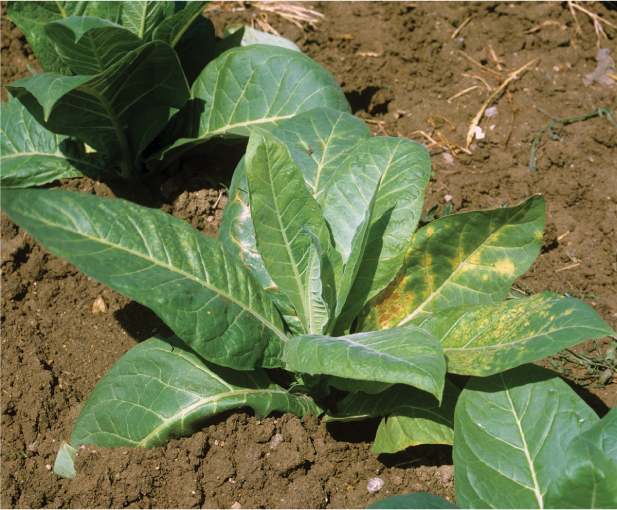
Late in the nineteenth century, researchers struggled to find the cause of a disease striking tobacco plants. They recognized that the pathogen was too small to be a bacterium. After many years of research, they were able to identify it as a virus, which they named tobacco mosaic virus. Plants that are infected exhibit a mosaic of colors in their leaves and may also have growths that resemble blisters (Figure 15.3). Although this virus was first discovered in tobacco, we now know that it can infect more than 150 species of plants. Viruses that infect plants can have a devastating effect on food production, which makes them a matter of great concern to farmers and consumers.
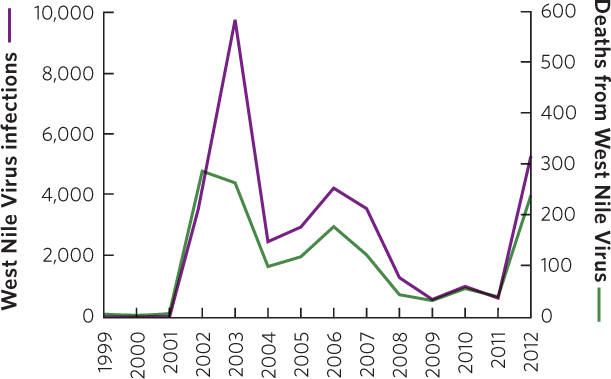
Animals can also be infected by many different pathogenic viruses. For example, there are different genera of the pox virus that can infect mammals: cowpox in cattle, monkeypox in primates and rodents, and smallpox in humans. Hoofed animals such as deer, cattle, and sheep are susceptible to bluetongue virus, which can cause high mortality rates. Similarly, the West Nile virus—named for the location of the first known human case in the West Nile region of Uganda in 1937—can be highly lethal to some species of birds. The mosquitoes that carry this virus usually transmit the disease to birds, but occasionally to horses and humans, both of which can die from the infection. The virus arrived in the United States in 1999 and spread rapidly. Several species of North American birds experienced high rates of death, including blue jays (Cyanocitta cristata), American robins, and American crows (Corvus brachyrhynchos). At the same time, horses and people started to become infected. In the decade following the introduction of the virus into the United States more than 1,000 people died. As a result, public health officials in the United States have made great efforts to reduce mosquito populations that spread the disease. These efforts, possibly combined with the evolution of reduced virulence by the pathogen, have caused the number of infections and deaths to decline dramatically from 2003 to 2011, as illustrated in Figure 15.4. However, in 2012 there was a sharp increase in the number of infections and deaths in the United States. The sharp increase is thought to be due to unusually warm and humid conditions combined with abundant rain that created a lot of standing water where mosquitoes can breed and subsequently build up a large adult population.
348
Both mammals and birds are susceptible to strains of influenza virus. For example, a strain of influenza known as the Spanish flu is caused by the H1N1 virus and normally infects only birds, but in 1918 it also infected humans. Researchers hypothesize that the virus experienced a mutation that allowed it to survive in humans and to infect other humans directly. They believe that humans initially contracted the disease by handling domesticated ducks and chickens. However, once the virus found its way into humans, it rapidly spread around the world and killed up to 100 million people. More recently, the H1N1 virus has been found on pig farms, which is why the pathogen also has the name swine flu.
In 2006, a similar strain of virus known as bird flu, or H5N1, infected and killed large numbers of birds and also jumped from domesticated birds to humans. Hundreds of thousands of domesticated birds were killed in an attempt to stop the spread of the virus (Figure 15.5). By 2012, the World Health Organization reported that 595 people had been infected with bird flu and more than half of them had died. The ability of pathogens to jump from traditional hosts to human hosts, as we will discuss in the next section, causes a great deal of concern for human health in the future.

Prions
Prions are a recently discovered category of pathogenic parasite. All prions begin as a beneficial protein in the brain of an animal, but occasionally a protein folds into an incorrect shape and becomes pathogenic. Prions do not contain any RNA or DNA; instead, they replicate by coming into contact with normal proteins and causing them to fold incorrectly, with the first prion serving as a template.
One of the best-known diseases caused by prions is bovine spongiform encephalopathy, commonly known as mad cow disease. This disease, which is always fatal, gets its name from the way that infected cows lose control of their body, as if they have gone mad. While cows cannot transmit prions to each other, in the 1980s it was a common practice to feed cows the ground remains of other cows. Cows that consumed the mutated prions from the dead cattle became infected and ultimately passed the infection on to humans. More than 150 people have died from mad cow disease. Today, new rules forbid feeding dead cattle to other cattle and the incidence of mad cow disease has declined.
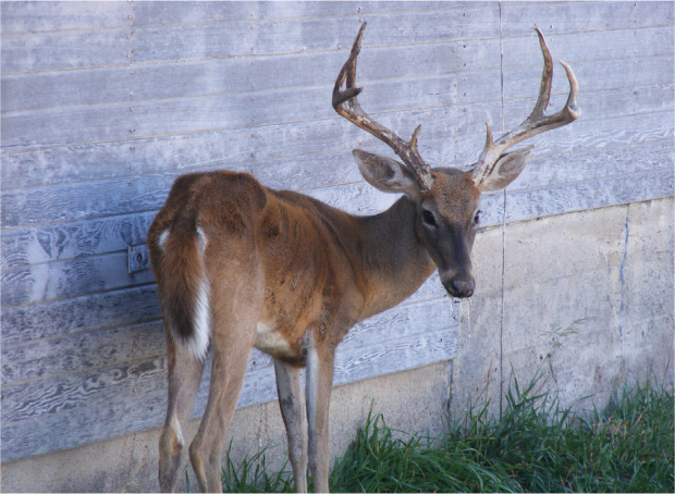
Chronic wasting disease is a more common prion disease. It infects members of the deer family, including white-tailed deer, mule deer (Odocoileus hemionus), elk, and moose. Individuals infected with chronic wasting disease excrete prions that are later consumed inadvertently by others. Infected individuals begin to lose weight and eventually die (Figure 15.6). Currently, chronic wasting disease is concentrated in Colorado and Wyoming, but small numbers of infected deer have been discovered in other regions of the United States and Canada.
349
Protozoans
Protozoans are a group of parasites that can cause a variety of diseases. For example, several protozoans cause diarrhea in humans and other animals. They are also the source of malaria in humans and avian malaria in birds. Malaria is carried by mosquitoes that acquire the protists when they feed on an infected individual and then transfer the protists to uninfected individuals they subsequently feed on. In Hawaii, as well as in some other parts of the world, there is no historic presence of avian malaria, so the birds of the Hawaiian Islands have not evolved defenses against the protists. Mosquitoes were accidentally introduced into Hawaii in the early 1800s and by the early 1900s the protists that cause avian malaria had also arrived, which has contributed to the decline and extinction of many native bird species in these islands.
Bacteria
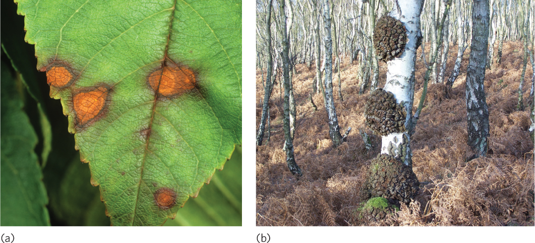
Bacterial parasites can cause a wide variety of plant and animal diseases. In plants, bacterial infections can cause spotted leaves, wilted stems, scabbed surfaces, and large abnormal tissue growths known as galls (Figure 15.7). Because bacteria must enter a plant through a wound in the plant’s tissues, they commonly require the assistance of herbivores that pierce a plant’s tissues.
Bacterial infection in animals can also be quite harmful to the host. One of the earliest discovered species of deadly bacteria was anthrax (Bacillus anthracis). Anthrax, originally isolated from cows and sheep, is also highly lethal to humans. After an incubation period of about 1 week, the growing bacterial population begins to release toxic compounds that cause internal bleeding, which often brings rapid death. Other common bacterial pathogens include those that cause plague, pneumonia, salmonella, leprosy, and many sexually transmitted diseases, all of which occur in a wide variety of animals.
Fungi
Fungal parasites have large ecological impacts on a wide range of plants and animals. Fungal diseases have devastated many dominant plant species, including critical food sources for humans. A number of tree species in North America have suffered tremendous declines as a result of pathogenic fungi that have been introduced from Europe and Asia. The American chestnut tree was once one of the tallest tree species in the temperate forests of the eastern United States and comprised up to 50 percent of all trees in the forest. It was also one of the most prized trees for lumber as well as for the crop of edible nuts that it produced every autumn. Around 1900, however, species of Asian chestnut trees imported to New York inadvertently carried a fungus (Cryphonectria parasitica). While Asian chestnut trees had a long evolutionary history with the fungus and were resistant to its harmful effects, American chestnut trees had no such history. As a result, the American chestnut rapidly succumbed to the disease known as chestnut blight that was caused by the fungus. The decline of the chestnut trees also likely had widespread effects on the many animal species that relied on the tree’s large crop of nuts for food. Today, the American chestnut tree is quite rare; young seedlings can emerge from seeds but they ultimately become infected with the fungus and die early in life.
350
Similar problems have occurred with other trees, including the American elm tree (Ulmus americana). The elm was widely planted along North American streets because of its attractive arching branches that provided shade (Figure 15.8a). In the 1930s, a fungus from Asia was introduced into Europe and North America that caused a rapid decline in elm trees. Because the fungal disease was first described by researchers in Holland, it is known as Dutch elm disease. The American elm had no evolutionary history with the Asian fungus, and 95 percent of the elm trees across North America have succumbed (Figure 15.8b). The small number of remaining trees appears to be resistant to the fungal disease, and researchers are currently working to plant these resistant genotypes back into eastern forests.

Some fungal parasites also damage crop plants. One of the best-known examples is a group of fungi that causes a disease known as rust. Throughout human history, rust diseases have affected many of our most important crops, including wheat, corn, rice, coffee, and apples. The effect of rust can range from reduced food production to a complete loss of the crop costing millions of dollars of damage. Widespread rust infections, such as wheat rust, have caused the starvation of millions of people over the past two centuries.
Animals can also be infected by fungi. One high profile animal fungal disease is caused by a species of chytrid fungus that infects amphibians (Batrachochytrium dendrobatidis). During the 1990s, scientists working in Central America began to notice massive die-offs of amphibians and subsequently determined that the dead animals were infected with the fungus. The fungus, which lives in the outer layers of the amphibian’s skin, causes an imbalance of ions in the body that ultimately causes the animal’s heart to stop.
During 2 decades of surveying amphibians from Costa Rica to Panama, researchers found that the fungus was moving from the northwest to the southeast, as shown in Figure 15.9a. Between 2000 and July 2004, at a site in El Copé, Panama, a team of researchers sampled more than 1,500 amphibians before the fungus arrived; not one individual tested positive. By October 2004, however, 21 of 27 sampled species were infected in at least 10 percent of their populations and by December 2004, 40 species tested positive for the fungus.
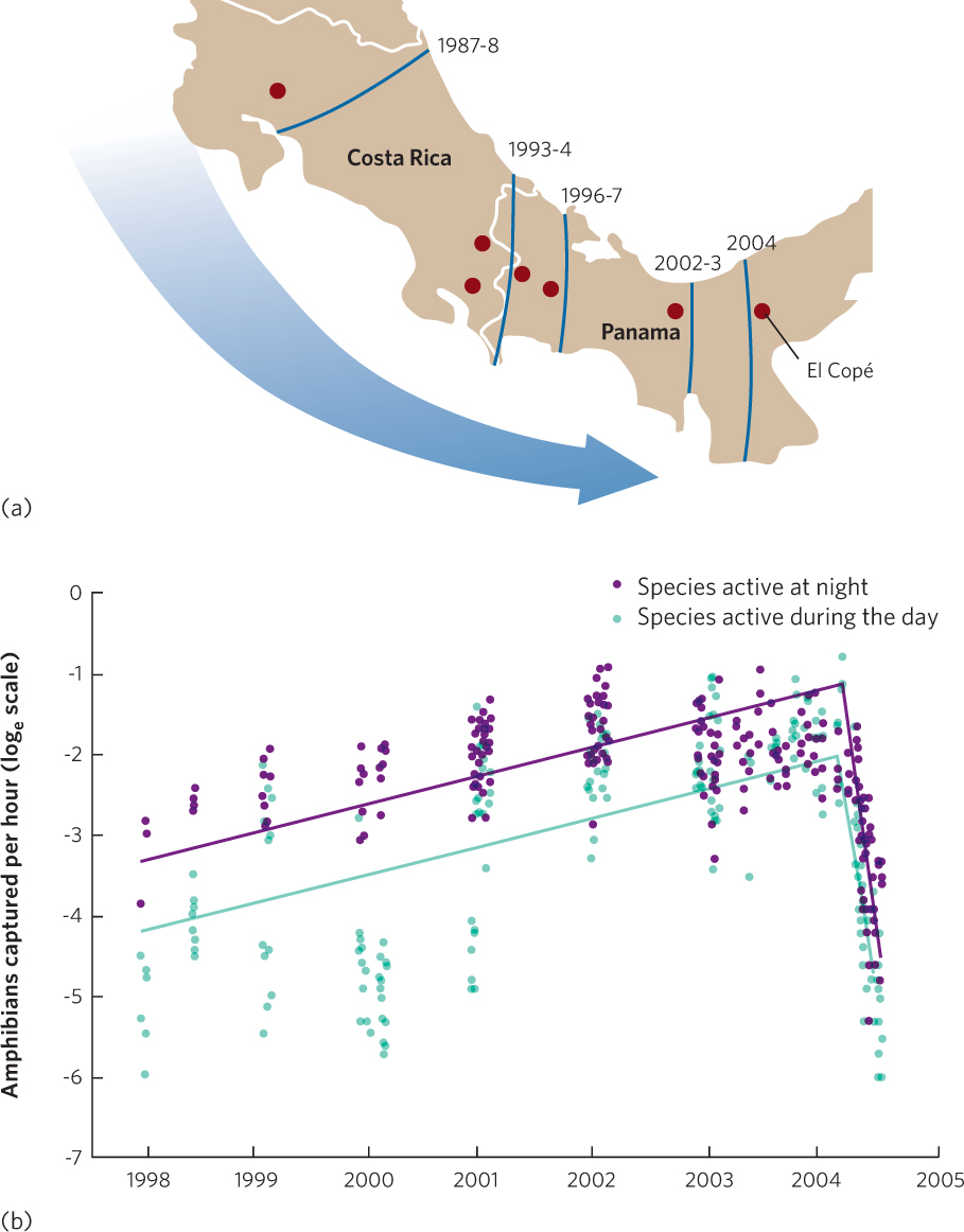
The researchers also estimated the sizes of amphibian populations in El Copé with transect surveys of amphibians that were active either during the day or during the night. After the fungus arrived in 2004, the number of live amphibians declined sharply (Figure 15.9b). The dead amphibians included 38 different frog species. Moreover, 99 percent of the 318 dead individuals collected had moderate to severe chytrid infections. In 2010, researchers reported that of the 63 species of amphibians that were present at the site before the fungus arrived, 30 species were gone. Relatively little is known about this fungus, although the massive die-offs suggest that amphibians have few defenses against it, which indicates that the fungus may have been introduced to Central America. The fungus is now suspected to have caused the extinction of dozens of species of amphibians around the world.
351
Helminths
Helminths include several groups of roundworms and flatworms that can cause serious diseases. We have already discussed the impact of nematodes as ectoparasites on plant roots. Nematodes can also live as endoparasites within the tissues of plants, where they damage the plant’s growth, reproduction, and survival. In animals, disease-causing helminths include hookworms that feed on the blood of intestines, lungworms that live in the lungs, and echinostome worms that live in the kidneys. In livestock, infection by liver flukes has been a problem for centuries. Livestock inadvertently consume these parasites when they drink water containing fluke-infected snails or when they eat grass that contains a stage of the parasite that is excreted by snails. Liver flukes are a particular problem in sheep where they cause liver damage, hemorrhaging, and sudden death. Left untreated, liver flukes can kill up to 10 percent of a sheep population; fortunately, drugs can cure infected animals.
Emerging infectious disease A disease that is newly discovered, or has been rare and then suddenly increases in occurrence.
Emerging Infectious Diseases
Many infectious diseases have been infecting hosts for thousands of years while others have only emerged recently. When a new disease is discovered, or a formerly common disease that has become rare suddenly increases in occurrence, it is called an emerging infectious disease. New diseases typically emerge when a mutation allows a pathogen to jump to a new host species. We have already mentioned several emerging infectious diseases, including the chytrid fungus that has decimated amphibians around the world, the H5N1 bird flu that jumped from birds to humans, and mad cow disease that infected cattle, sheep, and humans. Since the 1970s, the world has experienced an average of one new emerging infectious disease each year.
352
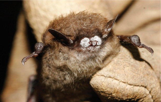
In 2006 researchers identified an emerging disease infecting bats in a cave near Albany, New York. These bats had a white-colored fungus (Geomyces destructans) growing on their noses, and they were dying in large numbers (Figure 15.10). Researchers named the disease white-nose syndrome. They hypothesized that the fungus causes bats hibernating in caves to come out of their normal torpor and to use up their fat reserves until they become emaciated and die. By 2012, the U.S. Fish and Wildlife Service reported that the fungus had spread to 16 states and 4 Canadian provinces, and killed more than 5 million bats. In caves containing infected bats, the mortality rate can reach 100 percent. This is important because bats provide many important services, including the consumption of a large number of insects. Therefore, it is plausible that the death of so many bats will have substantial effects on many communities and ecosystems.
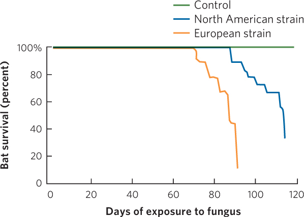
An interesting detail in the story of white-nose syndrome is that bats in Europe also carry the fungus but do not experience widespread death. This raises the possibility that the fungus is native to Europe and has only recently been introduced to North America. Alternatively, it could be that the fungus has always been in North America but recently mutated to become much more lethal. To test these alternative hypotheses, researchers isolated strains of the fungus from European and North American bats. They then exposed the little brown bat (Myotis lucifugus), a species from North America, to fungal spores from the European strain, fungal spores from the North American strain, or a control treatment containing no fungal spores. After the exposures, they monitored bat survival for 120 days. In 2012, the researchers reported their results, which you can see in Figure 15.11. In the control treatment, no bats died, but bats exposed to either fungal strain experienced high rates of death, which suggests that the fungus is a recent arrival from Europe and not a mutated North American native. Moreover, the bats died more slowly when exposed to the North American strain of fungus than the European strain, which may suggest that North American bats were already beginning to evolve some level of resistance to their local strain of fungus. Unfortunately, there is currently no treatment to prevent the continued death of bats from white-nose syndrome.