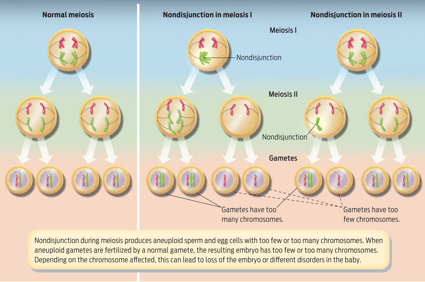
Nondisjunction
 Why does the risk of having a baby with Down syndrome go up as a woman ages?
ANSWER: At age 25, a woman’s risk of having a baby with Down syndrome is 1 in 1,250 births (that is, 1 out of every 1,250 babies born to women who are 25 is likely to have Down syndrome). At age 40 her risk skyrockets to 1 in 100 births.
As women age, the risk of giving birth to a baby with any chromosomal abnormality increases. That’s because as a woman ages, so do her eggs. All the eggs that a woman will ever have were formed before she was born, and they have been aging like the rest of the cells in her body. Until puberty, a woman’s eggs are “paused” in the middle of meiosis (at meiosis I); they haven’t yet completed their cell division. During a menstrual cycle, one egg resumes meiosis and is ovulated. In older women, when these eggs complete meiosis and are ovulated, they are more likely to have an error in chromosome segregation, leading to a chromosomal abnormality.
ANEUPLOIDY An abnormal number of one or more chromosomes (either extra or missing copies).
A chromosomal abnormality means that a developing fetus carries a chromosome number that differs from the usual 46. The most common abnormalities in humans are called aneuploidies, deviations from the normal number of chromosomes because single chromosomes are either duplicated or deleted. Most aneuploidies arise during meiosis in the parents’ sex cells. If chromosomes segregate improperly during meiosis, an occurrence called nondisjunction, the resulting gamete will either lack a chromosome or carry an extra copy. When that egg is fertilized by a normal gamete, the resulting zygote can have an abnormal number of chromosomes. In most cases, the abnormality is so severe the zygote spontaneously aborts (INFOGRAPHIC 12.12).
NONDISJUNCTION The failure of chromosomes to separate accurately during cell division; nondisjunction in meiosis leads to aneuploid gametes.
276
Birth defects can arise when chromosomes fail to separate normally during meiosis, a phenomenon called nondisjunction. The resulting gametes carry an abnormal number of chromosomes, a condition called aneuploidy.
TRISOMY 21 Carrying an extra copy of chromosome 21; also known as Down syndrome.
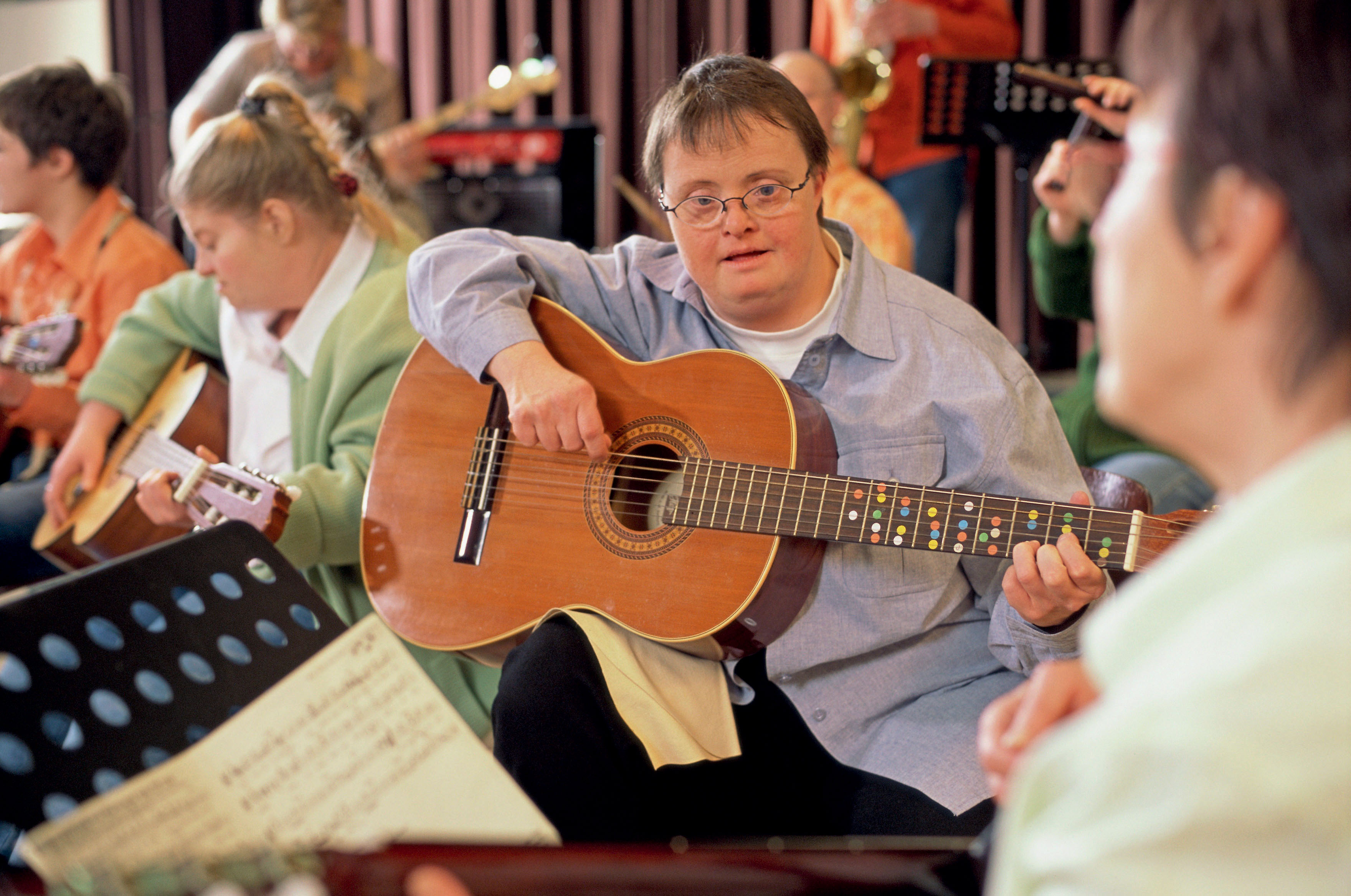
There are, however, cases in which the abnormality is not life threatening but does cause severe disability—the most common is trisomy 21, also called Down syndrome. Trisomy 21 results when an embryo inherits an extra copy of chromosome 21. Anyone can conceive a child with the abnormality, but older women are at much higher risk.
Most Down syndrome children have learning disabilities that range from mild to moderate, but some have profound mental disability. They are also at higher risk for other diseases and typically don’t live beyond 50 years of age.
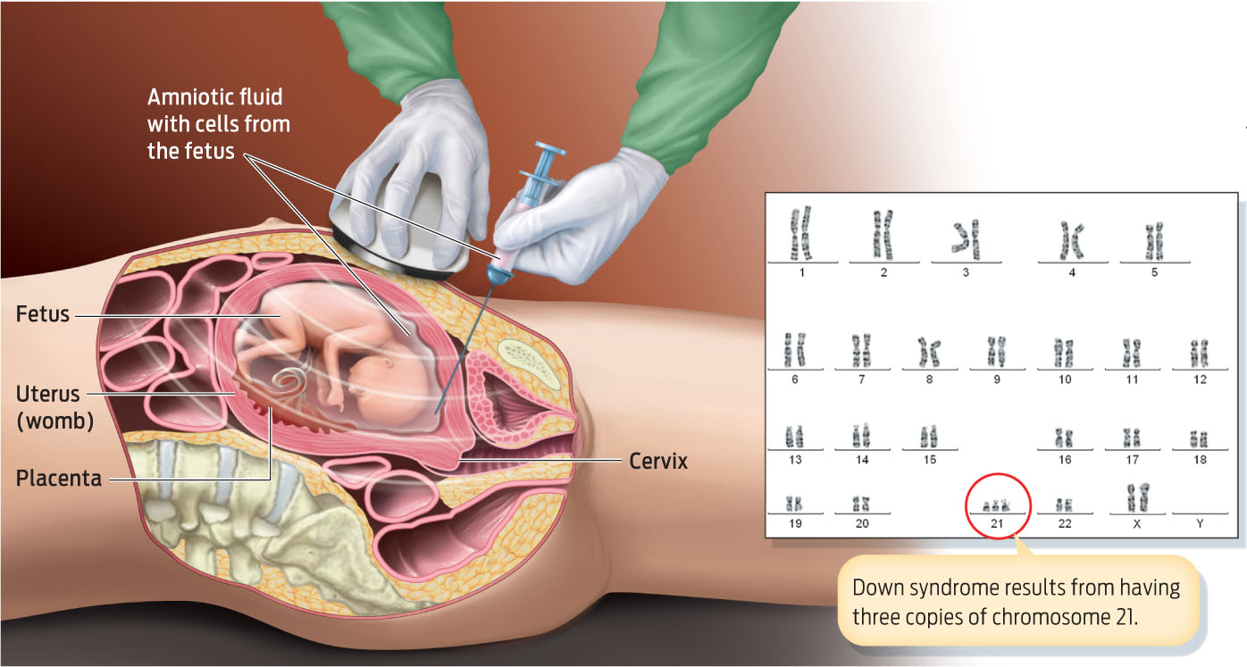
AMNIOCENTESIS A procedure that removes fluid surrounding the fetus to obtain and analyze fetal cells to diagnose genetic disorders.
Down syndrome, as well as other chromosomal abnormalities, can be diagnosed by amniocentesis. This procedure is usually performed between 14 and 20 weeks of pregnancy, although some medical centers may perform it as early as 11 weeks. The procedure is quick. A long, thin, hollow needle is inserted through a woman’s abdominal wall and into her uterus. Through the needle, the equivalent of 2 to 4 teaspoons of amniotic fluid, which surrounds the growing fetus, is removed. This fluid contains fetal cells that contain the fetus’s DNA. From that fluid, technicians analyze the fetal karyotype—that is, the chromosomal make-up in its cells (INFOGRAPHIC 12.13).
KARYOTYPE The chromosomal makeup of cells. Karyotype analysis can be used to detect trisomy 21 prenatally.
277
Doctors perform a procedure called amniocentesis to obtain fetal cells and diagnose chromosomal abnormalities such as Down syndrome. A karyotype analysis is done on the fetal cells to look for chromosomal abnormalities, in particular missing or extra chromosomes.
The reasons to undergo amniocentesis vary from couple to couple. But if a test comes back positive, couples have options: they can begin to plan for a disabled child, or make the decision not to carry the child to term.
Although scientists have linked some of the most obvious birth defects to the age of a woman’s eggs, recent research also shows that a man’s age affects his sperm quality. Men who father children after age 45 are more likely to have children with cognitive disorders such as autism, for example. Male fertility declines over time, too, although much more gradually than does female fertility. Research shows that the older the man, the more likely he is to produce sperm with genetic defects.
278