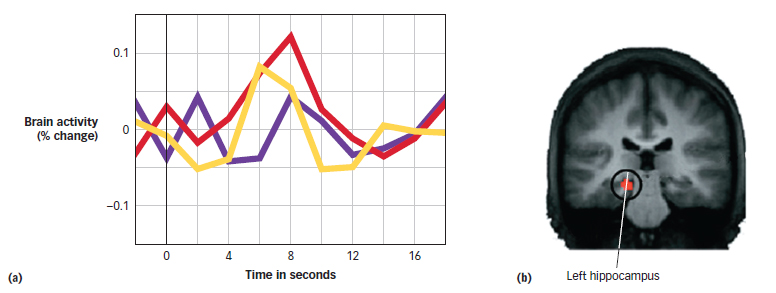
Hippocampal Activity during True and False Recognition Many brain regions show similar activation during true and false recognition, including the hippocampus. The figure shows results from an fMRI study of true and false recognition of visual shapes (Slotnick & Schacter, 2004). A plot of the activity level in the strength of the fMRI signal from the hippocampus over time (a) shows that after a few seconds, there is comparable activation for true recognition of previously studied shapes (red line) and false recognition of similar shapes that were not presented (yellow line). Both true and false recognition show increased hippocampal activity compared with correctly classifying unrelated shapes as new (purple line). The brain scan (b) shows a region of the left hippocampus.
SLOTNICK & SCHACTER, NATURE NEUROSCIENCE, 2004, 7(61), P. 669.