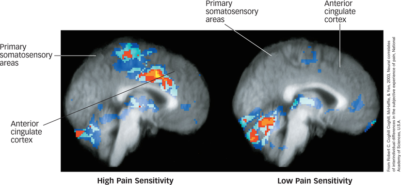
FIGURE 13.4 The Brain in Pain These are fMRI scans of brain activation in high- (left) and low-pain-sensitive (right) individuals during painful stimulation. The anterior cingulate cortex and primary somatosensory areas show greater activation in high-pain-sensitive individuals. Levels of activation are highest in yellow and red, then light blue and dark blue (Coghill, McHaffie, & Yen, 2003).
From Robert C. Coghill Coghill, McHaffie, & Yen, 2003, Neural correlates of interindividual differences in the subjective experience of pain, National Academy of Sciences, U.S.A