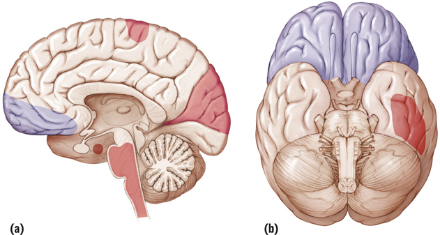
FIGURE 5.10 Brain Activation and Deactivation during REM Sleep Brain areas shaded red are activated during REM sleep; those shaded blue are deactivated. (a) The medial view shows activation of (starting at the top and going clockwise) the motor cortex, the visual association areas, the brain stem, and the amygdala and deactivation of the prefrontal cortex. (b) The ventral view shows activation of other visual association areas and deactivation of the prefrontal cortex (Schwartz & Maquet, 2002).