4.2 Vision I: How the Eyes and the Brain Convert Light Waves to Neural Signals
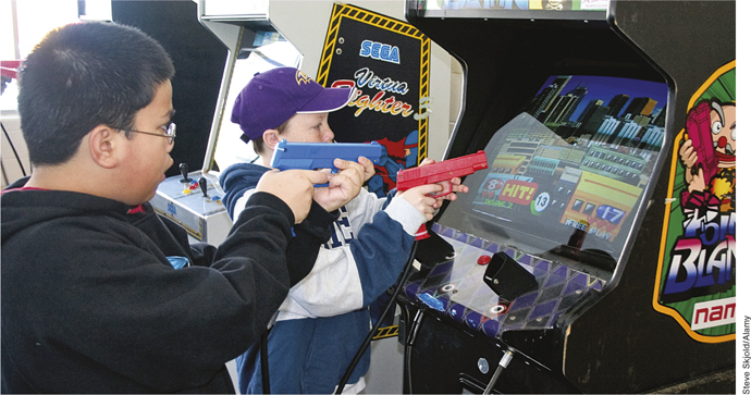
Though controversial for other reasons, research has shown that action-shooter games improve attention and even basic visual acuity (Green & Bavelier, 2007; Li et al., 2009).
Steve Skjold/Alamy
You might be proud of your 20/20 vision, even if it is corrected by glasses or contact lenses. The expression 20/20 refers to a measurement of visual acuity, the ability to see fine detail; it is the smallest line of letters that a typical person can read from a distance of 20 feet. But hawks, eagles, owls, and other raptors have up to eight times greater visual acuity than humans, or the equivalent of 20/2 vision (meaning that what the normal human can just see from 2 feet away can be seen by these birds at a distance of 20 feet away). Your sophisticated visual system has evolved to transduce visual energy in the world into neural signals in the brain. Understanding vision, then, starts with understanding light.
Sensing Light
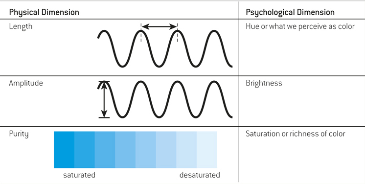
Visible light is simply the portion of the electromagnetic spectrum that we can see, and it is an extremely small slice of the pie (see FIGURE 4.2). You can think about light as waves of energy. Like ocean waves, light waves vary in height and in the distance between their peaks, or wavelengths. There are three physical properties of light waves, each of which produces a corresponding psychological dimension (see TABLE 4.3). The length of a light wave determines its hue, or what humans perceive as color. The intensity or amplitude of a light wave—how high the peaks are—determines what we perceive as brightness. Purity is the number of distinct wavelengths that make up the light, and it is purity that determines what we perceive as saturation, or the richness of colors.
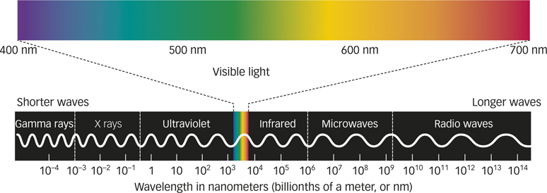
Figure 4.2: FIGURE 4.2 Electromagnetic Spectrum The sliver of light waves visible to humans as a rainbow of colors from violet-blue to red is bounded on the short end by ultraviolet rays, which honeybees can see, and on the long end by infrared waves, upon which night-vision equipment operates. Light waves are minute, but the scale along the bottom of this chart offers a glimpse of their varying lengths, measured in nanometers (nm; 1 nm = 1 billionth of a meter).
Table 4.3Properties of Light Waves
The Human Eye
Eyes have evolved as specialized organs to detect light. FIGURE 4.3 shows the human eye in cross-section. Light that reaches the eyes passes first through a clear, smooth outer tissue called the cornea and then continues through the pupil, a hole in the colored part of the eye. This colored part is the iris, which is a translucent, doughnut-shaped muscle that controls the size of the pupil and hence the amount of light that can enter the eye.
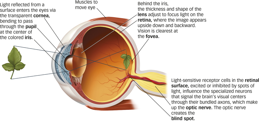
Figure 4.3: FIGURE 4.3 Anatomy of the Human Eye Specialized organs of the eye evolved to detect light.
Immediately behind the iris, muscles inside the eye control the shape of the lens to bend the light and focus it onto the retina, light-sensitive tissue lining the back of the eyeball. The muscles change the shape of the lens to focus objects at different distances, making the lens flatter for objects that are far away or rounder for nearby objects. This is called accommodation, the process by which the eye maintains a clear image on the retina. FIGURE 4.4a shows how accommodation works.
retina
Light-sensitive tissue lining the back of the eyeball.
accommodation
The process by which the eye maintains a clear image on the retina.
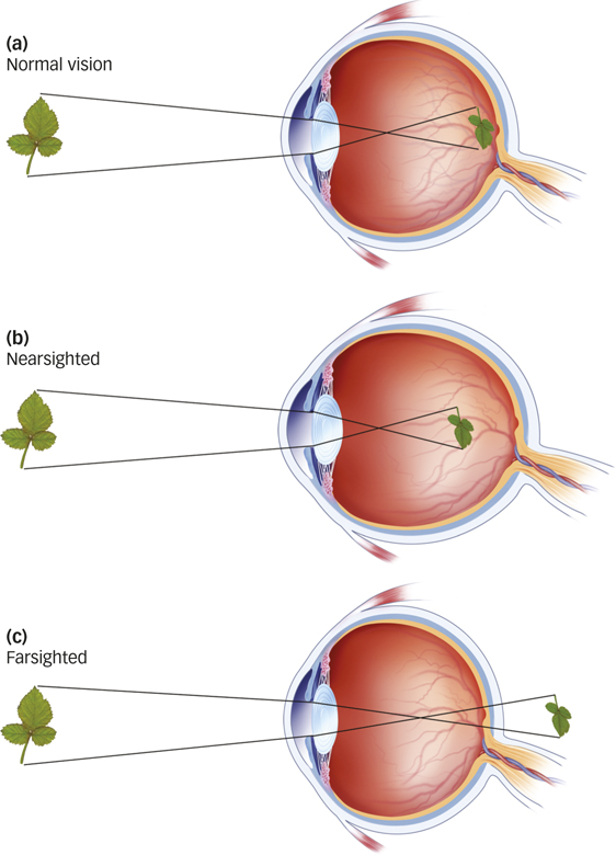
Figure 4.4: FIGURE 4.4 Accommodation Inside the eye, the lens changes shape to focus nearby or faraway objects on the retina. (a) People with normal vision focus the image on the retina at the back of the eye, both for near and far objects. (b) Nearsighted people see clearly what’s nearby, but distant objects are blurry because light from them is focused in front of the retina, a condition called myopia. (c) Farsighted people have the opposite problem: Distant objects are clear, but those nearby are blurry because their point of focus falls beyond the surface of the retina, a condition called hyperopia.
How do eyeglasses actually correct vision?
If your eyeballs are a little too long or a little too short, the lens will not focus images properly on the retina. If the eyeball is too long, images are focused in front of the retina, leading to nearsightedness (myopia), which is shown in FIGURE 4.4b. If the eyeball is too short, images are focused behind the retina, and the result is farsightedness (hyperopia), as shown in FIGURE 4.4c. Eyeglasses, contact lenses, and surgical procedures can correct either condition. For example, eyeglasses and contacts both provide an additional lens to help focus light more appropriately, and procedures such as LASIK physically reshape the eye’s existing lens.
From the Eye to the Brain
How does a wavelength of light become a meaningful image? The retina is the interface between the world of light outside the body and the world of vision inside the central nervous system. Two types of photoreceptor cells in the retina contain light-sensitive pigments that transduce light into neural impulses. Cones detect color, operate under normal daylight conditions, and allow us to focus on fine detail. Rods become active under low-light conditions for night vision (see FIGURE 4.5).
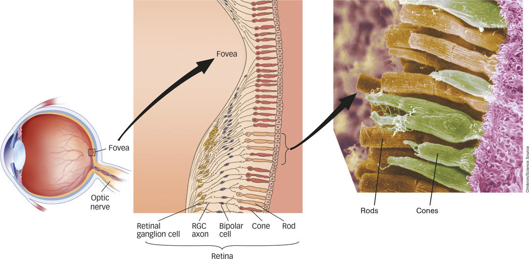
Figure 4.5: FIGURE 4.5 Close-up of the Retina The surface of the retina is composed of photoreceptor cells, the rods and cones, beneath a layer of transparent neurons, the bipolar and retinal ganglion cells (RGCs). The axons of the RGCs form the optic nerve. Viewed close up in this cross-sectional diagram is the area of greatest visual acuity, the fovea, where most color-sensitive cones are concentrated, allowing us to see fine detail as well as color. Rods, the predominant photoreceptors activated in low-light conditions, are distributed everywhere else on the retina.
cones
Photoreceptors that detect color, operate under normal daylight conditions, and allow us to focus on fine detail.
rods
Photoreceptors that become active under low-light conditions for night vision.

The image on the left was taken at a higher resolution than the image on the right. The difference in quality is analogous to light falling on the fovea versus the periphery of the retina.
Thinkstock
Rods are much more sensitive photoreceptors than cones, but this sensitivity comes at a cost. Because all rods contain the same photopigment, they provide no information about color and sense only shades of gray. Think about this the next time you wake up in the middle of the night and make your way to the bathroom for a drink of water. Using only the moonlight from the window to light your way, do you see the room in shades of gray? About 120 million rods are distributed more or less evenly around each retina except in the very center, the fovea, an area of the retina where vision is the clearest and there are no rods at all. The absence of rods in the fovea decreases the sharpness of vision in reduced light.
fovea
An area of the retina where vision is the clearest and there are no rods at all.
In contrast to rods, each retina contains only about six million cones, which are densely packed in the fovea and much more sparsely distributed over the rest of the retina, as you can see in FIGURE 4.5. This distribution of cones directly affects visual acuity and explains why objects off to the side, in your peripheral vision, aren’t so clear. The light reflecting from those peripheral objects is less likely to land in the fovea, making the resulting image less clear.
What are the major differences between rods and cones?
As seen in FIGURE 4.5, the photoreceptor cells (rods and cones) form the innermost layer of the retina. Above them, the bipolar cells collect neural signals from the rods and cones and transmit them to the retinal ganglion cells (RGCs), which organize the signals and send them to the brain. The bundled RGC axons form the optic nerve, which leaves the eye through a hole in the retina. Because it contains neither rods nor cones and therefore has no mechanism to sense light, this hole in the retina creates a blind spot, a location in the visual field that produces no sensation on the retina. Try the demonstration in FIGURE 4.6 to find the blind spot in each of your own eyes.

Figure 4.6: FIGURE 4.6 Blind Spot Demonstration To find your blind spot, close your left eye and stare at the cross with your right eye. Hold the book 6 to 12 inches (15 to 30 centimeters) away from your eyes and move it slowly toward and away from you until the dot disappears. The dot is now in your blind spot. At this point, the vertical lines may appear as one continuous line because the visual system fills in the area occupied by the missing dot. To test your left eye’s blind spot, turn the book upside down and repeat with your right eye closed.
blind spot
A location in the visual field that produces no sensation on the retina.
Perceiving Color
Sir Isaac Newton pointed out around 1670 that color is not something “in” light. In fact, color is nothing but our perception of wavelengths from the spectrum of visible light (see FIGURE 4.2). We perceive the shortest visible wavelengths as deep purple. As wavelengths increase, the color perceived changes gradually and continuously to blue, then green, yellow, orange, and, with the longest visible wavelengths, red. This rainbow of hues and accompanying wavelengths is called the visible spectrum, illustrated in FIGURE 4.7.
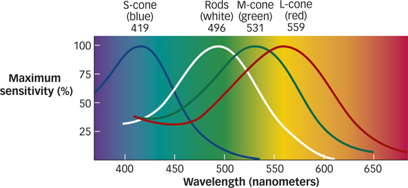
Figure 4.7: FIGURE 4.7 Seeing in Color We perceive a spectrum of color because objects selectively absorb some wavelengths of light and reflect others. Color perception corresponds to the summed activity of the three types of cones. Each type is most sensitive to a narrow range of wavelengths in the visible spectrum—short (bluish light), medium (greenish light), or long (reddish light). Rods, represented by the white curve, are most sensitive to the medium wavelengths of visible light but do not contribute to color perception.
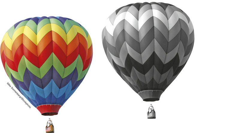
The full-color image on the left is what you’d see if your rods and cones are fully at work. The grayscale image on the right is what you’d see if only your rods are functioning.
Mike Sonnenberg/iStockphoto
Cones come in three types; each type is especially sensitive to either red (long-wavelength), green (medium-wavelength), or blue (short-wavelength) light. Red, green, and blue are the primary colors of light; color perception results from different combinations of the three basic elements in the retina. For example, lighting designers add primary colors of light together, such as shining red and green spotlights on a surface to create a yellow light, as shown in FIGURE 4.8. Notice that in the center of the figure, where the red, green, and blue lights overlap, the surface looks white. This demonstrates that a white surface really is reflecting all visible wavelengths of light.
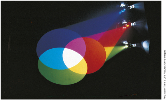
Figure 4.8: FIGURE 4.8 Color Mixing The millions of shades of color that humans can perceive are products not only of a light’s wavelength, but also of the mixture of wavelengths a stimulus absorbs or reflects. Colored spotlights work by causing the surface to reflect light of a particular wavelength, which stimulates the red, blue, or green photo-pigments in the cones. When all visible wavelengths are present, we see white.
Fritz Goro/Time & Life Pictures/Getty Images
What happens when the cones in your eyes get fatigued?
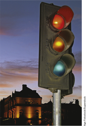
Many people (including about 5% of all males) inherit conditions in which either the “red” or the “green” photoreceptors do not transduce light properly. Such people have difficulty distinguishing hues that to typical individuals appear as red or green. Unfortunately, in the United States, traffic signals use red and green lights to indicate whether cars should stop or go through an intersection. Why do drivers with red–green blindness not risk auto accidents every time they approach an intersection?
Age Fotostock/Superstock
A genetic disorder in which one of the cone types is missing—and, in some very rare cases, two or all three—causes a color deficiency. This trait is sex-linked, affecting men much more often than women. Color deficiency is often referred to as color blindness, but in fact, people missing only one type of cone can still distinguish many colors, just not as many as someone who has the full complement of three cone types. You can create a kind of temporary color deficiency by exploiting the idea of sensory adaptation. Staring too long at one color fatigues the cones that respond to that color, producing a form of sensory adaptation that results in a color afterimage. To demonstrate this effect for yourself, follow these instructions for FIGURE 4.9:
- Stare at the small cross between the two color patches for about 1 minute. Try to keep your eyes as still as possible.
- After a minute, look at the lower cross. You should see a vivid color aftereffect that lasts for a minute or more. Pay particular attention to the colors in the afterimage.
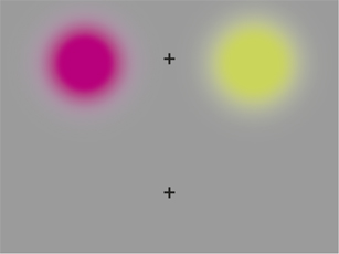
Figure 4.9: FIGURE 4.9 Color Afterimage Demonstration Follow the accompanying instructions in the text, and sensory adaptation will do the rest. When the afterimage fades, you can get back to reading the chapter.
Were you puzzled that the red patch produces a green afterimage and the green patch produces a red afterimage? When you view a color, let’s say, green, the cones that respond most strongly to green become fatigued over time. Now, when you stare at a white or gray patch, which reflects all the colors equally, the green-sensitive cones respond only weakly compared with the still-fresh red-sensitive cones, which fire strongly. The result? You perceive the patch as tinted red.
The Visual Brain
What is the relationship between the right and left eyes, and the right and left visual fields?
Streams of action potentials containing information encoded by the retina (neural impulses) travel to the brain along the optic nerve. Half of the axons in the optic nerve that leave each eye come from retinal ganglion cells (RGCs) that code information in the right visual field, whereas the other half code information in the left visual field. These two nerve bundles link to the left and right hemispheres of the brain, respectively (see FIGURE 4.10). The optic nerve travels from each eye to the thalamus. From there, the visual signal travels to the back of the brain, to a location called area V1, the part of the occipital lobe that contains the primary visual cortex. Here the information is systematically mapped into a representation of the visual scene.
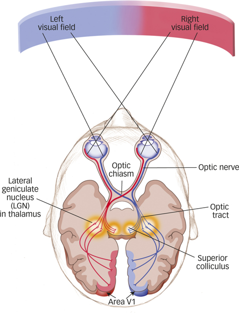
Figure 4.10: FIGURE 4.10 Visual Pathway from Eye through Brain Objects in the right visual field stimulate the left half of each retina, and objects in the left visual field stimulate the right half of each retina. The optic nerves, one exiting each eye, are formed by the axons of retinal ganglion cells emerging from the retina. Just before they enter the brain at the optic chiasm, about half the nerve fibers from each eye cross. The left half of each optic nerve (representing the right visual field) runs through the brain’s left hemisphere via the thalamus, and the right half of each optic nerve (representing the left visual field) travels this route through the right hemisphere. So, information from the right visual field ends up in the left hemisphere, and information from the left visual field ends up in the right hemisphere.
area V1
The part of the occipital lobe that contains the primary visual cortex.
Neural Systems for Perceiving Shape
One of the most important functions of vision involves perceiving the shapes of objects; our day-to-day lives would be a mess if we couldn’t reliably differentiate between a warm doughnut with glazed icing and a straight stalk of celery. Perceiving shape depends on the location and orientation of an object’s edges. As you read in the Neuroscience and Behavior chapter, neurons in the visual cortex selectively respond to bars and edges in specific orientations in space (Hubel & Wiesel, 1962, 1998). In effect, area V1 contains populations of neurons, each “tuned” to respond to edges oriented at each position in the visual field. This means that some neurons fire when an object in a vertical orientation is perceived, other neurons fire when an object in a horizontal orientation is perceived, still other neurons fire when objects in a diagonal orientation of 45° are perceived, and so on (see FIGURE 4.11). The outcome of the coordinated response of all these feature detectors contributes to a sophisticated visual system that can detect where a doughnut ends and celery begins.
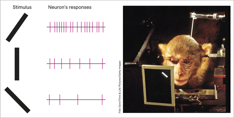
Figure 4.11: FIGURE 4.11 Single-Neuron Feature Detectors Area V1 contains neurons that respond to specific orientations of edges. Here a single neuron’s responses are recorded (left) as the monkey views bars at different orientations (right). This neuron fires continuously when the bar is pointing to the right at 45°, less often when it is vertical, and not at all when it is pointing to the left at 45°.
Fritz Goro/Time & Life Pictures/Getty Images
Pathways for What, Where, and How
Two functionally distinct pathways, or visual streams, project from the occipital cortex to visual areas in other parts of the brain (see FIGURE 4.12). One pathway, the ventral (below) stream, travels across the occipital lobes to the temporal lobe and includes brain areas that represent an object’s shape and identity—in other words, what it is, essentially a “what” pathway (Kravitz et al., 2013; Ungerleider & Mishkin, 1982). The other pathway, the dorsal (above) stream, travels up from the occipital lobe to the parietal lobes (including some of the middle and upper levels of the temporal lobes), connecting with brain areas that identify the location and motion of an object—in other words, where it is (Kravitz et al., 2011). Because the dorsal stream allows us to perceive spatial relations, researchers originally dubbed it the “where” pathway (Ungerleider & Mishkin, 1982). Neuroscientists later argued that because the dorsal stream is crucial for guiding movements, such as aiming, reaching, or tracking with the eyes, the “where” pathway should more appropriately be called the “how” pathway (Milner & Goodale, 1995).
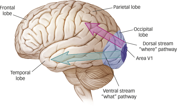
Figure 4.12: FIGURE 4.12 Visual Streaming The ventral pathway courses from the occipital visual regions into the lower temporal lobe and enables us to identify what we see. The dorsal pathway travels from the occipital lobe through the upper regions of the temporal lobe into the parietal regions and allows us to locate objects, to track their movements, and to move in relation to them.
What are the main jobs of the ventral and dorsal streams?
Some of the most dramatic evidence for the existence of these two pathways comes from studying patients with brain injuries. For example, a woman known as D.F. suffered permanent damage to a large region of the lateral occipital cortex, an area in the ventral stream (Goodale et al., 1991). Her ability to recognize objects by sight was greatly impaired, although her ability to recognize objects by touch was normal. This suggests that her visual representation of objects, not her memory for objects, was damaged. D.F.’s condition is called visual form agnosia, the inability to recognize objects by sight (Goodale & Milner, 1992, 2004). Conversely, other patients with brain damage to the parietal lobe, a section of the dorsal stream, have difficulty using vision to guide their reaching and grasping movements (Perenin & Vighetto, 1988). However, their ventral streams are intact, meaning they recognize what objects are. We can conclude from these two patterns of impairment that the ventral and dorsal visual streams are functionally distinct; it is possible to damage one while leaving the other intact.
visual form agnosia
The inability to recognize objects by sight.
SUMMARY QUIZ [4.2]
Question
4.4
| 1. |
The world of light outside the body is linked to the world of vision inside the central nervous system by the |
- cornea.
- lens.
- retina.
- optic nerve.
c.
Question
4.5
| 2. |
Light striking the retina, causing a specific pattern of response in the three cone types, leads to our ability to see |
- motion.
- colors.
- depth.
- shadows.
b.
Question
4.6
| 3. |
In which part of the brain is the primary visual cortex, where encoded information is systematically mapped into a representation of the visual scene? |
- the thalamus
- the lateral geniculate nucleus
- the fovea
- area V1
d.