2.3 Neurons and Neural Communication
In the next few pages, you will be introduced to some fairly tough biology concepts. Stay focused, do not give up, and reread the section as necessary. Communication among the cells in your nervous system underlies every behavior and mental process. In order to fully understand psychology, you need to understand the biology of the nervous system. So prepare to learn about something truly electrifying: the human neuron.
53
Just the Basics
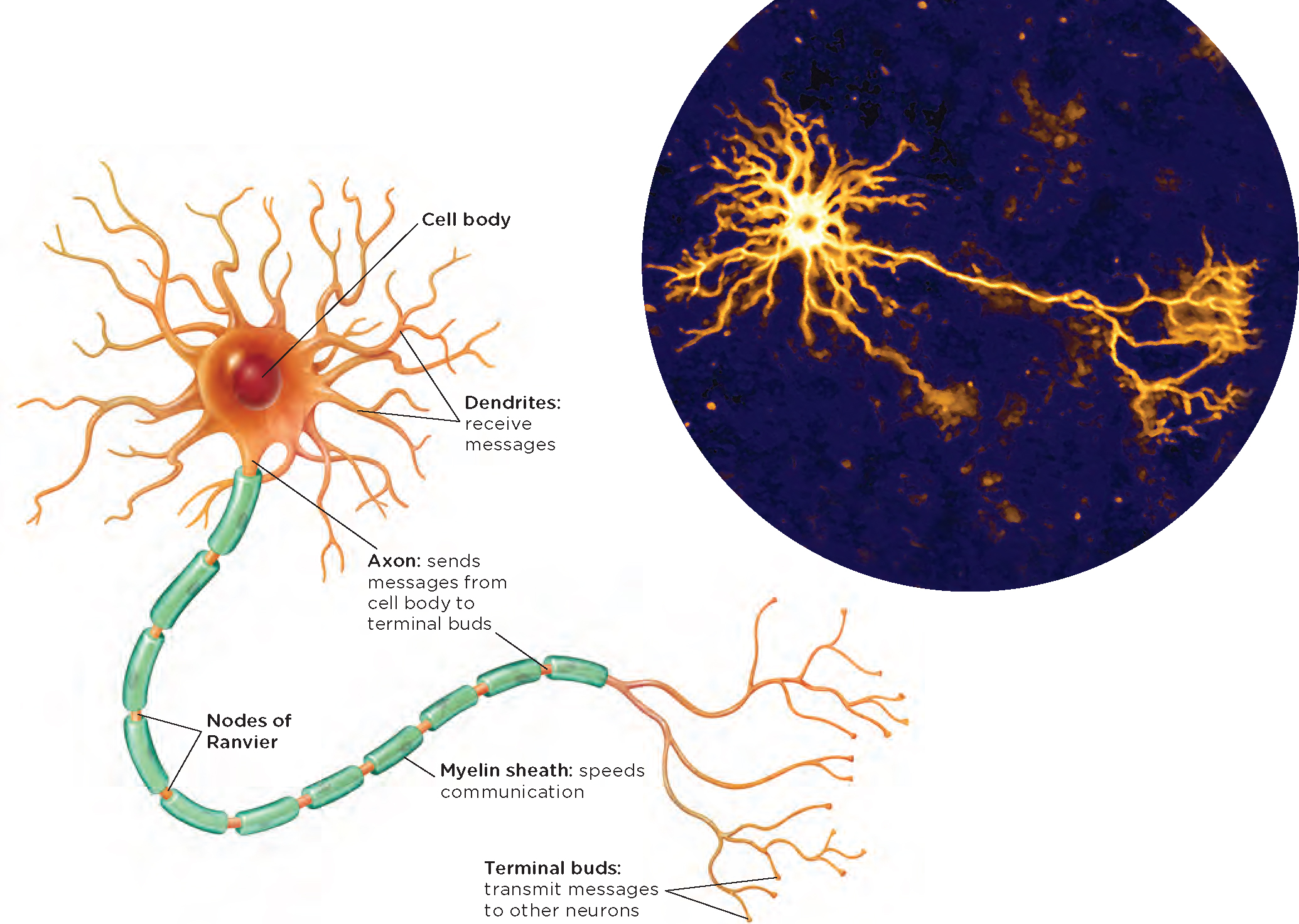
The Structure of a Typical Neuron
LO 2 Label the parts of a neuron and describe an action potential.
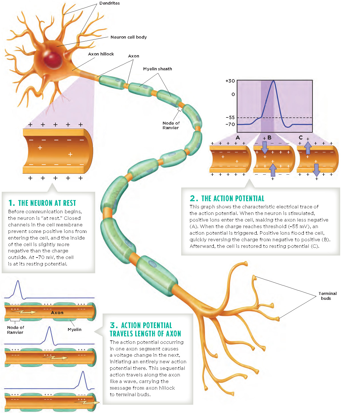
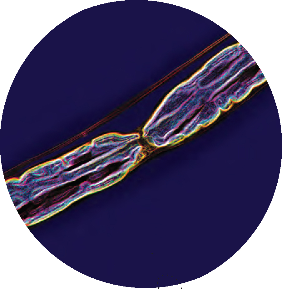
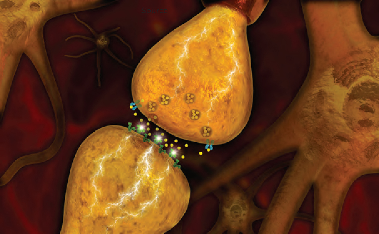
A typical neuron has three basic parts: a cell body, dendrites, and an axon (Figure 2.1). The cell body of a neuron contains the standard components found in most human cells: a nucleus containing DNA, protein-producing mechanisms, and other structures that nourish the cell. Extending from the cell body are many dendrites (den-drīts), which are tiny, branchlike fibers. Generally projecting in the opposite direction from the dendrites is a single axon, which is a long, skinny, tubelike extension of the cell body, with branches ending in terminal buds. Many axons are surrounded by a myelin sheath (mī-ə-lən shəth), a fatty substance that provides insulation. The axon is not entirely enclosed, but rather, covered in segments of myelin. The gaps between segments of the myelin are called nodes of Ranvier. The synapse (si-naps) is the tiny gap between a terminal bud of one axon and a neighboring dendrite of the next neuron (see photo). Just for perspective, the synaptic gap is only about 0.000127 millimeters (1.27 × 10⁴) wide, whereas a single sheet of printer paper is 0.1 millimeter thick.
Holding it Together: Glial Cells
The function of neurons is to transmit information up and down the body all day and night, and neurons need a little support and nurturing to get this tough job done. This is where the glial cells (glē-əl) come into play. In the human brain, glial cells outnumber neurons by approximately 50 to 1 (Yuhas & Jabr, 2012). They hold neurons together (glia means “glue” in Greek) and maintain the structure of the nervous system. For many years, scientists believed that glial cells simply kept things together, but we now know they do much more (Ndubaku & de Bellard, 2008).
Glial cells come to the rescue if the brain is injured. When Brandon was shot in the head, glial cells called microglia began multiplying and secreting substances to defend his brain from infection and inflammation (d’Avila et al., 2012; Streit, 2000). Another class of glial cells called astrocytes began restoring the barrier between his brain and blood (Gruber, 2009). Astrocytes have been found to support communication between neurons as well (Araque & Navarrete, 2010). Another type of glial cells, Schwann cells, make the myelin that envelops axons.
54
Communication Within and Between
Neurons have properties that allow them to communicate with other cells. But what information do they convey? In essence, the message is simple: “I have been activated.” Neurons are activated in response to sensations, thoughts, and other neurons, and this forms the basis for all that we think, feel, and do.
Processes Inside the Neuron
A neuron is surrounded by and contains electrically charged solutions (Infographic 2.1). If the total charge in each of these solutions is different, a voltage will be generated between the outside and the inside of the cell. This voltage is determined by the electrical characteristics of particles called ions. (Some ions are negatively charged; others are positively charged.) The difference in the charges inside and outside of the neuron determines its polarity, the degree to which it is positive or negative overall. Two processes direct the flow of positive and negative ions into or out of the cell. Diffusion is the natural tendency of the ions to spread out or disperse, and electrostatic pressure causes similarly charged ions to spread apart and oppositely charged ions to move toward each other (like the behavior of magnets). The concentrations, inside and outside of the cell, of positively charged ions (sodium and potassium) and negatively charged ions (protein molecules) determine the activity in most neurons. A neuron is encased in a membrane that is selectively permeable, allowing only some of the ions to pass in and out of its channels. The membrane is impermeable to positive sodium ions and negative protein ions (it does not allow these ions to enter or exit). Positive sodium ions move toward the membrane from the outside, and negative protein ions move toward the membrane from the inside; each moves closer to its side of the membrane wall (because the opposite charges are attracted to each other).
Inside the neuron, the concentration of potassium ions is greater than that outside the membrane. Also inside the cell are negatively charged protein ions, which do not exist outside the cell. These protein ions are attracted to the excess positive charge outside and move toward the membrane, but they are too big to go through. Because the protein ions can’t get out, the inside of the neuron is negatively charged when the neuron is not active. The concentration of sodium ions inside the cell is much less than its concentration on the outside of the cell. As a result, the sodium ions on the outside are attracted to the cell wall. This is due not only to the force of diffusion, but also to the force of attraction that occurs between oppositely charged ions. An electrical potential is created by the differences in charge between the outside and the inside of the neuron, which is its resting potential.
Resting Potential
The resting potential represents the electrical potential of a cell “at rest,” the condition of a cell when it is not activated. The solutions on either side of the membrane wall come into equilibrium with a slightly more negative charge inside. In the resting state, the voltage inside the cell is about −70 millivolts (mV) compared to the voltage on the outside. (Think of a resting neuron like a battery, which holds a charge. For comparison, one double A battery has a 1,500 mV charge.) This resting potential is only one part of the story. Let’s look at what happens when a neuron stops “resting” and goes into “action.”
Action Potential
Although the positive sodium ions are being pulled toward the inside of the cell, they cannot move into the cell until the neuron is stimulated by neighboring cells. When this happens, a signal instructs the channels in the cell membrane to open up. The channels, or gates, open, freeing the positive sodium ions to move into the cell, through the dendrites, to the cell body, and to finally reach the beginning of the axon, known as the axon hillock. The positive sodium ions cannot leave the cell, because the channels in the dendrites closed immediately after they entered.
55
INFOGRAPHIC 2.1: Communication Within Neurons
Neural communication involves different processes within and between neurons. In this infographic, we follow the electrical action that conveys messages within the neuron, from one end to the other.
56
The influx of the positive sodium ions at the axon hillock raises the internal cell voltage of the first segment of the axon from the resting voltage of −70 mV to the threshold potential of −55 mV relative to the outside. This change in voltage causes the sodium gates to open, and the positive sodium ions flood into the cell. The voltage inside that section of the axon rises rapidly, increasing from −55 mV to +30 mV, after which the sodium gates immediately close. This produces a spike in the value of the voltage within the cell, as the charge inside the cell becomes more positive than that outside of the cell. This is the action potential, or the spike in electrical energy that passes through the axon of a neuron.
The potassium gates open, and the positive potassium ions, now repelled by the much more positively charged cell interior, flow out of the cell as the similarly charged ions repel each other. This segment of the neuron returns to the resting potential. However, because the proportion of sodium and potassium ions inside and outside the cell is not the same as before (the ions are no longer at equilibrium), a sodium/potassium pump within the cell membrane brings them back to their original values by pumping the excess positive sodium ions back outside the cell and the positive potassium ions back in. In this way, the solutions inside and outside this segment of the axon return to equilibrium.
Moving Down the Axon
The firing of the first segment of the axon produces excess positive sodium ions on the inside of the cell, and these positive sodium ions diffuse to the next segment within the axon. This causes the voltage of the second segment of the axon to reach the threshold potential (−55 mV), opening its sodium gates and causing a spike of voltage there (+30 mV). Meanwhile, the first segment of the axon returns to its resting potential so that the electrical spike cannot go in that direction. This process repeats through each segment of the axon, like a row of dominos tumbling down. Every time a segment of the axon fires, the positive sodium ions flood in from the outside of the cell, while the prior segment returns to its resting potential, all along the length of the axon to its end. Each action potential takes about 1 millisecond to complete, and a typical neuron can fire several hundred times per second.
Excitatory and Inhibitory Signals
How do neighboring cells initially signal for the channels to open up to let the positive sodium ions move into the dendrites of a neuron? The message to fire begins at the dendrites. Chemical messages from surrounding neurons are sent to the cell body. If enough sending neurons signal the receiving neuron to pass along the message, their combined signal becomes excitatory and the neuron will fire. However, not all neighboring neurons send an excitatory signal. Some will send an inhibitory signal, instructing the neuron not to fire. For an excitatory signal to occur, there have to be more excitatory than inhibitory signals, and the difference between the two has to meet the threshold potential of −55 mV. If “enough” positively charged ions enter the cell, the potential of the neuron increases and reaches the threshold, or trigger point, and the cell “fires.”
All-or-None
Action potentials are all-or-none: They either happen or they don’t. Their strength remains the same no matter what occurs. A neuron conveys the strength of a stimulus by firing more often and delivering its message to more neurons. This is how we sense the difference between a quiet sound and a loud bang, for example. When we hear a loud bang: (1) The number of sensory neurons being fired is greater than the number that would have been fired for a quiet sound, and (2) each individual neuron fires more often than it would for a quiet sound. Thus, there is no such thing as a partial action potential, or a strong or weak action potential.
57
Myelin Sheath
The firing of a neuron is facilitated by the myelin sheath, which insulates and protects the tiny spikes in electricity happening inside the axon. Because myelin is such a good insulator, it does not allow the exchange of ion fluid between the inside and the outside of the cell membrane. However, the axon is not covered with myelin at the nodes of Ranvier (you can see this in the photograph to the right). The exchange of ion fluid can only happen at the nodes of Ranvier, where there is no myelin. The action potential thus appears to “jump” from node to node, as opposed to traversing the entire axon in one continuous movement, speeding the transmission of the signal. The speed of the action potential in an unmyelinated axon is approximately 1.1 to 4.5 miles per hour (mph), and a myelinated axon has transmission speeds of approximately 157 to 268 mph (Susuki, 2010). Unmyelinated axons, or those damaged from multiple sclerosis or other diseases, have slower transmission speeds, because the signal must make its way down the entire length of the axon. The damaged myelination caused by multiple sclerosis can lead to many symptoms, including fatigue, trouble with vision, and cognitive disabilities (Su, Banker, Bourdette, & Forte, 2009).
Communication Between
LO 3 Illustrate how neurons communicate with each other.
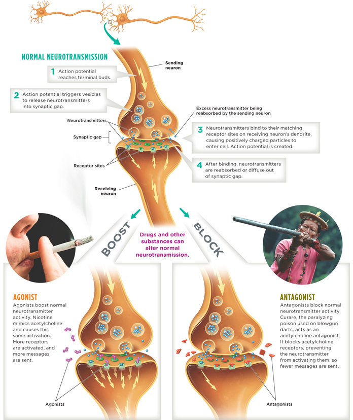
Neurons communicate with each other via chemicals called neurotransmitters. (Infographic 2.2 demonstrates communication of neurotransmitters in detail.) An action potential moves down the axon, eventually reaching the branches of the terminal buds. The signal to release neurotransmitters is the voltage change from the action potential, which results in vesicles (small fluid-filled sacs) that contain neurotransmitters attaching to the membrane on the terminal bud. This allows the neurotransmitter to be released into the synaptic gap. The majority of these neurotransmitters drift across the synaptic gap and come into contact with receptor sites of the receiving neuron’s dendrites. Because there are a variety of neurotransmitters, a variety of receptor sites also exist. Just as it takes the right key to unlock a door, it is necessary for the right neurotransmitter to fit a corresponding receptor site to convey the message.
When the neurotransmitters latch onto the receptors of the dendrites of the receiving neuron, tiny gates in the receiving cell’s membrane fly open, ushering positively charged particles into the cell and thus restarting the cycle of the action potential (if the threshold is met). Keep in mind that the firing of one neuron contributes to the potential for neighboring neurons to fire as a result of its chemical message.
Reuptake
When neurotransmitters are released into the synapse, many of them bind to receptors. Neurotransmitters can be reabsorbed by the sending terminal bud in a process known as reuptake. Those that are not reabsorbed drift out of the synaptic gap, through diffusion. This is how the synaptic gap is cleared of neurotransmitters, in preparation for the next release of a new batch of chemical messengers.
58
Major League Players: Neurotransmitters
LO 4 Summarize various neurotransmitters and the roles they play in human behavior.
As we have already mentioned, there are different types of neurotransmitters. Researchers have identified approximately 100 of them, with many more yet to be discovered. We already know that neurotransmitters secreted by one neuron under certain conditions can cause neighboring neurons to fire. Now let’s turn to how different neurotransmitters influence us in a variety of ways. The secretion of neurotransmitters can lead to a neuron firing, which can effect the regulation of mood, appetite, muscles, organs, arousal, and a variety of other outcomes. By examining what happens when neurotransmitters are not at their ideal levels, we can understand their effect at normal levels. We will describe only a handful of neurotransmitters, starting with the first neurotransmitter discovered, acetylcholine.
Acetylcholine
Acetylcholine is a neurotransmitter that relays messages from motor neurons to muscles, enabling movement. Any time you move some part of your body, whether dancing your fingers across a keypad or bouncing your head to one of your favorite songs, you have, in part, acetylcholine to thank for this. Too much acetylcholine leads to muscle spasms; too little causes paralysis. Acetylcholine is also known to be involved in memory. In particular, low levels of acetylcholine in the brain have been linked to Alzheimer’s disease (Kihara & Shimohama, 2004), which can lead to problems with memory, language, and thinking.
Glutamate and Gaba
Much of the transmission of information involves two neurotransmitters: glutamate and GABA (short for gamma-aminobutyric acid). Glutamate is an excitatory neurotransmitter, so its main job is to kick neurons into action (make them fire), whereas GABA is inhibitory (it puts the brakes on firing). Glutamate plays a central role in learning and memory (Riedel, Platt, & Micheau, 2003); its overactivity may be associated with strokes (Castellanos et al., 2008); and its underactivity is theorized to be involved in some of the symptoms of schizophrenia (Gordon, 2010).
Norepinephrine
Norepinephrine has a variety of effects in the nervous system, but one of its most important functions is to help prepare the body for stressful situations. Think about how Brandon was surrounded by gunfire on every side while serving in Fallujah. Norepinephrine was working to enable Brandon’s nervous system to initiate action. In the brain, norepinephrine is involved in regulating arousal and sleep (Jones, 2003).
Serotonin
Serotonin is a neurotransmitter that plays a key role in controlling appetite, aggression, and mood, and also regulates sleep and breathing. Abnormally low serotonin activity is thought to drive depression. Many of the popular antidepressants called serotonin reuptake inhibitors (SSRIs), including Prozac and Zoloft, work to boost the effects of this “feel good” neurotransmitter (Chapter 14). Normally, neurotransmitters that do not connect with receptor sites can be reabsorbed by the sending terminal bud in the reuptake process. SSRIs work to prevent the reabsorption of serotonin back into the sending neuron. The longer the neurotransmitters (in this case, serotonin) are available in the gap, the more time they have to attach to a receptor.
Dopamine
The neurotransmitter dopamine is known to play a role in problems with drug use. Repeated use of drugs overstimulates and damages the functioning of the neurons in the brain’s reward circuit, theoretically making it more difficult to enjoy non-drug-related activities that would otherwise be rewarding. Dopamine also plays a key role in learning through reinforcement, attention, and regulating body movements. Deterioration of neurons that produce dopamine is linked to Parkinson’s disease, an incurable disorder that causes trembling of the hands, arms, legs, and face, and difficulty with movement, coordination, and balance.
59
INFOGRAPHIC 2.2: Communication Between Neurons
Messages travel within a neuron using electrical currents. But communication between neurons depends on the movement of chemicals—neurotransmitters. Though they all work in the same way, there are many different types of neurotransmitters, each linked to unique effects on behavior. However, drugs and other substances, known as agonists and antagonists, can alter this process of communication between neurons by boosting or blocking normal neurotransmitter activity.
60
Endorphins
Endorphins are a group of naturally produced opioids (Chapter 4) that regulate the secretion of other neurotransmitters. The term endorphin is derived from the words “endogenous,” meaning it is created within, and “morphine.” With brisk exercise the production of endorphins is increased. This reduces the experience of pain and elevates mood. Endorphins are released in response to pain, thereby blocking pain receptor sites to prevent the pain message from being sent.
Agonists and Antagonists
Drugs and other substances influence behavior by interfering at the level of the synapse (Chapter 4 and Chapter 14). Certain substances can be used to act like neurotransmitters and others can be used to block normal neurotransmitter activity. Agonists are substances that increase the normal activity of the neurotransmitter (whether it normally sends an excitatory or inhibitory signal) and antagonists reduce the activity or block the release of the neurotransmitter (Infographic 2.2). For example, some substances, such as nicotine and muscarine (found in poisonous mushrooms), increase the secretion of acetylcholine, causing sweating, pupil constriction, nausea, and respiratory distress. These substances increase the normal activity of acetylcholine; thus, they are agonists. On the other hand, the popular anti-wrinkle treatment Botox is an antagonist because it blocks acetylcholine release, paralyzing the facial muscles so they can no longer wrinkle the overlying skin (Kim, Oh, & Paik, 2006).
Caffeine and Neurotransmitters

Did you jump-start your day with coffee? Or perhaps you sipped a caffe latte, tea, or soda this morning? Caffeine works its magic, perking you up at the crack of dawn or jolting you from that mid-afternoon daze by manipulating the nervous system. One way caffeine works is by blocking the receptors for a neurotransmitter called adenosine. (Thus, caffeine’s primary role is as an antagonist.) When adenosine latches onto receptors, it slows down their activity (making them less likely to fire), and this tends to make you feel drowsy. Caffeine resembles adenosine enough that it can dock onto the same receptors (“posing” as adenosine), which makes fewer receptors available for adenosine. With caffeine occupying its receptors, adenosine can no longer exert its calming effect on the brain (Julien, Advokat, & Comaty, 2011). The result: More neurons fire and you feel full of energy. But note that continued use of caffeine can lead to anxiety, restlessness, and headaches if you reduce your consumption (Ozsunger, Brenner, & El-Sohemy, 2009; Chapter 4).
The effects of caffeine do not stop at the brain. As anyone who has enjoyed a double latte can testify, caffeine kicks the body into high gear by increasing activity in the branch of the nervous system serving the body (Corti et al., 2002). In the next section, we will examine the nervous system running through your arms, legs, fingers, toes—and everywhere else.
show what you know
Question 2.4
1. Many axons are surrounded by a __________, which is a fatty substance that insulates the axon.
Question 2.5
2. When Brandon was injured, __________ played an important role in his recovery by defending against infection and inflammation of the brain, as well as holding neurons together and maintaining the structure of the nervous system.
- glial cells
- dendrites
- action potentials
- sodium ions
Question 2.6
3. __________ are released into the __________ when an action potential reaches the branches of the terminal buds.
- Sodium ions; synaptic gap
- Neurotransmitters; synaptic gap
- Potassium ions; cell membrane
- Neurotransmitters; sodium gates
Question 2.7
4. Neural communication is very complicated. Draw a diagram depicting the process of neural communication, then explain it to yourself while looking at what you have drawn.
 CHECK YOUR ANSWERS IN APPENDIX C.
61