3.2 Vision
THE EYES OF THE TRIPLETS
 When Liz became pregnant back in 1999, she was busy caring for her firstborn child, 3-year-old Sarah. Her marriage wasn’t working. In fact, she and her husband had already begun to contemplate divorce. It could not have been a worse time for Liz to discover she was going to have not one, not two, but three babies.

Many triplets are fraternal, meaning they come from three distinct egg–sperm combinations, each with its own genetic makeup. Liz was pregnant with identical triplets, which results when one fertilized egg splits three ways. The babies were born more than 3 months early by emergency cesarean delivery due to grave concerns about Zoe’s fetal heartbeat. Each of the three babies entered the world weighing less than 2 pounds.
101
Liz remembers feeling shocked and “scared to death” as she lay on the operating table watching her newborns wheeling by in incubators. She was well aware of the health dangers associated with prematurity (breathing problems, brain bleeds, visual impairment, and so on). All Liz could do was hope for the best.
After spending their first months of life in the hospital, hooked to feeding tubes, breathing tubes, and IVs, the triplets finally came home—Sophie first, Emma second, and finally Zoe. But 2 days after Emma arrived home, Liz realized that something was wrong. Emma’s eyes weren’t “tracking,” or following moving objects. Liz took Emma to a specialist, who said she was blind. Soon it became clear that Zoe and Sophie also had vision problems.
What happened to the triplets’ eyes? To understand how the girls lost their vision, we must first learn how the eye turns light into electrical and chemical impulses for the brain to interpret.
Light Is Sight
LO 4 Explain how electromagnetic energy is transduced into a sensation of vision.
When you look at a stop sign, would you believe you are sensing light waves bouncing off the stop sign and into your eyes? The eyes do not sense faces, objects, or scenery. They detect light. Remember, if you don’t have light, you don’t have sight. But what exactly is light? Light is an electromagnetic energy wave, composed of fluctuating electric and magnetic fields zooming from place to place at a very fast rate. And when we say “fast,” we mean from Atlanta to Los Angeles in a tenth of a second. Electromagnetic energy waves are everywhere all the time, zipping past your head, and bouncing off your nose. As you can see in Figure 3.1, light that is visible to humans falls along a spectrum or range of electromagnetic energy. TABLE 3.1 summarizes this range.
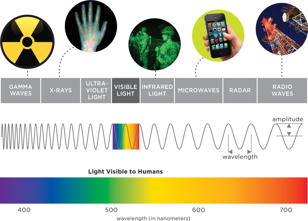
| EM Frequency | Real-World Examples |
| Gamma waves | Emitted by radioactive substances, such as the glucose tracers used in PET imaging |
| X-rays | X-ray machines (CT scanners included) used to generate pictures of the inside of the body |
| Ultraviolet | Sun rays that tan your skin (and can give you cancer) |
| Visible | What you see |
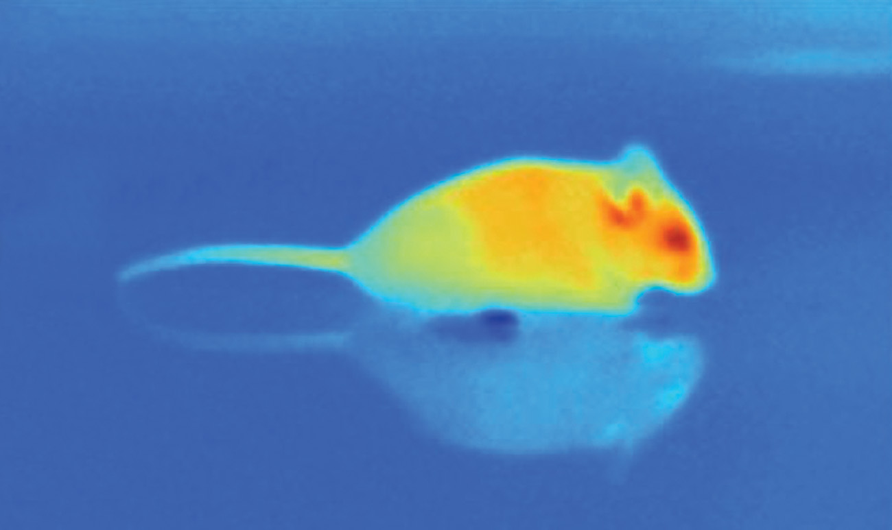
| Infrared | Heat emitted from human bodies |
| Microwaves | Microwave ovens, cell phones |
| Radio waves | AM and FM radios, televisions, wireless Internet |
| We use electromagnetic energy for a variety of purposes, from warming our dinners to carrying on digital conversations. Visible light is the only part of the spectrum detectable by human eyes. | |
| SOURCE: ADAPTED FROM BERKELEY LAB (N.D.). | |
102
Wavelength
The various types of electromagnetic energy can be distinguished by their wavelengths, which is the distance from one wave hump to the next (like the distance between the crests of waves rolling in the ocean; Figure 3.1). Gamma waves have short wavelengths and are located on the far left of the spectrum. At the opposite extreme (far right of the spectrum) are the long radio waves. The light humans can see falls in the middle of the spectrum, measuring between 400 to 700 nanometers (nm) or billionths of a meter (Brown & Wald, 1964). Wavelength also plays an important role in determining the colors humans and animals can detect.
The Colors We See
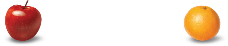
Although dogs can only see the world in blues, yellows, and grays (Coren, 2008, October 20), primates, including humans, can detect a wider spectrum of colors, including reds and oranges. This ability to see reds and oranges may be an adaptation to spot ripe fruits against the green backdrop of tree leaves (Rowe, 2002). Other creatures can see “colors” that we can’t. Snakes can detect infrared waves radiating off the bodies of their prey, and birds size up potential mates using the ultraviolet waves reflected by feathers (Bennett, Cuthill, Partridge, & Maier, 1996; Gracheva et al., 2010).
Features of Light
The colors you see result from light reflecting off of objects and reaching your eyes. Every color can be described according to three factors: hue, brightness, and saturation. The first factor, hue, is what we commonly refer to as “color” (blue jeans have a blue hue). Hue is determined by the wavelength reflecting off of an object: Violet has the shortest wavelength in the visible spectrum (400 nm), and red has the longest (700 nm). The brightness of a color represents a continuum from bright to dim in terms of the intensity of the color. Brightness depends on wave height, or amplitude, the distance from midpoint to peak (or from midpoint to trough; Figure 3.1). Just remember, the taller the height, the brighter the light. Saturation, or color purity, is determined by uniformity of wavelength. Saturated colors are made up of same-size wavelengths. Objects we see as pure violet, for instance, are reflecting only 700-nm light waves. We can “pollute” the violet light by mixing it with other wavelengths and the result will be a less saturated, pale lavender. Most colors in the environment are not pure. The red pigments in Angelina Jolie’s red lipstick probably reflect a mixture of waves in the 600–700 nm range, rather than pure 700-nm red. Combinations of these three basic features—hue, brightness, and saturation—can produce an infinite number of colors; our eyes are just not sensitive enough to tell them all apart. Amazingly, though, the average person sees some 2.3 million colors (Linhares, Pinto, & Nascimento, 2008).
103

The Perception of Colors
And now, the 2.3 million-dollar question: Most of us agree that stop signs are red and taxicabs are yellow, but can we be sure that one person’s perception of color is identical to another’s? Is it possible that your “red” may be someone else’s “yellow”? The colors we perceive do not actually exist in the world around us; they are the product of what is in our environment, including properties of the object and the wavelength of the light it reflects, and the brain’s interpretation of that light. Without getting inside the mind of another person and perceiving the world as he does, this question is very difficult to answer. When these factors change, so, too, do the colors we see.
try this
Go find the brightest, most saturated object you possess that has a yellow hue, and grab a strong flashlight while you are at it. Wait until it is dark outside. Now put the object on a table right in front of you and look at it with a dim light shining overhead. Next, shine the flashlight directly onto the yellow object and notice how your perception of the color changes. Finally, turn off all the lights in the room and notice again how your perception of the color changes.
Your three perceptions of this same yellow object will be very different—not because the object itself has changed, but rather because the light shining upon it (and thus, the light it reflects) has changed.
Color perception results from the interaction between light and brain activity. The eyes take light energy and transform it into neural code for the brain to interpret. The exquisite specificity of this code allows us to distinguish between electric blue and cobalt, lime green, and chartreuse. All this is made possible by the eye’s remarkable biology.
You Won’t Believe Your Eyes
The human eye is nothing short of an engineering marvel (Infographic 3.2). When you look in the mirror, you see only a fraction of each ping-pong-sized eyeball; the rest is hidden inside your head. Let’s explore this biological wonder, starting from the outside and working our way toward the brain.
The Cornea
The surface of the eye looks wet and glassy. This clear outer layer over the colored portion of the eye is called the cornea, and it has two important jobs: (1) shielding the eye from damage by dust, bacteria, or even a poke in the eye, and (2) focusing incoming light waves. About 65–75% of the focusing ability of the eye comes from the cornea, which is why imperfections in its shape can lead to blurred vision (National Eye Institute, 2012). Corneal flaws are primarily to blame for so many people (about a third of the American population) needing glasses or contact lenses. LASIK eye surgery, a popular alternative to corrective lenses, uses a laser to reshape the cornea so that it can focus light properly.
104
The Iris and the Pupil
Directly behind the cornea is the donut-shaped iris. When you say someone has velvety brown eyes, you are really talking about the color of her irises. The black hole in the center of the iris is called the pupil. In dim lighting, the muscles of the iris relax, widening the pupil to allow more light inside the eye. In bright sunlight, the iris muscles squeeze, constricting the pupil to limit the amount of light. At first glance, the pupils may seem empty and boring. But pupils are actually quite sexy. Studies show, for example, that men are more enticed by women with enlarged pupils (Hess, 1975; Tombs & Silverman, 2004). When looking at someone she finds attractive, a woman’s pupils dilate—and this response is magnified during the most fertile time of her monthly cycle (Laeng & Falkenberg, 2007).
The Lens and Accommodation
Behind the pupil is the lens, a tough, transparent structure that is similar in size and shape to an “M&M’s candy” (Mayo Clinic, 2011). Like the cornea, the lens specializes in focusing incoming light, but it can also change shape in order to adjust to images near and far, a process called accommodation. If you take your eyes off this page and look across the room, faraway objects immediately come into focus because your lens changes shape. As we age, the lens begins to stiffen, impairing our ability to focus on up-close images like the letters on a computer screen. You can tell if a person has this condition, called presbyopia, when she strains to read a book or restaurant menu at arm’s length. Most people develop some degree of presbyopia between 40 to 65 years old (Mayo Clinic, 2011).
The Retina
After passing through the cornea, pupil, and lens, light waves travel through the eyeball’s jellylike filling and land on the retina, a carpet of neurons covering the back wall of the eye. This area of the eye was the source of the triplets’ visual impairments. Emma, Zoe, and Sophie all suffered from retinopathy of prematurity (ROP), a condition in which blood vessels in the retina grow incorrectly. The last 3 months of pregnancy are critical for fetal eye development, when webs of blood vessels rapidly branch from the center of the retina outward, delivering crucial oxygen and nutrients to the developing tissue. The triplets were born over 3 months early, long before the blood vessel development was complete. The vessels began to branch abnormally, eventually pulling the retina from the back of the eye (Chawla et al., 2012).
The retina is responsible for the transduction of light energy into neural activity; that is, sensing light and relaying a message to the brain. Without the retina, vision is impossible.
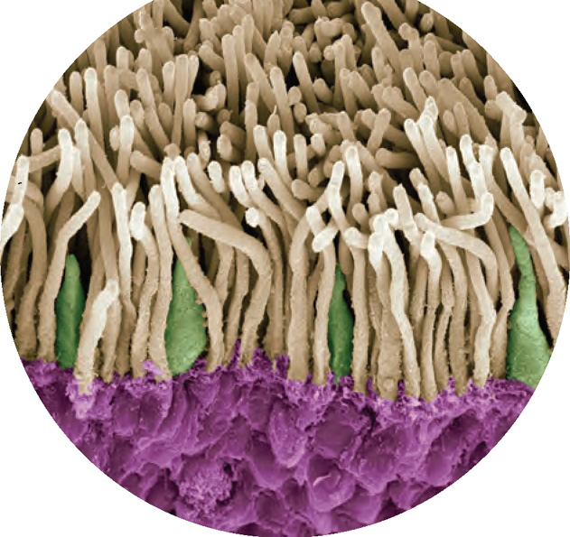
Photoreceptors and Other Neurons
LO 5 Describe the function of rods and cones.
The retina is home to millions of specialized neurons called photoreceptors, which absorb light energy and turn it into chemical and electrical signals for the brain to process. Two types of photoreceptors are located along the back of the retina, rods and cones, which get their names from their characteristic shapes. Rods are extremely sensitive, firing in response to even a single photon, the smallest possible packet of light (Rieke & Baylor, 1998). If rods were all we had, the world would look something like an old black-and-white movie. Cones enable us to enjoy a visual experience more akin to HDTV (except for those of us with color deficiencies, which we will discuss later). In addition to providing color vision, cones allow us to see fine details, such as the small print on the back of a shampoo bottle.
CONNECTIONS
In Chapter 2, we noted that neurons are activated in response to sensations. In the case of vision, the sensation is light and the neurons being stimulated start with the photoreceptors in the retina. When a photoreceptor fires, its message is sent on its way to the brain.
105
INFOGRAPHIC 3.2: Seeing
“Seeing” involves more than simply looking at an object. Vision is a complex process in which light waves entering the eye are directed toward the retina where they are transduced into messages the brain can understand. The triplets all experienced vision loss from retinopathy of prematurity (ROP), a condition in which blood vessels in the retina grow incorrectly. Damage to this crucial structure has a severe impact on vision.
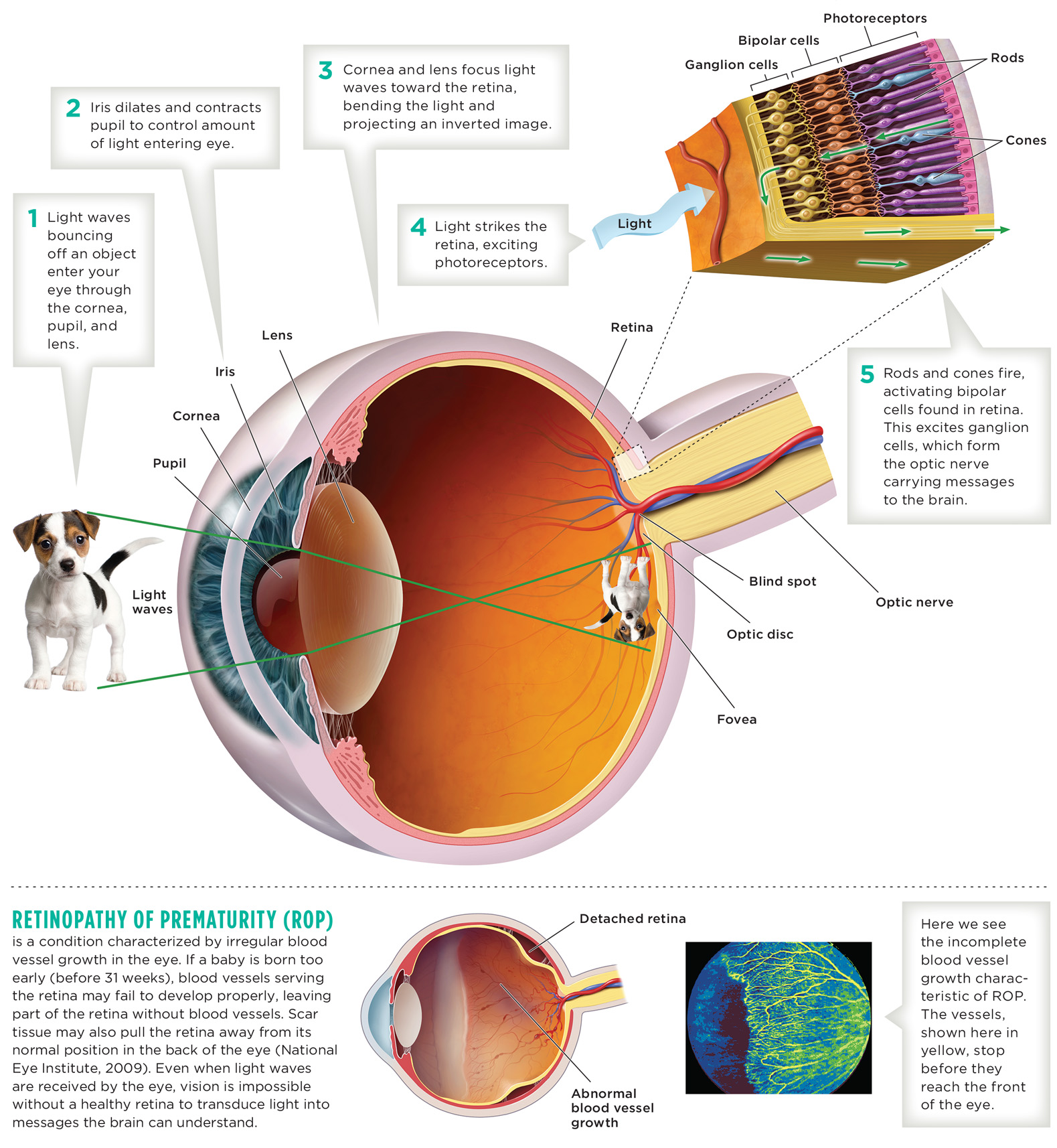
106
The rods and cones are just the first step in a complex signaling cascade involving many neurons, which ultimately leads to a visual experience in the brain. The rods and cones are in close proximity to bipolar cells, which are also a specialized type of neuron located approximately in the middle of the retina. When a rod or cone is stimulated by light energy, it conveys its signal to nearby bipolar cells. These bipolar cells in turn convey their signal to ganglion cells. The ganglion cells, which comprise another type of neuron, are located toward the front of the retina. Axons of the ganglion cells bundle together in the optic nerve, which is like an electrical cable hooking the retina to the brain. The axons of the ganglion cells form one bundle for each eye in the optic nerves, which exit the retina at the optic disc, causing a blind spot, since it lacks rods or cones. You can find your blind spot by following the instructions in the Try This.
try this
The Fovea
The retina in each eye is home to some 120 million rods and 6 million cones (Amesbury & Schallhorn, 2003). Rods are found everywhere in the retina except for a tiny central spot called the fovea. Although cones are packed most densely in the fovea, they can also be found sprinkled throughout the rest of the retina. When you need to study something in great detail (like the tiny serial number on the back of a computer), hold it under a bright light and stare at it straight-on. The cones in the fovea excel at sensing detail and operate best in ample light. If, however, you want to get a look at something in the dim light, focus your gaze slightly to its side, stimulating the super-light-sensitive rods outside the fovea.
Is that Oprah Winfrey Over There?
Let’s stop for a moment and examine how information flows through the visual pathway (Infographic 3.2). Suppose you are looking at Oprah Winfrey’s face. Remember, you’re actually seeing the light that her face reflects. Normally, light rays bouncing off Oprah would continue moving along a straight-line path, but they encounter the bulging curvature of the cornea covering your pupil, which bends them. (The light is later bent by the lens, but to a lesser extent.) The cornea redirects light rays on their way to the retina. Rays entering the top bend downward and those striking the base bend upward. The result is an inverted, or flip-flopped, projection on your retina. It’s like your eye is a movie theater, the retina is its screen, and the feature film being played is “Your World, Turned Upside Down” (Kornheiser, 1976; Stratton, 1896). The neurons in the retina respond to the stimulus, sending signals from the photoreceptor cells to the bipolar cells, and then on to the ganglion cells, which bundle into the optic nerves.
107
Feature Detectors
The optic nerves (one from each eye) intersect at a place in the brain called the optic chiasm. From there, information coming from each eye gets split, with about half traveling to the same-side thalamus and half going to the opposite-side thalamus. Interneurons then shuttle the data to the visual cortex, located in the occipital lobes in the back of your head. Neurons in the visual cortex called feature detectors specialize in detecting specific features of your visual experience, such as angles, lines, and movements. How these features are pieced together into a unified visual experience (I see Oprah!) is complex. Hubel and Wiesel (1979) proposed that visual processing begins in the visual cortex, where teams of cells respond to specifically oriented lines (as opposed to just pixel-like spots of light), and then continues in other parts of the cortex, where information from both eyes is integrated. Scientists have now identified at least 30 different areas in the brains of humans and other primates that play a role in visual processing (Ramachandran & Rogers-Ramachandran, 2009).
CONNECTIONS
Interneurons are found in the central nervous system (the brain and the spinal cord). As described in Chapter 2, the interneurons receive and help process signals from sensory neurons.
Now that we have outlined the general flow of information from eyes to brain, let’s learn how the visual system adapts to the ever-changing environment.
Dark and Light Adaptation
The eye has an amazing ability to adjust to drastic fluctuations in light levels. This process starts with the pupil, which rapidly shrinks and expands in response to light changes, and then continues with the rods and cones, which need more time to adjust to changes in lighting. When you walk into a dark movie theater after being outside in the bright sun, you can barely see an inch in front of your face. After a few minutes, your eyes start to adjust to the dark in a process called dark adaptation, which takes about 8 minutes for cones and 30 minutes for rods (Hecht & Mandelbaum, 1938; Klaver, Wolfs, Vingerling, Hoffman, & de Jong, 1998). Cones respond more quickly, so they are more useful in the first few minutes of dark adaptation. Then the rods kick into action, allowing you to make out silhouettes of objects and people. Why are these photoreceptors so sluggish in their responses to darkness? To restore their sensitivity to light, rods and cones must undergo a chemical change associated with protein molecules, and this takes time (Caruso, 2007, August 13; Ludel, 1978). Also keep in mind that, for most of human evolution, dark adaptation meant adjusting to the gradual setting of the sun (Caruso, 2007, August 13).
What happens when you leave the dark theater and return to the blinding light of day—do the eyes also adjust then? The answer is yes. With light adaptation, the pupil constricts to reduce the amount of light flooding the retina, and the rods and cones become less sensitive to light. Light adaptation occurs relatively quickly, lasting at most 10 minutes (Ludel, 1978). Dark and light adaptation demonstrate the eye’s ability to accommodate to changes in brightness—an amazing feat. But the eye is capable of much more; remember, it can discern among millions of colors.
In Living Color
Although we have discussed the various features of color, we have not yet explained how waves of electromagnetic energy result in our perception of colors. How does the brain know red from maroon, green from turquoise, grey from charcoal? Two main theories explain human color vision—the trichromatic theory and the opponent-process theory—and you need to understand both because they address different aspects of the phenomenon.
The Trichromatic Theory
LO 6 Compare and contrast the theories of color vision.
Proposed in the 1800s by an English physician-scientist, Thomas Young (1773–1829), and then expanded upon several decades later by Hermann von Helmholtz (1821–1894), a Prussian physicist, the trichromatic theory (trī-krō-′ma-tik) suggests that we have three types of cones. Red cones are excited by electromagnetic energy with wavelengths in the red range (about 620–700 nm); green cones fire in response to electromagnetic energy with wavelengths in the green realm (500–575 nm); and blue cones are activated by electromagnetic energy with wavelengths corresponding to blues (about 450–490 nm; Mollon, 1982).
108
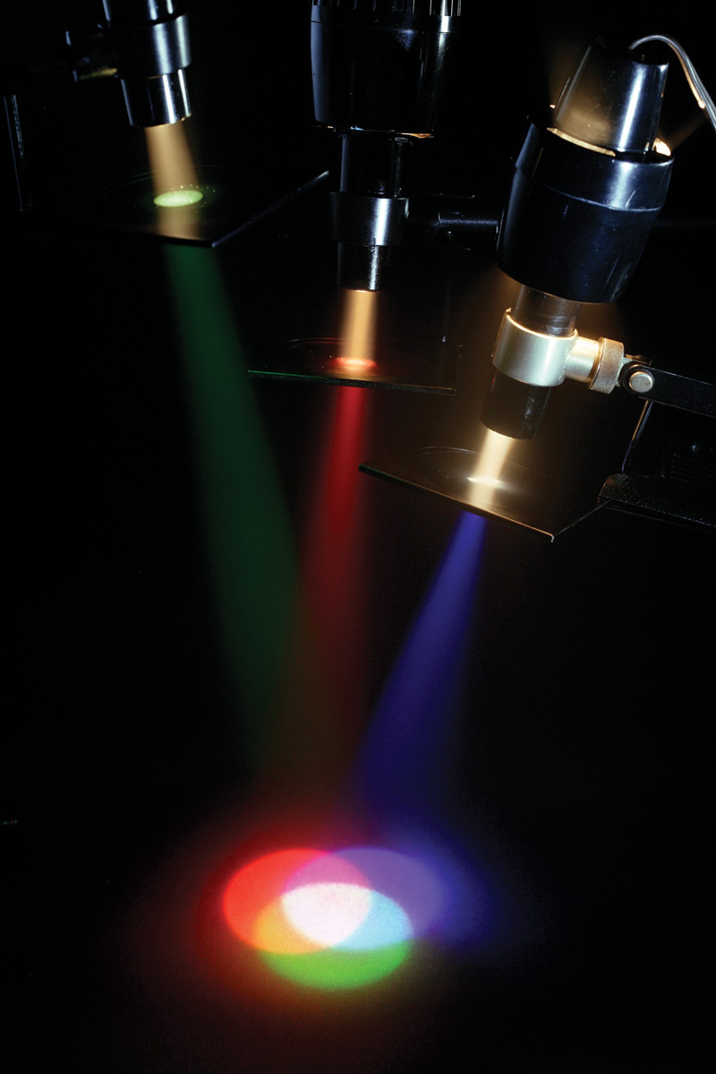
So how is it that we can detect millions of colors when our cones are only sensitive to red, green, and blue? The primary colors of light are also red, green, and blue, and when mixed together in equal proportions, they appear as white light (see the photograph to the right). According to the trichromatic theory, the brain identifies a precise hue by calculating patterns of excitement among the three cone populations. When you look at a yellow banana, for example, both the red and green cones fire, but not the blue ones. The brain interprets this pattern of red and green activation as “yellow.” And because white light is actually a mixture of all the wavelengths in the visible spectrum, it excites all three cone types, creating a sensation of “white” in the mind’s eye. Thus, it is the relative activity of the three types of cones that the brain uses to make its color calculations.
Color Deficiency and Color Blindness
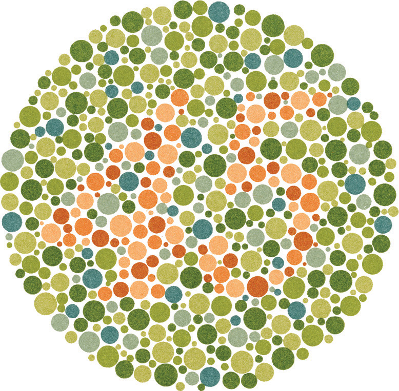
Loss or damage to one or more of the cone types leads to color deficiency, more commonly known as “color blindness.” These terms are often used interchangeably, but true color blindness is extremely rare. Sometimes color blindness is accompanied by extreme sensitivity to light and poor vision for detail, both resulting from deficient or missing cones (Tränkner et al., 2004). The condition associated with red–green color defects occurs in about 8% of men and less than 1% of women with European ancestors (Deeb, 2005). Color deficiencies may stem from problems with the cones. Most people with color deficiencies have trouble distinguishing between red and green because they are missing or deficient in the red or green receptors, but can see other colors very well. You can get an indication of red–green deficiency by looking at the Ishihara color plate below. If you can’t see the number shown, then you may have a red–green deficiency (Wong, 2011).
Ample research backs up the trichromatic theory, but there are some color-related phenomena it cannot explain. A prime example is the afterimage effect. An afterimage is an image that appears to linger in your visual field after its stimulus, or source, is gone.
try this
Opponent-Process Theory
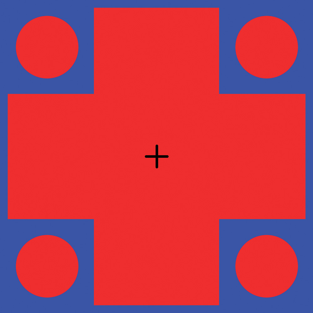
German physiologist Ewald Hering (1834–1918) realized that the trichromatic theory could not explain the visual phenomenon of the afterimage effect and thus developed the opponent-process theory of color vision. Hering proposed that in addition to the color-sensitive cones, a special group of neurons responds to opponent colors—pairs of colors such as red–green and blue–yellow that cannot be perceived simultaneously (there is no such thing as reddish-green or bluish-yellow light). One neuron in an opponent pair fires when you look at red, but is inactive when you see green. The other neuron gets excited by green but turned off by red. If you spend enough time staring at the red and blue picture in the Try This, the neurons excited by these colors become exhausted and stop responding. When you shift your gaze to the white surface, the opponent neurons fire in response to the green and yellow wavelengths. Meanwhile, the red and blue neurons are too fatigued to rally (when not fatigued, they would normally respond). You end up seeing an afterimage of the box and a plus sign in green and yellow. Research has provided strong support for the opponent-process theory, identifying particular types of neurons located in a region of the thalamus (DeValois & DeValois, 1975; Jameson & Hurvich, 1989).
109

As it turns out, we need both the trichromatic and opponent-process theories to clarify different aspects of color vision. Color perception occurs in the light-sensing cones in the retina and in the opponent cells serving the brain. The ability to perceive color is not based on processing at a single point along the visual pathway, or even one area of the brain (Gegenfurtner, 2003; Solomon & Lennie, 2007).
Ultimately, these color perceptions lead to cognitive and emotional responses. You may believe blue is a sad color, and yellow is warm and comforting. What you think and feel about a particular color depends on many factors, including your culture.
across the WORLD
Colors and Culture
 Red is a lucky color in China but a sign of bad fortune in Nigeria. People in the United States see white as a symbol of virginity and cleanliness; those in East Asia associate it with dying and bereavement (Sable & Akcay, 2010). Black is considered upscale and influential across cultures (Madden, Hewett, & Roth, 2000). And blue, the most popular “favorite color” in the world, carries different meanings for various groups: sanctity in the Jewish religion, warmth for the Dutch, and the color of the god Krishna for Hindus (Akcay, Dalgin, & Bhatnagar, 2011; Sable & Akcay, 2011).
SEEING RED IN CHINA
How do you explain the similarities and differences in these responses to color? Like many questions in psychology, we can use the biopsychosocial perspective to explore this. Seeing certain colors can elicit measurable biological responses. In one study, for example, people exposed to blue light—but not red light—experienced significant drops in blood pressure and breathing rate (Visweswaraiah & Telles, 2006). In addition, some scholars suggest that learning is a key factor shaping the cultural meanings of color (Sable & Akcay, 2011). If a color is repeatedly presented in a certain context (black clothes at a funeral, pink clothes on a girl baby), mental associations begin to take hold (black is for grieving; pink is for girls). It appears our perception of color is the result of a complex mixture of biological, psychological, and sociocultural factors.
CONNECTIONS
In Chapter 1, we mentioned that psychologists use the biopsychosocial perspective to understand behavior. Here, we look at how biology, learning, and culture influence differences in color perception.
Perception of color may vary significantly across cultures, and even between individuals, but the early stages of sensation are virtually the same for everyone with normal vision. The fact that the complex visual system works so well for so many people is really quite amazing. Equally intricate is the auditory system. Next we’ll discover how hearing works—and how easily it can be dismantled—using the triplets as our example.
110
show what you know
Question 3.4
1. The hue of a color is determined by the __________ of the light reflecting off an object.
Question 3.5
2. Cells contained in the retina that absorb light energy and turn it into chemical and electrical signals are called __________.
- opponent-processing
- photoreceptors
- fovea
- feature detectors
Question 3.6
3. Explain the two theories of color vision presented in the chapter and how they differ.
Question 3.7
4. It’s dark in your house, and you are struggling to see what time it is without turning on the light. You notice that if you turn your gaze slightly to the side of your watch, you can make out the large numbers. The ability to see these large details in the dark is due to your:
- presbyopia.
- optic disc.
- cones.
- rods.
 CHECK YOUR ANSWERS IN APPENDIX C.