6.5 The Biology of Memory
What did you do today? Did you have breakfast, brush your teeth, put your clothes on, drive your car, read an assignment, text a friend? Whatever you did, we are sure of one thing: It required a whole lot of memory. Every task described here, and probably every task you completed today, draws on a vast fund of memories residing in your brain. You could not send a text message without knowing how to spell, read, and use a cell phone—all things you had to learn and remember. Likewise, you could not drive without remembering how to unlock your car, start the engine, use the pedals. Memory is involved in virtually everything you do.
If memory is behind all your daily activities, important processes must be occurring in the brain to make this happen: both on the macro (large) and micro (small) scale. But as we learned from Clive’s experiences, the process is fragile and can be profoundly disrupted. Exploring the causes of memory failure in cases of amnesia can help us understand the biological basis of memory.
Amnesia
LO 13 Compare and contrast anterograde and retrograde amnesia.
In the months and years following Clive’s illness, researchers administered many tests to assess his cognitive functioning. They found his IQ to be within an average range but his ability to remember past events deeply impaired. When prompted to name as many musical composers as possible in 1 minute, Clive—a man who had devoted his career to the study of music—could only produce four: Mozart, Beethoven, Bach, and Haydn. He denied that dragonflies have wings and claimed he had never heard of John F. Kennedy (Wilson et al., 1995).
Clive was even more disabled when it came to developing new memories. Initially, he could not hold onto incoming information for more than a blink of an eye. If his wife Deborah left the room, even for a short trip to the restroom, he would welcome her back as if she had been away for years—embracing, celebrating, sometimes weeping. “How long have I been ill?” he would ask, forgetting the answer and repeating himself within seconds (Wearing, 2005, p. 181).
268
Amnesia, or memory loss, can result from either a physical or psychological condition. There are different types and degrees of amnesia, ranging from extreme (losing decades of autobiographical memories) to mild (temporarily forgetting names of acquaintances and other details following a concussion).
Anterograde Amnesia
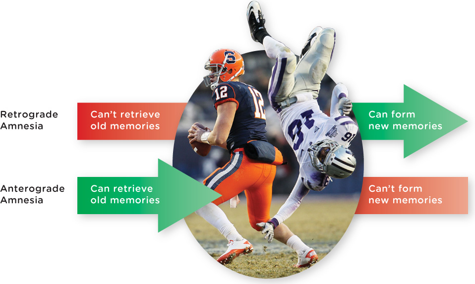
According to researchers, Clive suffers from “a more severe anterograde amnesia than any other patient previously reported” (Wilson et al., 1995, p. 680). Anterograde amnesia (an-tə-ˌrō-grād) is the inability to “lay down” or create new long-term memories (Figure 6.12), and it is generally caused by damage or injury to the brain, resulting from surgery, alcohol, head trauma, or illness. Someone with anterograde amnesia cannot form memories of events and experiences that occur following the damage to the brain, regardless of its cause. People affected by anterograde amnesia may be incapable of holding down a job, as their inability to lay down new memories affects their capacity to remember daily tasks. For Clive, his short-term memory still functioned to a certain extent, but he could only absorb and process information for several seconds before it was lost. From his perspective, every experience was fresh, and every person (with the exception of some he knew well from the past) a total stranger.
Retrograde Amnesia
A second type of memory loss is retrograde amnesia, an inability to access memories created before brain injury or surgery (Brandt & Benedict, 1993; Figure 6.12). With retrograde amnesia, a person has difficulty retrieving old memories, though how “old” depends on the extent of the injury or trauma to the brain. People with retrograde amnesia generally remember who they are and the most important events of their earlier lives (Manns, Hopkins, & Squire, 2003; Squire & Wixted, 2011). Keep in mind that retrograde refers to the inability to access old memories, and anterograde refers to the inability to create new memories.
Clive suffered from retrograde amnesia in addition to his anterograde amnesia. While he appeared to retain a vague outline of his past (hazy information about his childhood, the fact that he had been a choral scholar at Clare College, Cambridge, and so on), he could not retrieve the names of his children unless prompted. And although Clive’s children were all adults when he developed encephalitis, he came out of the illness thinking they were young children. The retrograde amnesia has improved, but only minimally. In 2005, for example, Clive asked his forty-something son what subjects he was studying in grammar school (equivalent to American high school). Nowadays when inquiring about his children, Clive simply asks, “What are they doing?” (D. Wearing, personal communication, June 18 and 25, July 11, 2013).
269
In spite of the severe retrograde and anterograde amnesia, some of Clive’s memory functions continued to operate quite well. At one point, Deborah arranged for Clive to be reunited with the singers from the London Lassus Ensemble, a group he had conducted for more than a decade before his illness. At first Clive paused and looked at the musicians with uncertainty, but then he raised his hands and began conducting, leading them through the music with precision and grace. Remembering the piece (which he had edited himself), Clive mouthed its words in Latin and employed the same tempo and conducting style he had used in the past (D. Wearing, personal communication, July 11, 2013). After the performance, the musicians left and Clive sat in the empty chapel wondering what had gone on there earlier (Wearing, 2005). When shown a video of himself leading the chorus, he remarked, “I wasn’t conscious then” (Wilson et al., 2008). Clive’s explicit memory of the event vanished in seconds, but his implicit memory—knowing how to conduct—was intact.
How is it possible that some of Clive’s long-term memories were blotted out, while others, such as how to conduct music, remained fairly clear? The evidence suggests that different types of long-term memories travel distinct processing routes in the brain. Thus, damage to one area of the brain may impair some types of memory but not others. Let’s take a closer look at where memories seem to be stored in the brain.
Where Memories Live in the Brain—A Macro Perspective
LO 14 Identify the brain structures involved in memory.
A few years after the onset of Clive’s illness, doctors evaluated his brain using an MRI scan. A troubling picture emerged; the virus had destroyed many parts of his brain, notably the hippocampus, which plays a vital role in the creation of new memories (Wilson et al., 2008).
CONNECTIONS
In Chapter 2, we described the hippocampus as a pair of seahorse-shaped structures buried deep within the temporal lobes. The hippocampus is primarily responsible for processing and making new memories, but is not where memories are permanently stored. It is also one of the brain areas where neurogenesis occurs, that is, where new neurons are generated.
Only in the last half-century have scientists come to appreciate the role of the hippocampus in memory (Infographic 6.3). Back in the 1920s, psychologist Karl Lashley set out to find a memory trace: the physical spot where memories are etched in the brain, also called an engram. Lashley selected a group of rats that had learned the layout of specific mazes, and then made large cuts at different places in their cortices to see how this affected their memory of the mazes. No matter where Lashley sliced, the rats still managed to maneuver their way through the mazes (Costandi, 2009, February 10; Lashley, 1950). These findings led Lashley and other scientists to believe that memory is spread throughout the brain rather than localized in a particular region (Costandi, 2009, February 10; Kandel & Pittenger, 1999). Connectionism is a model that suggests our memories are distributed throughout the brain in a network of interlinked neurons.
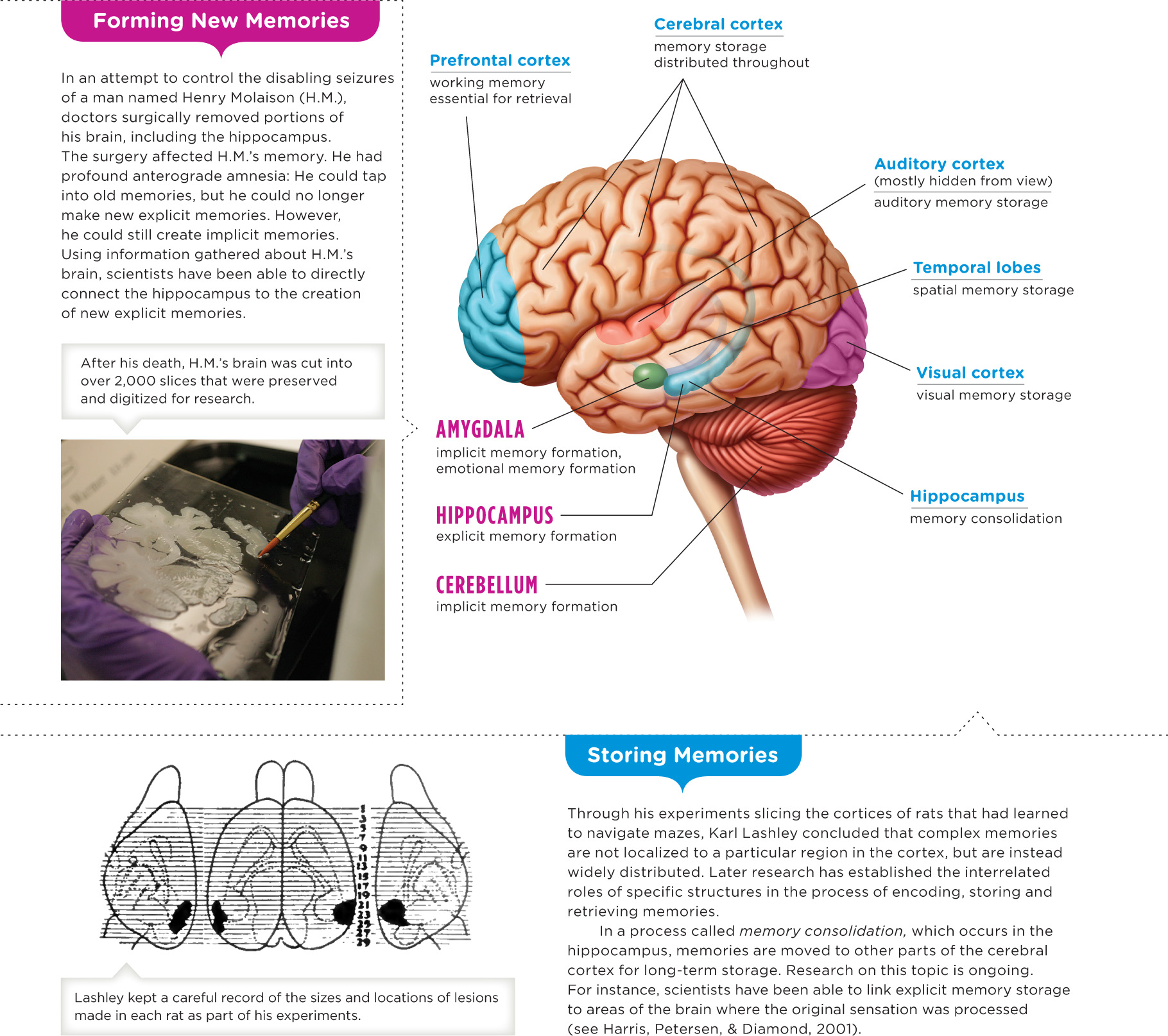
The Case of H.M.
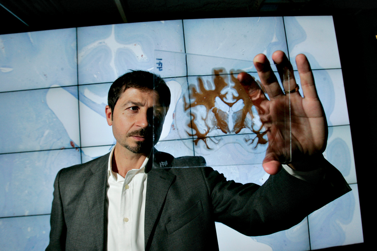
Henry Molaison (better known as “H.M.”) forced scientists to completely reevaluate their understanding of the brain’s memory system. From the onset of his amnesia in 1953 until his death in 2008, H.M. served as a research participant for some 100 scientists (Corkin, 2002), making him the most extensively studied amnesic patient.
270
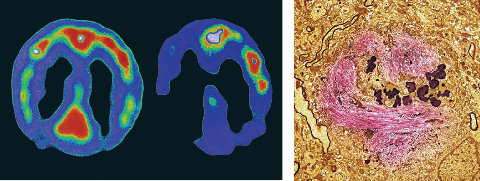
INFOGRAPHIC 6.3: Tracking Memory in the Brain
Whether with lab rats or case studies, psychologists have spent decades tracking the location of memory in the brain. What they’ve found so far should be no surprise: Memory is a complex system involving multiple structures and regions of the brain. Memory is formed, processed, and stored throughout the brain, and different types of memory have different paths. So to find memory in the brain, it helps to know your way around the brain’s structures. Remembering the amygdala’s role in processing basic emotion, for instance, can help you understand its role in processing the emotional content of memories.
271
H.M.’s brain troubles began at the age of 10, a year or so after being knocked unconscious in a bicycle accident. He began to experience seizures, which worsened with age and eventually became so debilitating that he could no longer hold a steady job. Antiseizure medications were unsuccessful in controlling his seizures, so at the age of 27, H.M. opted for an experimental surgery to remove parts of his brain: the temporal lobes (just beneath the temples), including the hippocampus (Scoville & Milner, 1957).
H.M.’s surgery succeeded in reining in his epilepsy but left his memory in shambles. Upon waking from the operation, he could no longer find his way to the bathroom or recognize the hospital workers caring for him. He played the same jigsaw puzzles and read the same magazines day after day as if he were seeing them for the first time (Scoville & Milner, 1957). Like Clive, H.M. suffered from profound anterograde amnesia, the inability to encode new long-term memories, and a milder form of retrograde amnesia, trouble retrieving existing memories from storage. Although H.M. had difficulty recalling what occurred during the few years leading up to his surgery (Scoville & Milner, 1957), he did remember events from the more distant past, for example, the 1929 stock market crash and the events of World War II (Carey, 2008).
H.M. maintained a working implicit memory, which he demonstrated in an experiment involving the complex task of tracing a pattern reflected in a mirror. With repeated practice sessions (none of which he remembered), H.M. improved his performance on the drawing task, learning it as well as someone without amnesia (Gabrieli, Corkin, Mickel, & Growdon, 1993). Clive can also acquire new implicit memories, but his ability is very limited. According to Deborah, it took years for Clive to learn how to get to his bedroom in the small community residence where he moved after leaving the hospital (Wearing, 2005).
The Role of the Hippocampus
Imagine you are a scientist trying to figure out exactly what role the hippocampus plays in memory. Consider the facts you know about H.M.: (1) He has virtually no hippocampus; (2) he has lost the ability to make new explicit memories, yet can create implicit memories; and (3) he can still tap into memories of the distant past. So what do you think the hippocampus does? Evidence suggests that the hippocampus is essential for creating new explicit memories but not implicit memories. Researchers have also shown that explicit memories are processed and stored in other parts of the brain, including the temporal lobes and areas of the frontal cortex (García-Lázaro, Ramirez-Carmona, Lara-Romero, & Roldan-Valadez, 2012).
As in H.M.’s case, Clive’s ability to form explicit memories is profoundly compromised, which is largely a result of the destruction of his hippocampus. Yet Clive also struggles with implicit memories—not surprising given the extensive damage to other regions of his brain, such as the amygdala and temporal lobes (Wilson et al., 2008). Studies have zeroed in on other brain areas, such as the cerebellum and amygdala, as processing hubs for implicit memory (Thompson & Kim, 1996; Thompson & Steinmetz, 2009). The amygdala also plays a central role in the processing of emotional memories (García-Lázaro et al., 2012). See Infographic 6.3 for more information about memory processing in the brain.
272
So although the hippocampus plays a central role in laying down new memories, it does not appear to serve as their ultimate destination. This process of memory formation, which moves a memory from the hippocampus to other areas of the brain, is called memory consolidation (Squire & Bayley, 2007). The consolidation that begins in the hippocampus allows for the long-term storage of memories. According to Kandel and Pittenger (1999): “The final locus of storage of memory is widely assumed to be the cerebral cortex, though this is a difficult assertion to prove”. As for retrieval, the hippocampus appears to be in charge of accessing young memories, but then passes on that responsibility to other brain regions as memories grow older (Smith & Squire, 2009).
This idea that the hippocampus is essential for creating explicit memories (as opposed to implicit memories) is supported by what we know about infantile amnesia, that is, the inability to remember events from our earliest years. Most adults cannot remember events before the age of 3, though it is not clear why. Some researchers suggest that it is because the hippocampus and frontal cortex, both important for the creation of long-term explicit memories, are not fully developed in children (Bauer, 2006; Willoughby, Desrocher, Levine, & Rovet, 2012).
This macrolevel perspective allows us to see the “big picture” of memory, but what’s going on microscopically? The next section focuses on the important changes occurring in and between neurons, but keep in mind that neurons are not the only cells in the brain. Neighboring glial cells may also play a role in memory.
from the pages of SCIENTIFIC AMERICAN
What Marijuana Reveals About Memory
Glial cells, not neurons, are responsible for marijuana-induced forgetfulness
Until recently, most scientists believed that neurons were the all-important brain cells controlling mental functions and that the surrounding glial cells were little more than neuron supporters and “glue.” Now research published in March in Cell reveals that astrocytes, a type of glia, have a principal role in working memory. And the scientists made the discovery by getting mice stoned.
Marijuana impairs working memory—the short-term memory we use to hold on to and process thoughts. Think of the classic stoner who, midsentence, forgets the point he was making. Although such stupor might give recreational users the giggles, people using the drug for medical reasons might prefer to maintain their cognitive capacity.
To study how marijuana impairs working memory, Giovanni Marsicano of the University of Bordeaux in France and his colleagues removed cannabinoid receptors—proteins that respond to marijuana’s psychoactive ingredient THC—from neurons in mice. These mice, it turned out, were just as forgetful as regular mice when given THC: they were equally poor at memorizing the position of a hidden platform in a water pool. When the receptors were removed from astrocytes, however, the mice could find the platform just fine while on THC.
The results suggest that the role of glia in mental activity has been overlooked. Although research in recent years has revealed that glia are implicated in many unconscious processes and diseases [see “The Hidden Brain,” by R. Douglas Fields: Scientific American Mind, May/June 2011], this is one of the first studies to suggest that glia play a key role in conscious thought. “It’s very likely that astrocytes have many more functions than we thought,” Marsicano says. “Certainly their role in cognition is now being revealed.”
Unlike THC’s effect on memory, its pain-relieving property appears to work through neurons. In theory, therefore, it might be possible to design THC-type drugs that target neurons—but not glia—and offer pain relief without the forgetfulness. Ruth Williams. Reproduced with permission. Copyright © 2012 Scientific American, a division of Nature America, Inc. All rights reserved.
273
Where Memories Live in the Brain—A Micro Perspective
LO 15 Describe long-term potentiation.
How does your brain change when you learn a new driving route to school? If we could peer into your skull, we might see a change in your hippocampus. As one study found, London taxicab drivers with greater time spent on the job had structural changes in some regions of the hippocampus, particularly to an area that processes “spatial knowledge” (Maguire, Woollett, & Spiers, 2006). Zooming in for a closer look, we might actually see changes at the level of the neuron. If you are looking for a memory imprint, the best place to look is the synapse.
CONNECTIONS
In Chapter 2, we introduced the synapse: the tiny gap between two neurons. Neurons communicate with each other via chemicals called neurotransmitters, which are released into the synapse. Here, we see how the activities at the neural level are related to the formation and maintenance of memories.
Long-Term Potentiation
As it turns out, the more neurons communicate, the better the connections between them. Long-term potentiation occurs when sending neurons release neurotransmitters more effectively, and receiving neurons become more sensitive, boosting synaptic strength for days or even weeks (Lynch, 2002; Malenka & Nicoll, 1999; Whitlock, Heynen, Shuler, & Bear, 2006). In other words, long-term potentiation refers to the increased efficiency of neural communication over time, resulting in learning and the formation of memories. Researchers suggest long-term potentiation may be the biological basis of many kinds of learning. As you learn a new skill, for example, the neurons involved in performing that skill increase their communication with each other. It might start with a somewhat random firing of neurons, but eventually the neurons responsible for the new skill develop pathways through which they communicate more efficiently. Having trouble visualizing the process? Imagine this scenario: Your college has opened a new campus with an array of brand-new buildings, but it has yet to construct the sidewalks connecting them. In order to go from one class to the next, students have to wade through tall grass and weeds. All the trampling eventually gives way to a system of well-worn paths linking the buildings and a multitude of efficient paths develops among the buildings. Long-term potentiation of neural connections occurs in a similar fashion; over time, the communication among neurons improves and strengthens, allowing for the skill to develop and become more natural (Whitlock et al., 2006). This is where memory comes in; these paths represent how a skill, whether tying your shoes, learning to drive a stick shift, or mastering a skateboarding trick, is learned and thus becomes a memory.
Aplysia
Amazingly, we have learned much about long-term potentiation from the sea slug Aplysia. Why the sea slug? One reason is that the sea slug only has about 20,000 neurons (Kandel, 2009)—a little easier to work with than the 100 billion neurons of a human brain. The sea slug’s neural simplicity, that is, the fact that the synapses are relatively easy to examine individually, also allows for the intensive study of habituation, conditioning, and other processes involved in classical conditioning. Studies using sea slugs as their subjects indicate that long-term potentiation, or increases in synaptic “strength,” is associated with learning and memory. What can a sea slug learn? They can be classically conditioned to retract their gills in response to being squirted with water. Researchers report that when the sea slugs are conditioned in this way, there are structural changes in both pre-synaptic and postsynaptic cells, including changes to connections between neurons (Kandel, 2009)—evidence of long-term potentiation. So never, ever complain to your instructor that you cannot learn: If a sea slug can do it, so can you!
274
CONNECTIONS
In Chapter 5, we discussed classical conditioning and how a neutral stimulus can be paired with an unconditioned stimulus, ultimately leading to a conditioned stimulus resulting in a conditioned response. In the case of the sea slug, the squirt of water is the conditioned stimulus and its involuntary response of retracting its gills is the conditioned response.
Alzheimer’S Disease
On a less positive note, disruptions in long-term potentiation appear to be at work in Alzheimer’s disease, a progressive, devastating brain illness that causes cognitive decline, including memory, language, and thinking problems. Alzheimer’s affects as many as 5.1 million Americans (National Institute on Aging, 2013). The disease was first discovered by Alois Alzheimer, a German neuropathologist, in the early 1900s. He had a patient with severe memory problems whose later autopsy revealed that the neurons in her brain were tangled up like wires of your earbud headphones. These neurofibrillary tangles, as they came to be called, were eventually shown to result from twisted protein fibers accumulating inside brain cells. In addition to the tangles, the other distinctive sign of Alzheimer’s is the presence of amyloid plaques, protein clumps that build up between neurons, blocking their lines of communication (Vingtdeux, Davies, Dickson, & Marambaud, 2011).
There is no cure for Alzheimer’s disease, and current treatments focus only on reducing the severity of symptoms rather than correcting the brain damage responsible. But there is also reason to be hopeful. Promising new drugs are coming down the pipeline, and there is some preliminary evidence suggesting that simple lifestyle changes, like becoming more physically active and pursuing intellectually and socially stimulating activities, may actually decrease the speed and severity of cognitive decline (Hertzog, Kramer, Wilson, & Lindenberger, 2009; Wilson & Bennett, 2003). Additional good news comes from recent research suggesting that we should not think of cognitive decline as inevitable in aging. We have more control of aging than previously thought, especially when we focus on the lifelong possibilities of learning and autonomy (Hertzog et al., 2009). Furthermore, it’s not only the strengthening of synapses that makes for enduring memories, but also the activation of new ones (Yu, Ponomarev, & Davis, 2004), thus ameliorating, or offsetting, cognitive decline (TABLE 6.2).
| Memory loss does not have to be an inevitable part of aging. Occasionally forgetting where you put your keys does not mean you have a degenerative memory disorder. This table provides some facts you should know about memory loss. |
|---|
| In the United States, 1 out of every 8 people who are 65 or older suffers from Alzheimer’s disease, which leads to loss of memory and other cognitive functions (Alzheimer’s Association, 2010). |
| The prevalence of dementia will double every 20 years, reaching 81 million by the year 2040 (Ferri et al., 2005). |
| There is no definitive way to know whether you or a family member will suffer from a neurocognitive disorder; most cases result from a complex combination of genetic, environmental, and lifestyle factors. |
| Studies of both animals and people have linked physical exercise to a variety of positive changes in the brain, including enhanced blood flow, increased thickness of the cortex, and less age-related deterioration of the hippocampus (Polidori, Nelles, & Pientka, 2010). |
| Some research suggests that people who begin exercising in their thirties (and stick with it) experience less cognitive decline than their sedentary peers by the time they reach their forties and fifties (Hertzog et al., 2009), although consistent exercise at any age has lasting cognitive benefits (Cotman & Berchtold, 2002; Kramer, Erickson, & Colcombe, 2006). |
| Intellectually engaging activities such as reading books and newspapers, writing, drawing, and solving crossword puzzles have been associated with a lower risk of memory loss (Hertzog et al., 2009; Wang, Karp, Winblad, & Fratiglioni, 2002). |
| Being socially active and hooked into social networks may reduce the risk of developing dementia (Fratiglioni, Paillard-Borg, & Winblad, 2004). |
275
Like many topics psychologists study, the biological mechanisms that give rise to memory remain somewhat mysterious. We know we have memories; we know they are formed in the brain; and we know the brain is a physical entity, yet we still don’t know exactly how we go from a bunch of firing neurons to a vivid recollection of your 21st birthday bash, your high school prom, or the image of Dave Roberts stealing second base to start the Red Sox’s improbable comeback. Studies attempting to test the various theories of memory formation are inconclusive, often generating more questions than answers. But one thing seems certain: Memory researchers have plenty of exciting work ahead.
show what you know
Question 6.18
1. __________ refers to the inability to lay down new long-term memories, generally resulting from damage to the brain.
- Anterograde amnesia
- Retrograde amnesia
- Infantile amnesia
- Long-term potentiation
Question 6.19
2. The __________ is a pair of seahorse-shaped structures in the brain that play a central role in memory.
- engram
- temporal lobe
- hippocampus
- aplysia
Question 6.20
3. __________ is the process of memory formation, which moves a memory from the hippocampus to other areas of the brain.
- Long-term potentiation
- Memory consolidation
- Priming
- The memory trace
Question 6.21
4. Infantile amnesia makes it difficult for people to remember events that occurred before the age of 3. What is your earliest memory and how old were you when that event occurred?
 CHECK YOUR ANSWERS IN APPENDIX C.
276
FINAL THOUGHTS
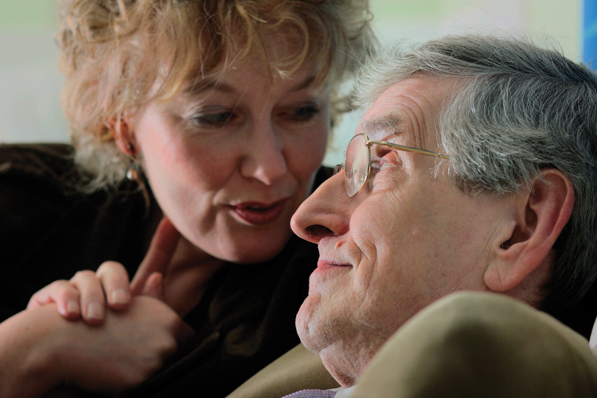
 At this point, you may be wondering what became of the two people featured in this chapter, Clive Wearing and Dorothea Seitz. After living in the hospital 7 years, Clive moved to a country residence specially designed for people suffering from brain injuries. As he left the hospital, some of the staff members offered him a farewell and said they would miss him. Addressing them with a polite bow, Clive exclaimed, “You’re the first people I’ve seen!” When Deborah would visit Clive in his new home, she found him happy and relaxed, spending much of his time on walks through gardens and the local village (Wearing, 2005, p. 293). In 2002 Clive and Deborah renewed their marriage vows. Clive participated fully in the service, reciting scripture he had memorized during his career as a professional singer decades before (D. Wearing, personal communication, June 10, 2013). After the ceremony, he had no recollection of what had taken place but nevertheless was very happy, laughing and devouring sponge cake (Wearing, 2005).
 As for Dorothea, she succeeded in defending her title of Junior World Memory Champion in 2009 and finished the competition in 11th place overall. After finishing high school, she spent a year doing volunteer work in Peru. She is currently studying political science and sociology in Freiburg, Germany.

Dorothea (right) became the Junior World Memory Champion for the second time in 2009. Although she no longer competes in memory sport, she continues to exercise her intellect as a student of political science and sociology.
© Ilona Huege