Hemispherectomy
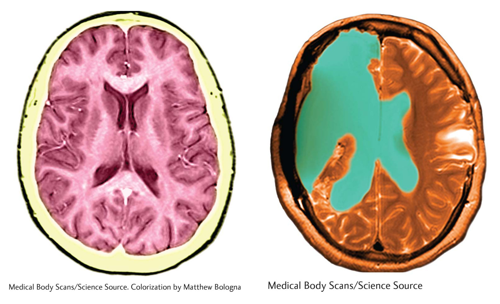
On the left is an MRI scan of a brain with both hemispheres intact. The scan on the right shows the brain of a person who has undergone a hemispherectomy. The green area, once occupied by the removed hemisphere, is now filled with cerebrospinal fluid.
On the left is an MRI scan of a brain with both hemispheres intact. The scan on the right shows the brain of a person who has undergone a hemispherectomy. The green area, once occupied by the removed hemisphere, is now filled with cerebrospinal fluid.
left: Medical Body Scans/Science Source. Colorization by Matthew Bologna; right: Medical Body Scans/Science Source