2.2 Neurons and Neural Communication
Just the Basics
52
LO 3 Label the parts of a neuron and describe an action potential.
cell body The region of the neuron that includes protein-
dendrites (den-
axon Skinny tubelike structure of a neuron that extends from the cell body, and which sends messages to other neurons.
myelin sheath (mī-ə-lən shēth) Fatty substance that insulates the axon and speeds the transmission of neural messages.
synapse (si-
Synonyms
cell body soma
terminal buds axon buds, synaptic knobs, terminal buttons
synapse synaptic cleft, synaptic gap
THE STRUCTURE OF A TYPICAL NEURON A typical neuron has three basic parts: a cell body, dendrites, and an axon (Figure 2.1). The cell body of a neuron contains the standard components found in most human cells: protein-

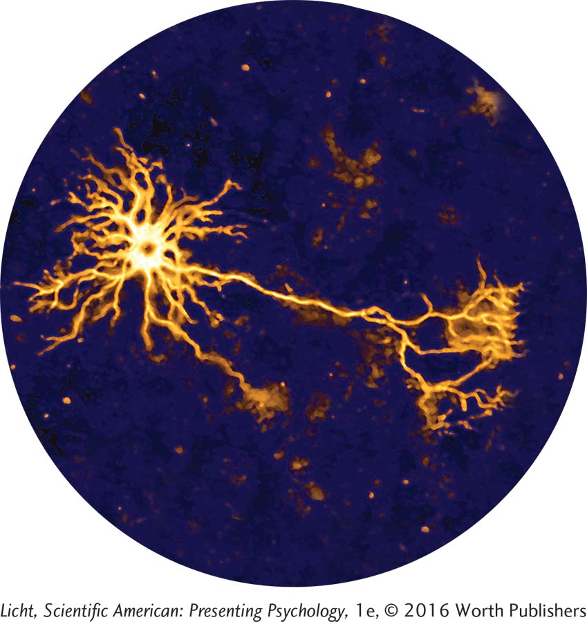
A scanning electron micrograph shows neurons (green) and glia (orange). Glial cells serve as the “glue” of the nervous system, providing cohesion and support for the neurons.
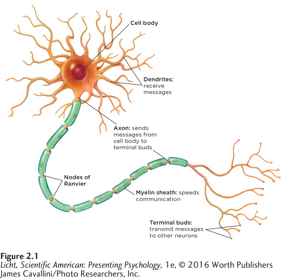
The neuron, the basic building block of the nervous system, has three main components: (1) the cell body, which contains vital cellular structures; (2) bushy dendrites that receive messages from neighboring neurons; and (3) a long, thin axon that sends messages to other neurons through its branchlike terminal buds.
glial cells (glē-əl) Cells that support, nourish, and protect neurons; produce myelin that covers axons.
HOLDING IT TOGETHER: GLIAL CELLS The function of neurons is to transmit information up and down the body all day and night, and neurons need a little support and nurturing to get this tough job done. This is where the glial cells (glē-əl) come into play. In the human brain, glial cells outnumber neurons by approximately 50 to 1 (Yuhas & Jabr, 2012), holding neurons together and maintaining the structure of the nervous system. (Glia means “glue” in Greek.)
53
For many years, scientists believed that glial cells simply kept things together, but we now know they do much more (Ndubaku & de Bellard, 2008). For example, glial cells come to the rescue if the brain is injured. When Brandon was shot in the head, glial cells called microglia worked to defend his brain from infection and inflammation (d’Avila et al., 2012; Streit, 2000). Another class of glial cells called astrocytes began to restore the barrier between his brain and blood (Gruber, 2009). Astrocytes have been found to support communication between neurons as well (Araque & Navarrete, 2010). Another type of glial cells, Schwann cells, produce the myelin that envelops axons.
Processes Inside the Neuron
Neurons have properties that allow them to communicate with other cells. But what information do they convey? In essence, the message is simple: “I have been activated.” Neurons are activated in response to sensations, thoughts, and other neurons, and this forms the basis for all that we think, feel, and do.
A neuron is surrounded by and contains electrically charged solutions (Infographic 2.2 below). If the total charge in each of these solutions is different, a voltage will be generated between the outside and the inside of the cell. This voltage is determined by the electrical characteristics of particles called ions. Some ions are negatively charged; others are positively charged. The difference in the charges inside and outside of the neuron determines its polarity, the degree to which it is positive or negative overall. Two processes direct the flow of positive and negative ions into or out of the cell. Diffusion is the natural tendency of the ions to spread out or disperse, and electrostatic pressure causes similarly charged ions to spread apart and oppositely charged ions to move toward each other (like the behavior of magnets). The concentrations, inside and outside of the cell, of positively charged ions (sodium and potassium) and negatively charged ions (protein molecules) determine the activity in most neurons.
54
INFOGRAPHIC 2.2
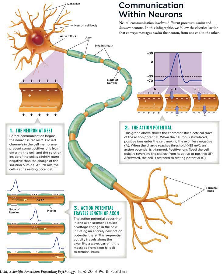
A neuron is encased in a membrane that is selectively permeable, allowing only some of the ions to pass in and out of its channels. The membrane is impermeable to positive sodium ions and negative protein ions (it does not allow these ions to enter or exit). Positive sodium ions move toward the membrane from the outside, and negative protein ions move toward the membrane from the inside; each moves closer to its side of the membrane wall (because the opposite charges are attracted to each other).
Inside the neuron, the concentration of potassium ions is greater than that outside the membrane. Also inside the cell are negatively charged protein ions, which do not exist outside the cell. These protein ions are attracted to the excess positive charge outside and move toward the membrane, but they are too big to go through. Because the protein ions can’t get out, the inside of the neuron is negatively charged when the neuron is not active. The concentration of sodium ions inside the cell is much less than its concentration on the outside of the cell. As a result, the sodium ions on the outside are attracted to the cell wall (because of diffusion and electrostatic pressure). An electrical potential is created by the differences in charge between the outside and the inside of the neuron, which is its resting potential.
55
resting potential The electrical potential of a cell “at rest”; the state of a cell when it is not activated.
RESTING POTENTIAL The resting potential represents the electrical potential of a cell “at rest,” the condition of a cell when it is not activated. The solutions on either side of the membrane wall come into equilibrium with a slightly more negative charge inside. In the resting state, the voltage inside the cell is about −70 millivolts (mV) compared to the voltage on the outside. (Think of a resting neuron like a battery, which holds a charge. For comparison, one double A battery has a 1,500-
ACTION POTENTIAL Although the positive sodium ions are being pulled toward the inside of the cell, they cannot move into the cell until the neuron is stimulated by neighboring cells. When this happens, a signal instructs the channels, or gates, in the cell membrane to open up. The channels in the dendrites open, freeing the positive sodium ions to enter the cell through the dendrites, move into the cell body, and to finally reach the beginning of the axon, known as the axon hillock.
Synonyms
threshold potential stimulus threshold
action potential spike potential
action potential The spike in electrical energy that passes through the axon of a neuron, the purpose of which is to convey information.
The influx of the positive sodium ions at the axon hillock raises the internal cell voltage of the first segment of the axon from the resting voltage of −70 mV to the threshold potential of −55 mV relative to the outside. This change in voltage causes the sodium gates to open, and the positive sodium ions flood into the cell. The voltage inside that section of the axon rises rapidly, increasing from −55 mV to +30 mV, after which the sodium gates immediately close. This produces a spike in the value of the voltage within the cell, as the charge inside the cell becomes more positive than that outside of the cell. This is the action potential, or the spike in electrical energy that passes through the axon of a neuron.
The potassium gates open, and the positive potassium ions, now repelled by the much more positively charged cell interior, flow out of the cell. However, because the proportion of sodium and potassium ions inside and outside the cell is not the same as before (the ions are no longer at equilibrium), a sodium/potassium pump within the cell membrane brings them back to their original values by pumping the excess positive sodium ions back outside the cell and the positive potassium ions back in. In this way, the solutions inside and outside this segment of the axon return to equilibrium. In other words, it returns to resting potential.
MOVING DOWN THE AXON The firing of the first segment of the axon produces excess sodium ions on the inside of the cell, and these positive sodium ions diffuse to the next segment within the axon. This causes the voltage of the second segment of the axon to reach the threshold potential (−55 mV), opening its sodium gates and causing a spike of voltage there (+30 mV). Meanwhile, the first segment of the axon returns to its resting potential, so that the electrical spike cannot travel back in that direction. This process repeats through each segment of the axon, like a row of dominos tumbling down. Every time a segment of the axon fires, the positive sodium ions flood in from the outside of the cell, while the prior segment returns to its resting potential, all along the length of the axon to its end. Each action potential takes about 1 millisecond to complete, and a typical neuron can fire several hundred times per second.
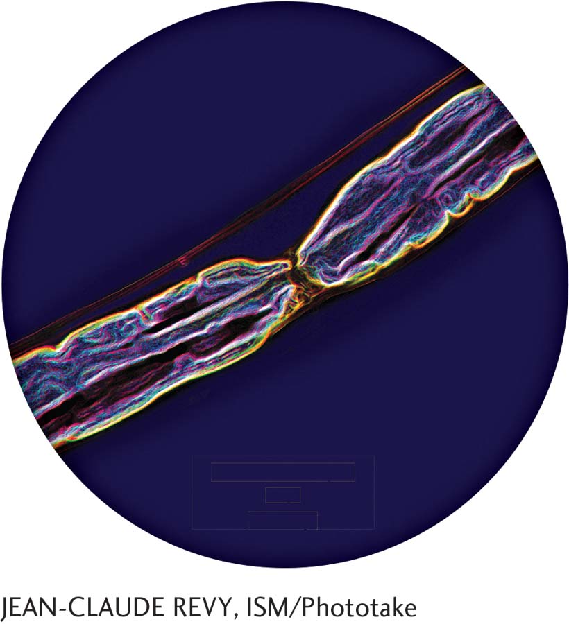
Action potentials may travel as fast as 268 miles per hour through a myelinated axon (Susuki, 2010). Myelin is a protein that envelops and insulates the axon, facilitating faster transmission of the impulse. The action potential “skips” over the segments of myelin, hopping from one node of Ranvier to the next (see small space in the center), instead of traversing the entire length of the axon.
EXCITATORY AND INHIBITORY SIGNALS How do neighboring cells initially signal for the channels to open up to let the positive sodium ions move into the dendrites of a neuron? The message to fire begins at the dendrites. Chemical messages from surrounding neurons are sent to the cell body. If enough sending neurons signal the receiving neuron to pass along the message, their combined signal becomes excitatory and the neuron will fire. However, not all neighboring neurons send an excitatory signal. Some will send an inhibitory signal, instructing the neuron not to fire. For an excitatory signal to occur, there have to be more excitatory than inhibitory signals, and the difference between the two has to meet the threshold potential of −55 mV. If “enough” positively charged ions enter the cell, the potential of the neuron increases and reaches the threshold, or trigger point, and the cell “fires.”
all-
ALL-
ROLE OF MYELIN SHEATH The firing of a neuron is facilitated by the myelin sheath, which insulates and protects the tiny spikes in electricity happening inside the axon. Because myelin is such a good insulator, it does not allow the exchange of ion fluid between the inside and the outside of the cell membrane. However, the axon is not covered with myelin at the nodes of Ranvier (you can see this in the photograph to the left). The exchange of ion fluid can only happen at these nodes. The action potential thus appears to “jump” from node to node, as opposed to traversing the entire axon in one continuous movement, speeding the transmission of the signal. The speed of the action potential in an unmyelinated axon is approximately 1.1 to 4.5 miles per hour (mph), and a myelinated axon has transmission speeds of approximately 157 to 268 mph (Susuki, 2010). Unmyelinated axons, or those damaged from multiple sclerosis or other diseases, have slower transmission speeds, because the signal must make its way down the entire length of the axon. The damaged myelination caused by multiple sclerosis can lead to many symptoms, including fatigue, trouble with vision, and cognitive disabilities (Su, Banker, Bourdette, & Forte, 2009).
Communication Between Neurons
LO 4 Illustrate how neurons communicate with each other.
neurotransmitters Chemical messengers that neurons use to communicate at the synapse.
receptor sites The location where neurotransmitters attach on the receiving side of the synaptic gap.
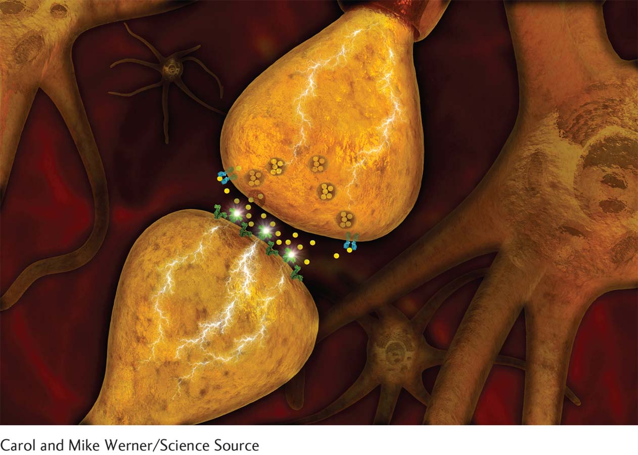
The terminal bud of a sending neuron (top) interacts with a dendrite of a receiving neuron by releasing chemical messengers (neurotransmitters) into the synapse. Once the neurotransmitters migrate across the gap and latch onto the dendrite’s receptor sites, the message has been conveyed.
Neurons communicate with each other via chemicals called neurotransmitters. (Infographic 2.3 illustrates communication between neurons in detail.) An action potential moves down the axon, eventually reaching the terminal buds. The signal to release neurotransmitters is the voltage change from the action potential, which results in vesicles (small fluid-
57
INFOGRAPHIC 2.3
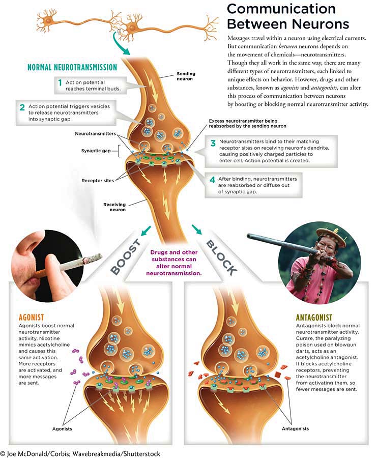
When the neurotransmitters latch onto the receptors of the dendrites of the receiving neuron, the gates in the receiving cell’s membrane fly open, ushering positively charged particles into the cell and thus restarting the cycle of the action potential (if the threshold is met). Keep in mind that the firing of one neuron contributes to the possibility for neighboring neurons to fire as a result of its chemical message.
reuptake Process by which neurotransmitters are reabsorbed by the sending terminal bud.
When neurotransmitters are released into the synapse, many of them bind to receptors. Ultimately, they may be reabsorbed by the sending terminal bud in a process known as reuptake. Those that are not reabsorbed drift out of the synaptic gap, through diffusion. This is how the synaptic gap is cleared of neurotransmitters, in preparation for the next release of chemical messengers.
Neurotransmitters and Behavior
LO 5 List various neurotransmitters and summarize their involvement in human behavior.
As mentioned, there are different types of neurotransmitters. Researchers have identified approximately 100 of them, with many more yet to be discovered. We already know that neurotransmitters secreted by one neuron under certain conditions can cause neighboring neurons to fire, which can affect the regulation of mood, appetite, muscles, organs, arousal, and a variety of other functions. Here we will describe only a handful of neurotransmitters, starting with the first neurotransmitter discovered, acetylcholine.
ACETYLCHOLINE Acetylcholine is a neurotransmitter that relays messages from neurons to muscles, thus enabling movement. Any time you move some part of your body, whether dancing your fingers across a keypad or bouncing your head to one of your favorite songs, you have, in part, acetylcholine to thank. Too much acetylcholine leads to muscle spasms; too little causes paralysis. Acetylcholine is also involved in memory. In particular, low levels in the brain have been linked to Alzheimer’s disease (Kihara & Shimohama, 2004), which can lead to problems of memory, language, and thinking.
GLUTAMATE AND GABA Much of the communication within the nervous system involves two neurotransmitters: glutamate and GABA (short for gamma-
Synonyms
norepinephrine noradrenaline
NOREPINEPHRINE Norepinephrine has a variety of effects in the nervous system, but one of its most important functions is to help prepare the body for stressful situations. Think about how Brandon was surrounded by gunfire during the battle of Fallujah. Norepinephrine was working to enable Brandon’s nervous system to initiate action. In the brain, norepinephrine is involved in regulating arousal and sleep (Jones, 2003).
59
SEROTONIN Serotonin plays a key role in controlling appetite, aggression, and mood, and also regulates sleep and breathing. Abnormally low serotonin activity is thought to drive depression. Antidepressants called serotonin reuptake inhibitors (SSRIs), including Prozac and Zoloft, boost the effects of this “feel good” neurotransmitter (Chapter 13). Normally, neurotransmitters that do not connect with receptor sites can be reabsorbed by the sending terminal bud in the reuptake process. SSRIs work to prevent this reabsorption. The longer the serotonin is available in the gap, the more time it has to attach to a receptor.
DOPAMINE The neurotransmitter dopamine is known to play a role in problems with drug use. Repeated use of drugs overstimulates and damages the functioning of the neurons in the brain’s reward circuit, theoretically making it more difficult to enjoy non-
ENDORPHINS Endorphins are a group of naturally produced opioids (Chapter 4) that regulate the secretion of other neurotransmitters. The term endorphin is derived from the words “endogenous,” meaning it is created within, and “morphine.” Released in response to pain, endorphins block pain receptor sites. Brisk exercise increases their production, reducing the experience of pain and elevating mood.
Agonists and Antagonists
Drugs and other substances influence behavior by interfering at the level of the synapse (Chapter 4 and 13). Certain substances can be used to act like neurotransmitters and others can be used to block normal neurotransmitter activity. Agonists are substances that increase the normal activity of the neurotransmitter (whether it normally sends an excitatory or inhibitory signal) and antagonists reduce the activity or block the release of the neurotransmitter (Infographic 2.3). For example, some substances, such as nicotine and muscarine (found in poisonous mushrooms), increase the secretion of acetylcholine, causing sweating, pupil constriction, nausea, and respiratory distress. These substances increase the normal activity of acetylcholine; thus, they are agonists. On the other hand, the popular anti-
60
Apply This
Where’s My Morning Antagonist?

Did you jump-
What are the health consequences of regular caffeine consumption? When used in moderation, caffeine may boost your ability to form long-![]()
Now sit back, relax, and sip on a beverage, caffeinated or not. It’s time to examine the nervous system running through your arms, legs, fingers, toes—
show what you know
Question 1
1. Many axons are surrounded by a ____________, which is a fatty substance that insulates the axon.
myelin sheath
Question 2
2. When Brandon was injured, ____________ played an important role in his recovery by defending against infection and inflammation of the brain, as well as holding neurons together and maintaining the structure of the nervous system.
glial cells
dendrites
action potentials
sodium ions
a. glial cells
Question 3
3. ____________ are released into the ____________ when an action potential reaches the branches of the terminal buds.
Sodium ions; synaptic gap
Neurotransmitters; synaptic gap
Potassium ions; cell membrane
Neurotransmitters; sodium gates
b. Neurotransmitters; synaptic gap
Question 4
4. Neural communication is very complicated. Draw a diagram depicting the process of neural communication, then explain it to yourself while looking at what you have drawn.
A diagram could include the following information: Neural communication involves different processes within and between neurons. Using Infographic 2.2, you can follow the electrical action within the neuron. Messages received by the dendrites from neighboring neurons are passed down the axon, which sends messages to other neurons through its branchlike terminal buds.
Infographic 2.3 demonstrates how neurons communicate with each other via chemicals called neurotransmitters. The signal to release neurotransmitters is the voltage change from the action potential, which results in vesicles that contain neurotransmitters attaching to the membrane on the terminal bud. This allows the neurotransmitter to be released into the synaptic gap. The majority of these neurotransmitters drift across the synaptic gap and come into contact with receptor sites of the receiving neuron's dendrites, which may (or may not) lead to an action potential in the receiving neuron.