2.4 The Amazing Brain
67
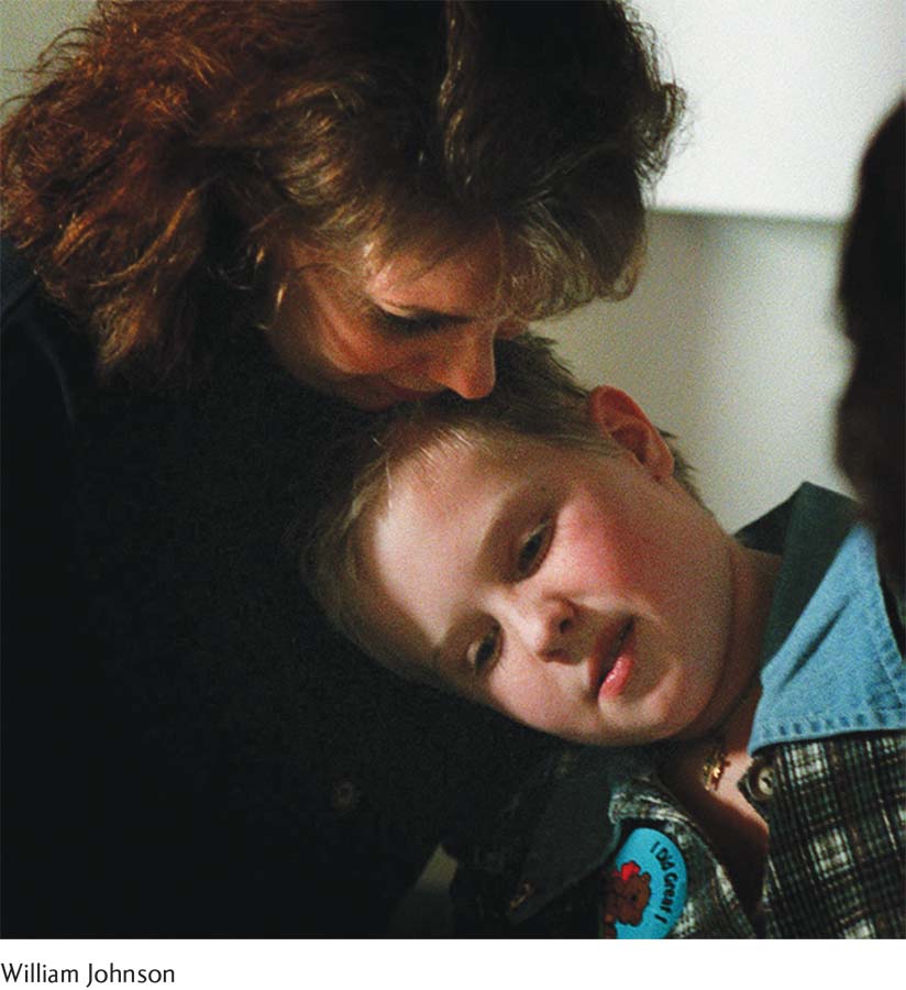
Christina Santhouse relaxes with her mother at Johns Hopkins, where she had a dramatic brain surgery known as a hemispherectomy. Prior to the operation, Christina experienced hundreds of seizures a day.
 THE GIRL WITH HALF A BRAIN As Brandon Burns began his long journey to recovery, a 17-
Christina’s remarkable story began when she was 7 years old. She was a vibrant, healthy child who loved soccer and playing outside with her friends. Barring an occasional ear infection, she basically never got sick—

As the days and weeks wore on, the tremors in Christina’s ankle moved up her left side and eventually spread throughout her body. In time, she was having seizures every 3 to 5 minutes. Doctors suspected she had Rasmussen’s encephalitis, a rare disease that causes severe swelling in one side of the brain, impairing movement and thinking and causing seizures that come as often as every few minutes (National Institute of Neurological Disorders and Stroke, 2011b).
Christina and her mother decided to seek treatment at The Johns Hopkins Hospital in Baltimore, the premiere center for treating children with seizure disorders. They met with Dr. John Freeman, a pediatric neurologist and an expert in hemispherectomy, a surgery to remove nearly half of the brain. A rare and last-
Why did Dr. Freeman recommend this drastic surgery to remove nearly half of Christina’s brain? And what side of the brain did he suggest removing? Before addressing these important questions, we need to develop a general sense of the brain’s geography. 
Right Brain, Left Brain: The Two Hemispheres
LO 9 Describe the functions of the two brain hemispheres and how they communicate.
cerebrum The largest part of the brain, includes virtually all parts of the brain except primitive structures; has two distinct hemispheres.
If you look at a photo or an illustration of the brain, you will see a walnut-
68
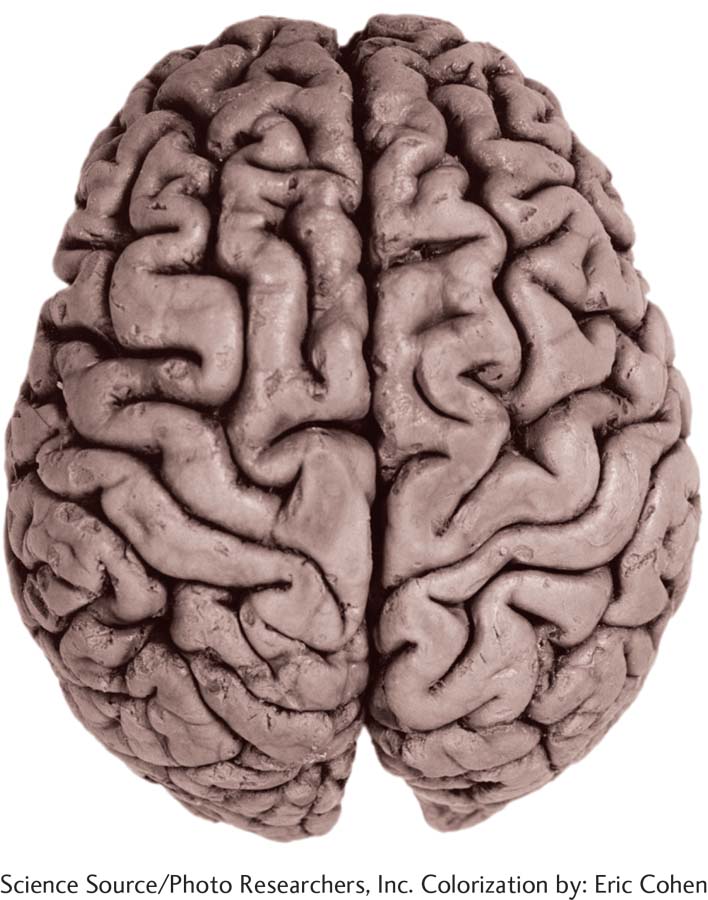
The cerebrum looks like a walnut with its two wrinkled halves. Regions of the left and right hemispheres specialize in different activities, but the two sides of the brain are constantly communicating and collaborating.
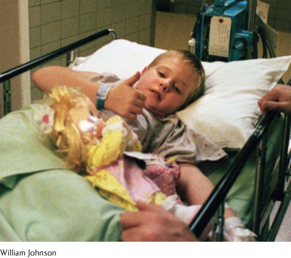
Christina is wheeled into the operating room for her 14-
CHRISTINA MAKES THE DECISION Within 2 months, Christina’s seizures were occurring every 3 minutes, hundreds of times a day. She was unable to play soccer or go outside during school recess, and she sat on a beanbag chair in class so she wouldn’t hurt herself when overcome with a seizure. “I couldn’t do anything anymore,” Christina says. “I wasn’t enjoying my life.”
In February 1996 the doctors at Johns Hopkins removed the right hemisphere of Christina’s brain. The operation lasted some 14 hours. When Christina emerged from the marathon surgery, her head was pounding with pain. “I remember screaming and asking for medicine,” she recalls. The migraines persisted for months but eventually tapered off, and ultimately the surgery served its purpose: Christina no longer experienced debilitating seizures.
The Split-
LO 10 Explain lateralization and how split-
Removing nearly half of a brain may sound barbaric, but hemispherectomies have proven to be effective for eliminating and reducing seizures. In a study of the 111 children who had hemispherectomies at Johns Hopkins between 1975 and 2001, 65% no longer suffered seizures at all, and 21% experienced infrequent, “nonhandicapping” seizures. The remaining 14% still had seizures described as “troublesome” (Kossoff et al., 2003).
split-
Synonyms
split-
corpus callosum (kȯr-
Hemispherectomies are exceptionally rare, used only when seizures occur many times a day, cannot be tempered with drugs, and stem from problems in one hemisphere (Choi, 2007, May 24). A less extreme, last-
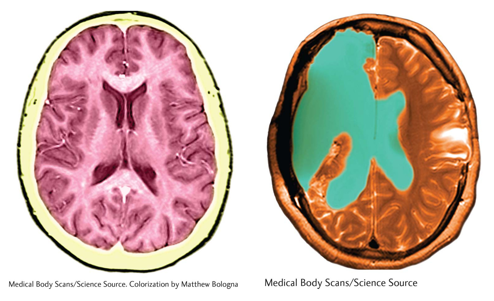
On the left is an MRI scan of a brain with both hemispheres intact. The scan on the right shows the brain of a person who has undergone a hemispherectomy. The green area, once occupied by the removed hemisphere, is now filled with cerebrospinal fluid.
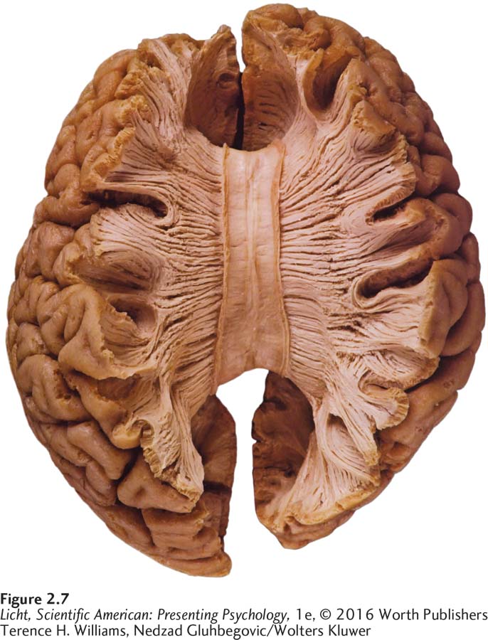
The image to the left shows a top view of the corpus callosum, the bundle of neurons linking the right and left hemispheres. When the corpus callosum is severed, we can see clear functional differences between the two sides of the brain. Studies of people who have undergone this procedure are known as “split-
STUDYING THE SPLIT BRAIN In addition to helping many patients with severe, drug-
69
Equipped with this knowledge, American neuropsychologist Roger Sperry (1913–

Brandon works on his pronunciation in front of a mirror during a speech therapy session at the Memphis VA hospital. You can see the extent of his injury on the left side of his head. Upon awaking from his coma, Brandon could not articulate a single word. Today, he can hold his own in complex conversations.
70
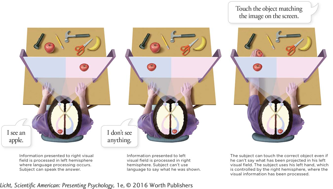
Because the hemispheres are disconnected through the surgery, researchers can study each hemisphere separately to explore its own unique capabilities (or specializations). Imagine that researchers flashed an image (let’s say an apple) on the right side of a screen, ensuring that it would be processed by the brain’s left hemisphere. The split-

Christina, as a teenager, walking her dog. The left side of her body is partially paralyzed, but her gait is quite natural. She wears a device on her left leg that activates her nerves, causing her muscles to contract at the appropriate time.
lateralization The idea that each cerebral hemisphere processes certain types of information and excels in certain activities.
LATERALIZATION The split-
The split-
The Special Roles of the Left and the Right
Armed with this new knowledge of the split-
71
HANDEDNESS AND LANGUAGE DOMINANCE Brandon’s difficulties with language are fairly typical for someone with a brain injury to the left hemisphere, because regions on the left side of the brain tend to predominate in language. This is not true for everyone, however. In a study examining handedness and language dominance, researchers measured participants’ degree of handedness (righty or lefty) and used brain scan technology to determine their predominant side for language processing. They found that around 27% of strongly left-
LO 11 Identify areas in the brain responsible for language production and comprehension.
Broca’s area (brȯ-kəz) A region of the cortex that is critical for speech production.
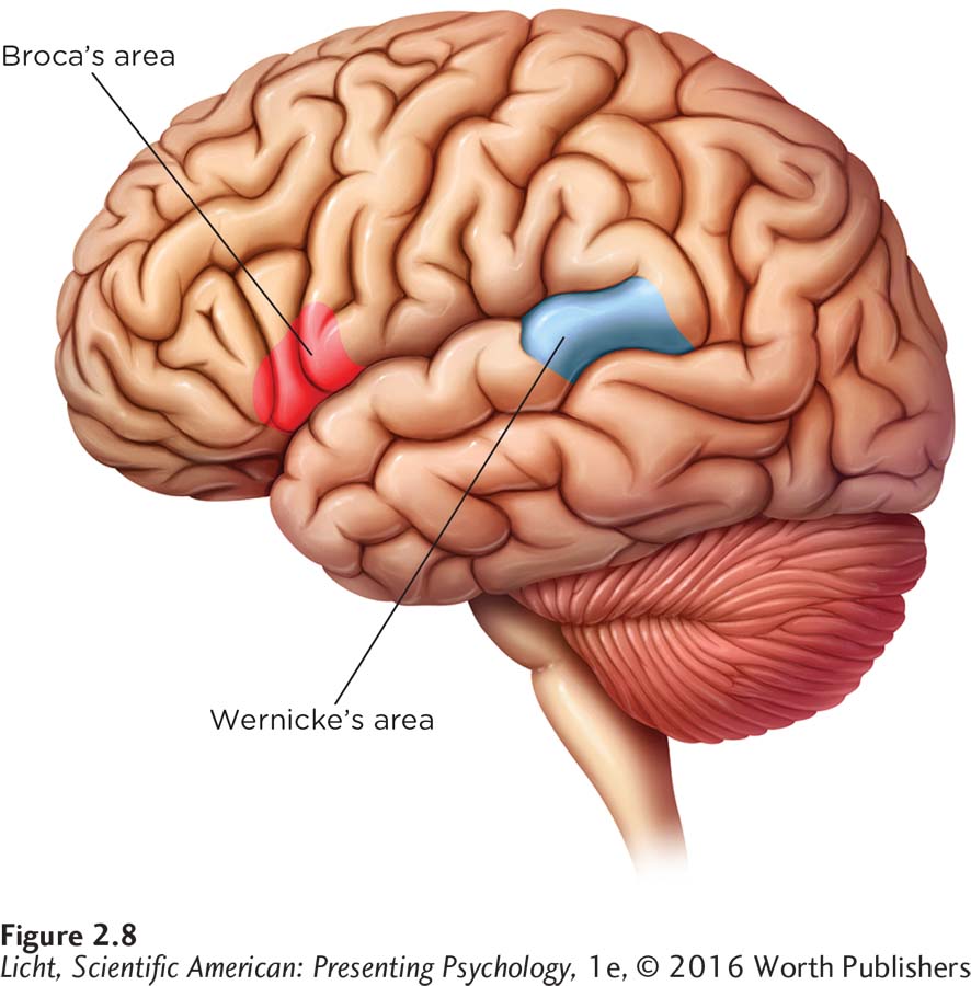
For most people, the left hemisphere controls language. Broca’s area plays a critical role in language production, and Wernicke’s area in language comprehension.
BROCA’S AREA Evidence for the “language on the left” notion appeared as early as 1861, when a French surgeon by the name of Pierre Paul Broca (1824 –1880) encountered two patients who had, for all practical purposes, lost the ability to talk. One of the patients could only say the word “tan,” and the other had an oral vocabulary of five words. When Broca later performed autopsies on the men, he found that both had sustained damage to the same area on the side of the left frontal lobe (right around the temple; Figure 2.8). Over the years, Broca identified several other speech-
Wernicke’s area (ver-
WERNICKE’S AREA Around the same time Broca was doing his research, a German doctor named Karl Wernicke (1848–
Broca’s and Wernicke’s work, along with other early findings, highlighted the left hemisphere’s critical role in language. Scientists initially suspected that Broca’s area was responsible for speech creation and Wernicke’s area for comprehension, but it is now clear the use of language is far more complicated. These areas may perform additional functions, such as processing music and interpreting hand gestures (Koelsch et al., 2002; Xu, Gannon, Emmorey, Smith, & Braun, 2009), and they cooperate with multiple brain regions to allow us to produce and understand language (Tate et al., 2014). Furthermore, some speech processing appears to occur in the right hemisphere.
THE ROLE OF THE RIGHT Research involving two split-
Neuroplasticity
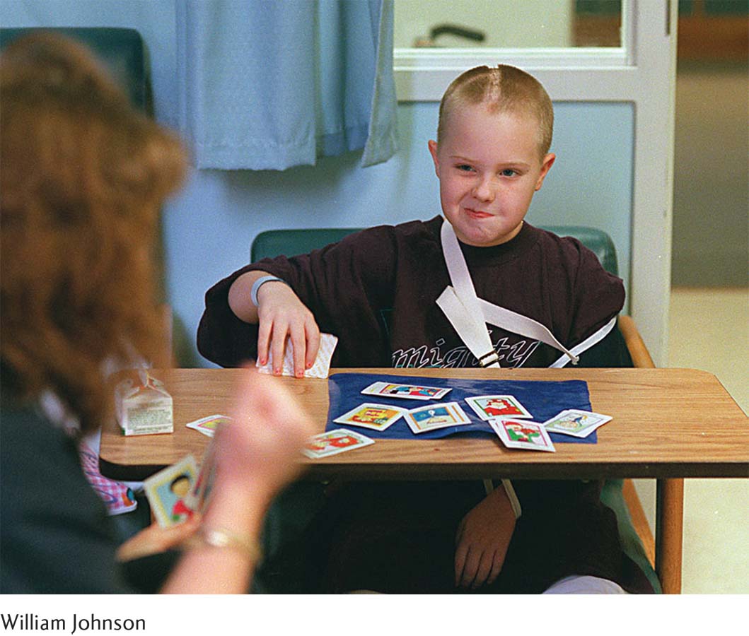
Christina’s dramatic recovery was facilitated by physical, occupational, vision, and speech therapy. “The more therapy,” says Christina, “the better chance of recovery.”
72
When Christina was wheeled out of surgery, her mother approached, grabbed hold of her right hand, and asked her to squeeze. Christina squeezed, demonstrating that she could understand and respond to language. Remember, she still had her left hemisphere.
Losing the right side of her brain did come at a cost, however. We know that Christina suffers partial paralysis on the left side of her body; this makes sense, because the right hemisphere controls movement and sensation on the left. We also know that it took Christina extra time to do her schoolwork. But if you ask Christina whether she has significant difficulty with any of the “right-
In addition to making the honor roll and leading the bowling team, Christina managed to get her driver’s license (even though some of her doctors said she never would), graduate from high school, and go to college. These accomplishments are the result of Christina’s steadfast determination, but also a testament to the brain’s amazing ability to heal and regenerate.
LO 12 Define neuroplasticity and recognize when it is evident in the brain.
neuroplasticity The brain’s ability to heal, grow new connections, and reorganize in order to adapt to the environment.
The brain undergoes constant alteration in response to experiences and is capable of some degree of physical adaptation and repair. Its ability to heal, grow new connections, and make do with what is available is a characteristic we refer to as neuroplasticity. New connections are constantly forming between neurons, and unused ones are fading away. Vast networks of neurons have the ability to reorganize in order to adapt to the environment and an organism’s ever-
CONNECTIONS
In Chapter 1, we described the guidelines psychologists use to ensure the ethical treatment of humans and animals. In order to conduct the experiment described here, the researchers had to get pre-
In one study, researchers removed the eyes of newborn opossums and found that brain tissues normally destined to become visual processing centers took a developmental turn. Instead, they became areas that specialized in processing other types of sensory stimuli, such as sounds and touch (Karlen, Kahn, & Krubitzer, 2006). The same appears to happen in humans. Brain scans reveal that when visually impaired individuals learn to read Braille early in life, a region of the brain that normally specializes in handling visual information becomes activated, suggesting it is used instead for processing touch sensations (Burton, 2003; Liu et al., 2007).
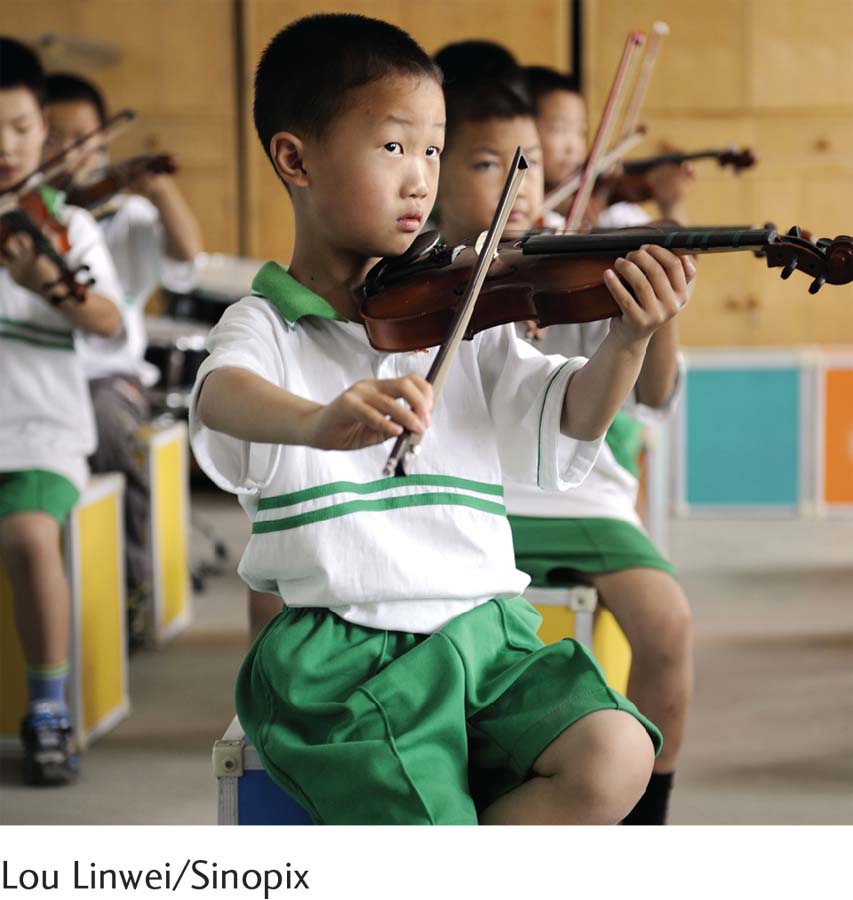
For millions of Chinese children, learning to play an instrument like the violin or piano begins early. Children with musical training outperform their untrained peers on tests of IQ and language skills (Schellenberg, 2011; Schellenberg & Winner, 2011). Is this because music lessons make children smarter, perhaps by stimulating positive changes in the brain? Or is it because smart children are more likely to take lessons? Researchers are still trying to get to the bottom of this conundrum.
Remarkably, this plasticity is evident even with the loss of an entire hemisphere. Researchers report that the younger the patient is when she has a hemispherectomy, the better her chances are for recovery. In fact, even with the loss of an entire left hemisphere (the primary location for language processing), speech is less severely impacted (though some impact is inevitable) in young patients. The younger the person undergoing the procedure, the less disability is evident in speech (Choi, 2008).

Because stem cells can differentiate into any type of cell in the body, they have great therapeutic potential. The cells pictured here are derived from a human embryo, but stem cells also reside in various adult tissues such as the brain and bone marrow.
neurogenesis The generation of new neurons in the brain.
73
STEM CELLS Scientists once thought that people were born with all the neurons they would ever have. Brain cells might die, but no new ones would crop up to replace them. Thanks to research beginning in the 1990s, that dismal notion has been turned on its head. In the last few decades, studies with animals and humans have shown that some areas of the brain are constantly generating new neurons, a process known as neurogenesis, which might be tied to learning and creating new memories (Eriksson et al., 1998; Gould, Beylin, Tanapat, Reeves, & Shors, 1999; Jessberger & Gage, 2014; Reynolds & Weiss, 1992).
stem cells Cells responsible for producing new neurons.
The cells responsible for churning out these new neurons are known as stem cells, and they are quite a hot topic in biomedical research. Scientists hope to harness these little cell factories to repair tissue that has been damaged or destroyed. Imagine that you could use stem cells to bring back all the neurons that Brandon lost from his injury or replace those that Christina lost to surgery. Cultivating new brain tissue is just one potential application of stem cell science. These cellular cure-
The Cortex: A Peek Beneath the Skull
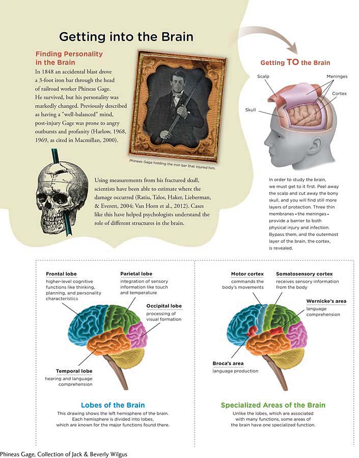
Imagine you were one of the surgeons performing Christina’s hemispherectomy. What exactly would you see when you peeled away the scalp and cut an opening into the skull? Before seeing the brain, you would come upon a layer of three thin membranes, the meninges, which envelop and protect the brain and spinal cord (Infographic 2.4 below). Perhaps you have heard of meningitis, a potentially life-
74
INFOGRAPHIC 2.4
cerebral cortex (sə-rē-brəl) The wrinkled outermost layer of the cerebrum, responsible for higher mental functions, such as decision making, planning, and processing visual information.
As Christina’s surgeon, your main task would be to remove part of the cerebrum’s outermost layer, the cerebral cortex (sə-rē-brəl). The cerebral cortex processes information and is the layer of cells surrounding nearly all the other brain structures. You’ll remember our earlier comment that the cerebrum looks like a wrinkled walnut. This is because the cortex is scrunched up and folded onto itself to fit inside a small space (the skull). This outermost section of the brain is also the part that is “newest,” or most recently evolved compared to the “older” structures closer to its core. We know this because researchers have compared the brains of humans with other primates. The structures we share with our primate relatives are considered more primitive, or less evolved, than the structures that are unique to humans.
LO 13 Identify the lobes of the cortex and explain their functions.
frontal lobes The area of the cortex that organizes information among the other lobes of the brain and is responsible for higher-
parietal lobes (pə-rī-ə-təl) The area of the cortex that receives and processes sensory information such as touch, pressure, temperature, and spatial orientation.
occipital lobes (äk-
temporal lobes The area of the cortex that processes auditory stimuli and language.
The cortex overlying each hemisphere is separated into different sections, or lobes. The major function of the frontal lobes is to organize information among the other lobes of the brain. The frontal lobes are also responsible for higher-
The Lobes: Up Close and Personal
75
Prior to her hemispherectomy, Christina was extroverted, easygoing, and full of energy. “I had absolutely no worries,” she says, recalling her pre-
The transformation of Christina’s personality may be a result of many factors, including the stress of dealing with a serious disease, undergoing a major surgery, and readjusting to life with disabilities. But it could also have something to do with the fact that she lost a considerable amount of brain tissue, including her right frontal lobe. Networks of neurons in the frontal lobes are involved in processing emotions, making plans, controlling impulses, and carrying out a vast array of mental tasks that each person does in a unique way (Williams, Suchy, & Kraybill, 2010). The frontal lobes play a key role in the development of personality (Stuss & Alexander, 2000). A striking illustration of this phenomenon involves an unlucky railroad foreman, Phineas Gage.
PHINEAS GAGE AND THE FRONTAL LOBES The year was 1848, and Phineas Gage was working on the railroad. An accidental explosion sent a 3-
Modern scientists have revisited Gage’s case, using measurements from his fractured skull and brain-
motor cortex A band of tissue toward the rear of the frontal lobes that works with other brain regions to plan and execute voluntary movements.
DOGS, CARTOONS, AND THE MOTOR CORTEX Toward the rear of the frontal lobes is a strip of the brain known as the motor cortex, which works with other areas to plan and execute voluntary movements (Infographic 2.4). Evidence for this region’s involvement in muscle movement first came from a study of dogs by Gustav Fritsch (1838–
North American neurosurgeon Wilder Penfield (1891–
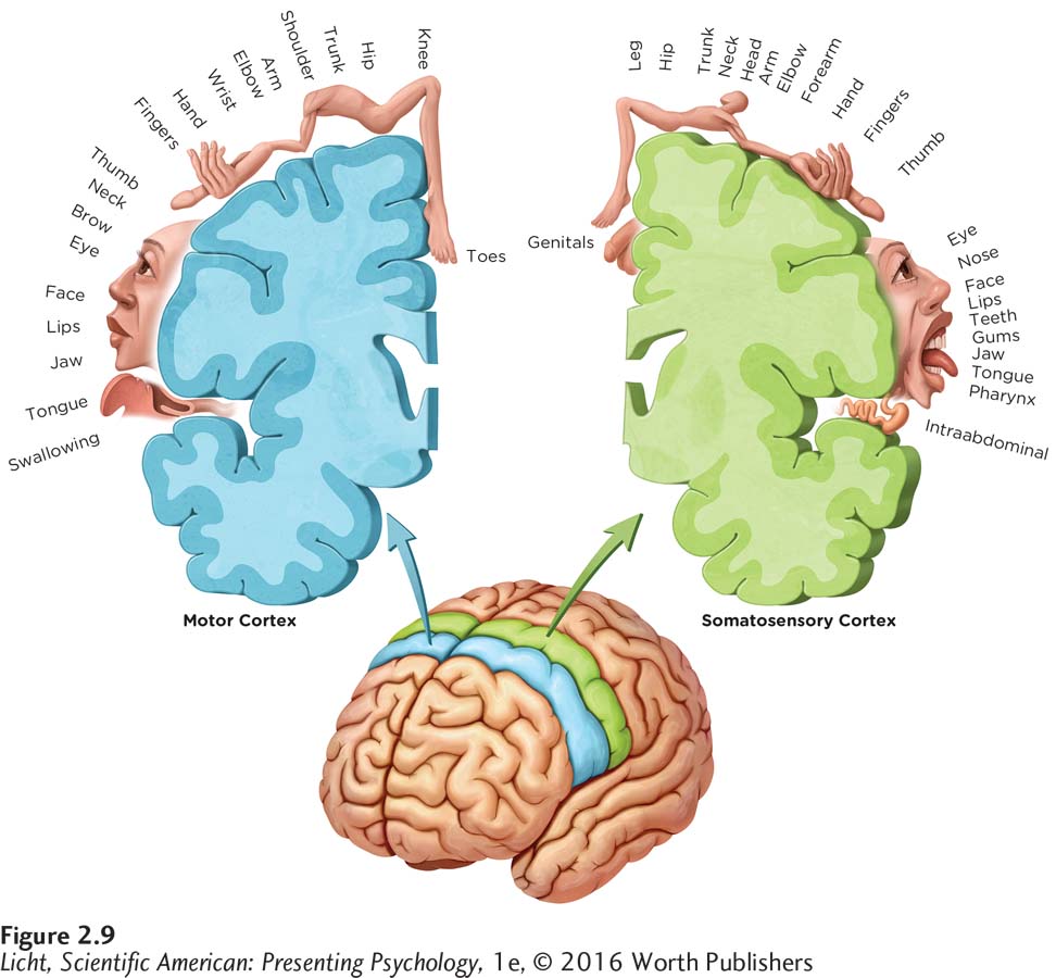
This drawing shows how the motor and somatosensory cortex correspond to the various regions of the body. Parts of the body that are shown larger, such as the face and hands, indicate areas of greater motor control or sensitivity. The size of each body part reflects the amount of cortex allocated to it.
CONNECTIONS
This research compared one man’s brain to a control group of 35 men. In Chapter 1, we discussed the importance of having a large enough sample size to give us reliable findings, which might be a consideration here. Also, we discussed the potential problems with using case studies for making generalizations to the population, and here, the use of Einstein as a single participant to compare to the control group could be considered a type of case study.
76
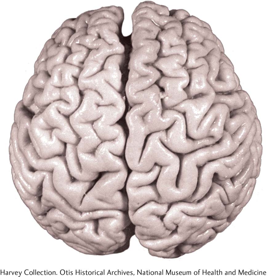
A photo of Einstein’s brain shows what researchers have referred to as his “extraordinary prefrontal cortex” (Falk, Lepore, & Noe, 2012, p. 1). The irregularities in Einstein’s parietal lobes may explain some of his spectacular mathematical and visual-
ALBERT EINSTEIN AND THE PARIETAL LOBES Directly behind the frontal lobes on the crown of your head are the parietal lobes (Infographic 2.4). The parietal lobes help orient the body in space, are involved in tactile processing (for example, interpreting sensations related to touch, such as pain and pressure), and may play a role in mathematical reasoning associated with spatial cognition (Desco et al., 2011; Grabner et al., 2007; Wolpert, Goodbody, & Husain, 1998). A study published in 1999 compared the brain of Albert Einstein to a control group of 35 brain specimens from men who had donated their bodies for use in research. Prior to their deaths, these men had normal cognitive functioning, average intelligence, and no mental health issues. The researchers reported that Einstein’s brain did not weigh more than the average brain of the control group, but a region of his parietal lobe believed to be important for visual–
77
somatosensory cortex A band of tissue running parallel to the motor cortex that receives and integrates sensory information from all over the body.
PENFIELD AND THE SOMATOSENSORY CORTEX The parietal lobes are home to the somatosensory cortex, a strip of brain running parallel to the motor cortex, which receives and integrates sensory information from all over the body (pain and temperature, for example). Penfield, the neurosurgeon who created the homunculus for the motor cortex, mapped the somatosensory cortex in the same way (Penfield & Boldrey, 1937; Figure 2.9). As you might expect, the most sensitive areas of the body like the face and tongue are oversized on the homunculus, whereas areas less sensitive to stimulation, such as the forearm and the calf, are smaller.

If you have ever been struck or fallen on the back of the head, you may recall perceiving bright blobs or dots floating by. The occipital lobes at the rear of the brain are home to the visual processing centers.
THE TEMPORAL LOBES AND THE AUDITORY CORTEX Below the parietal lobes, on the sides of your head, are the temporal lobes, which process auditory stimuli, recognize visual objects, especially faces, and play a key role in language comprehension and memory (Hickok & Poeppel, 2000; Infographic 2.4 on page 74). The temporal lobes are home to the auditory cortex, which receives information from the ears and allows us to “hear” sounds. In particular, researchers have supported the notion, based on studies of primate vocalizations, that the ability to recognize language has evolved over time and is processed within the temporal lobes (Scott, Blank, Rosen, & Wise, 2000; Squire, Stark, & Clark, 2004).
THE OCCIPITAL LOBES AND THE PRIMARY VISUAL CORTEX Visual information is initially processed in the occipital lobes, in the lower back of the head (Infographic 2.4). If you have ever suffered a severe blow to the rear of your head, you may remember “seeing stars,” probably because activity in the occipital lobes was disrupted (hopefully only for a few seconds). It is here, where the optic nerve connects to the primary visual cortex, that visual information is received and interpreted, and where we process that information (for example, about color, shape, and motion). (See Chapter 3 for more on the visual process.) Even for a person with healthy eyes, if this area were damaged, severe visual impairment could occur. At the same time, the individual would still be able to “see” vivid mental images (Bridge, Harrold, Holmes, Stokes, & Kennard, 2012).
LO 14 Describe the association areas and identify their functions.
association areas Regions of the cortex that integrate information from all over the brain, allowing us to learn, think in abstract terms, and carry out other intellectual tasks.
Synonyms
association areas intrinsic processing areas
THE ASSOCIATION AREAS In addition to the specialized functions of the different lobes described above, the cortex contains association areas whose role is to integrate information from all over the brain. The association areas are located in all four lobes; however, they are much harder to pinpoint than are the motor and sensory areas. The association areas allow us to learn (just as you’re doing now), to have abstract thoughts (for example, 2 + 2 = 4), and to carry out complex behaviors like texting and tweeting. The language-
78
SOCIAL MEDIA and psychology
Facebook in the Brain
 A major theme of this chapter is localization of function, the idea that certain areas of the brain tend to specialize in performing certain tasks. When we say “tasks,” we mean just about any activity you can imagine, from riding a bicycle to managing friend networks on Facebook.
The average number of Facebook “friends” is 338, but the friend tally varies significantly from one person to the next, ranging from zero to 5,000, the maximum allowed by Facebook (Facebook Help Center, 2015; Pew Research Center, 2014, February 3). What does a Facebook friend number reveal about a person—
FACEBOOK FRIENDS: A GRAY MATTER.
Using MRI technology, researchers studied the brain structures of a sample of Facebook users. They discovered a correlation between the number of Facebook friends and the density of gray matter (the primary tissue of the cerebral cortex) in areas of the brain important for social interaction. One of those regions, the superior temporal sulcus, is thought to be important for detecting socially meaningful movements such as hand gestures and eye shifts. Another, known as the entorhinal cortex, appears to play a key role in matching faces to names, a critical skill for Facebookers (Kanai, Bahrami, Roylance, & Rees, 2012). As anyone with a few hundred friends can testify, keeping track of all those names and faces can be challenging.
We should point out that this study is correlational. So it cannot reveal whether the number of friends causes changes in brain structure, or whether the characteristics of the brain structures determine the number of friends. Perhaps some other variable is responsible for both. This single study needs replication, but it has generated intriguing questions for researchers to tackle in the future. 
show what you know
Question 1
1. The left hemisphere excels in language and the right hemisphere excels in visual-
split-
brain. callosotomy.
hemispherectomy.
lateralization.
d. lateralization.
Question 2
2. A man involved in a car accident suffered severe brain trauma. As he recovered, it became clear he was having difficulty producing speech, even though he could understand what people were saying. It is very likely he had suffered damage to the left frontal lobe in a part of the brain referred to as:
Wernicke’s area.
Broca’s area.
the visual field.
the corpus callosum.
b. Broca’s area.
Question 3
3. The corpus callosum enables information sharing between the two hemispheres. In some cases, surgeons sever the corpus callosum to prevent seizures from spreading between the two hemispheres. How would you explain to a middle school student why this surgical procedure is used and how it is helpful?
The corpus callosum is a bundle of nerve fibers that allows the two halves of the brain to communicate and work together to process information. This same band of nerve fibers can also serve as a passageway for electrical storms responsible for seizures. With the split-
Question 4
4. The brain is constantly undergoing alterations in response to experiences and is capable of a certain degree of physical adaptation and repair. This ability is known as:
neuroplasticity.
phrenology.
ablation.
lateralization.
a. neuroplasticity
Question 5
5. The major function of the ____________ is to organize information among the other lobes of the brain.
parietal lobes
frontal lobes
corpus callosum
temporal lobes
b. frontal lobes
Question 6
6. The ____________ integrate information from all over the brain, allowing us to learn, have abstract thoughts, and carry out complex behaviors.
association areas