Chapter 1. Chapter 29: Central Nervous System
1.1 Introduction
Welcome to the Interactive Study Guide for Chapter 29: Central Nervous System! This Study Guide will help you master your understanding of the chapter's Driving Questions, using interactive Infographics and activities, as well as targeted assessment questions. Click "Next" to get started, or select a Driving Question from the drop-down menu to the right.
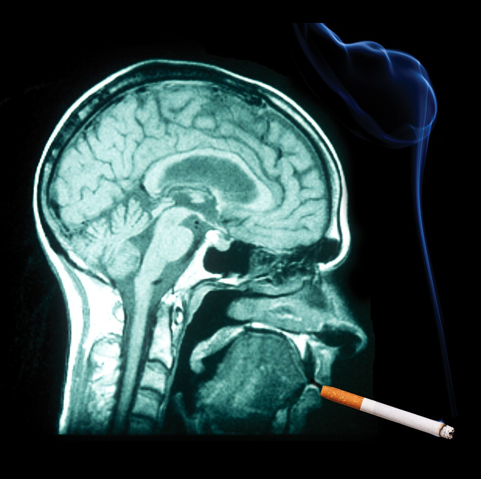
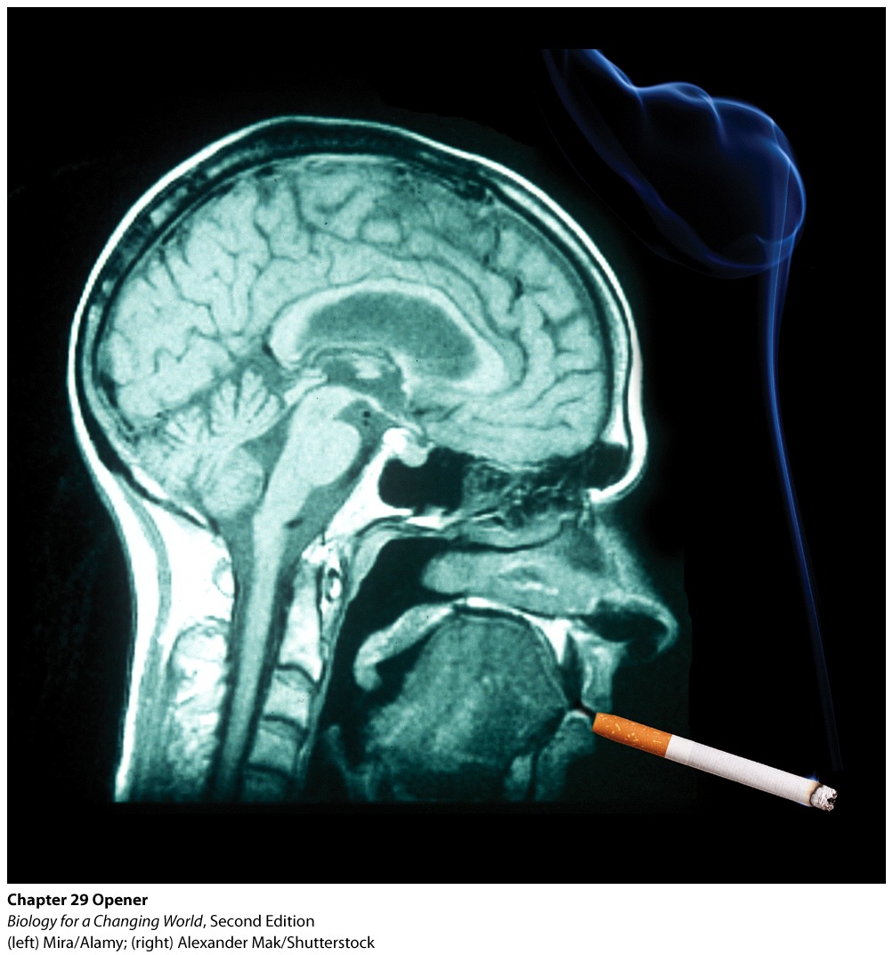
Smoke on the Brain:
Neuroscience explains why nicotine and other drugs are hard to kick
DRIVING QUESTIONS
- How is the nervous system organized?
- How do cells in the nervous system transmit signals?
- Why are some drugs (and some behaviors) addictive?
1.2 Driving Question 1:
Driving Question 1
How is the nervous system organized?
Why should you care?
The nervous system is the primary way we interact with both our internal and external environment. It allows us to receive, process, and act on information, sometimes consciously and sometimes subconsciously. As with all other organisms that possess one, our nervous system has been shaped by millions of years of evolution to maximize our ability to respond in adaptive ways to the information our environment provides us.
What should you know?
To fully answer this Driving Question, you should be able to:
- Distinguish between the central nervous system (CNS) and peripheral nervous system (PNS).
- Trace the flow of information through the nervous system from the time a stimulus is received to the action that results from the stimulus.
- Identify the major parts of the brain and describe their functions.
- List the parts of the brain primarily involved in addiction.
Infographic Focus
The Infographics most pertinent to the Driving Question are 29.2 and 29.5.
Question Test Your Vocabulary
Choose the correct term for each of the following definitions:
| Term | Definition |
|---|---|
| yHiQht3dVQ4o8m4iIfrMMXwEw9tdHtXHH1iFHe+iN326wab6NpSjZmGxvr3Omyfc2xoxxCmhIL16PvlpYTL0WpTOfuDBRjZMiphWNCsMsDsjGRRomquexJRu4yd0Hpt7BrCcxU1B+BJIkdX9aqxMnhhXpPl5c/tZ5JR8mthUlEsjFOsqJhC+7voqgTnLw0C99AAjkrkrh07egyuO0bw6IsmEbs1Uk4w6O9lcqQaIriRKnRVdbJSXLk6zlYPXwVOVMmqjYsMUeI1jb1/49DzOkDdMgv21Zwqc | Neurons that control the contraction of skeletal muscle. |
| u/p5k4VxFVsTNy58AlKQcU9zjnVvue8opvoz7jQ3laQc5NEhbvb34Z7siQP1oPigDjh5YcjzcsJM8f27N+pJrCcTdP8CRfndzjyFOfOJLVCGJRM3aQRJj3B+EXPkOhXya0wNoFk8AOA20a6txk9Y+6qdkqxmiFfiAwlBUDtjM0RLbdhlnzeEUIBKLdcUwybawjwdYG0k3i2VUAZ1Vi+PJmlGTC8A9Ri3FCJw7PbH0aw3bXenZ10XBUM6OzMWzlPpoqEfKrAYZElpSw7ZJR73LFOTRZWEFBLm | The outer layer of the cerebrum; the cerebral cortex participates in many advanced brain functions. |
| dmmmYGWPax932ks1yG6EnrFgmlARuNo1MDsOVI27FX4Dz0a6nFa5RGN2s7yCfy8zJ6+H8amn2mLSeNdepIU09V+/X/jdhS8aaY19JP/s1q8bABq+o4DYGs7QPlnpzoJmh/qlayg28LmbiZ8Yi5wwHoJLxUhiAnyOhX1QxlZf0PryJLLvG+mtwbX31RTLRGajBnmYQ/8pc/5Hxc493mpdpCnxpGvW4KI2KVMzt191+KjYIohu4/3EuSVyitkpDQpjfSXCXqPj6fT/5WN2CtIefQjkY/RBA+wA | A bundle of nerve fibers in the bony spinal column that transmits information between the brain and the rest of the body. |
| 9b06kYBNO3Yo4xFGyjAKwHwb3oLvlrwpUHy3QwA9HB0X/cztLeBXR6myvLT0y8jYzco2eePx+CQgnHCjQ86HhjGfKXd43LN69EDbzIJnvhllpRXPnjI1BBnQdhYmLlC72KzLJh9Op79mubSCJ8s/VMR7jlHnH9gVoxAJfZXAxNV78frU6VQRT8QHg3BEjPgmrzczTAwJHYnO8ExfjxXILB3PQJ2rTBoIl4Hsw60pYHx8A2n/ZJEd27awumgvsaCZpqtszhuuIY1ryOf4JTJj8Qxw43oSft/T | A neurotransmitter that participates in conveying a sense of pleasure in the brain. |
| ar9DuRgoDpNTFr2b3uraKENOGeAAiZuSCxEICG2OgsevBfaRsa3QlRGrXd5+z/zHFSyk7jH8XqwUuO25os7nrm75qN8DOjc9HKOxoPg4hPjP5mcNUw/s1fR842yv1WvcBYxqKv+60iTMW4S1HQ+pUWGTcVZKmPWntjbQPvIJex5FZPo77NMd1ZyeITzSBua+53bNcrraeSQAFBPioDaap0yq3sfjLDxTZxnLIiOBNJg7RYkH2L0dfJrDxj82KvSvAbQxWtVM2W/WDBRMLIl8Dy+kKxMuYA6C | A bundle of specialized cells that transmit information. |
| bFTw2lgnEZoNEQA6KP6KQoftzujOlHOFMHpiMPPJ0BmhYdWvgoY+gm48C2pv9Sk3vIhiU7ZHPR/uV6XF6jBg8Q0bzEtKilTeyOE3Sr0Chp2ktjcRjiP4QjLFKuXe8H/499QLL6rz+/t/wedGogvEbEve1kV6qJAJPbnB013KiGlkQXgGG06jPJSwWDS6yipDar1NZ2QIFpVNedJAwfma3wCTbbp9Y3FkkOBKJx1KbtxZHOUG8D6aByaVLE3HVypZwBTBxfEILCeX+18w4ZtxiOupBqbp4Bkc | A brain region between the brain stem and the cerebrum; it includes the thalamus and hypothalamus and regulates homeostatic functions such as body temperature, hunger, thirst, and the sex drive. |
| hVxsxLBZsh2QiiGvtRJgAY3JhaTMi/9MAWEZqEiyaa7LdVt3kIfCJWNf+9rn7d/EQ5Rr3kyQ6EngqgbHTuOQVT7SOCuM0w4n2byKYN54d2rqd7zWPypxP7mfUp1xdrs4xc/DYrcVAOsvrr54XCBOdpf7KrGk2CpyE7+FZWX+bRIMfpb7nvL/o6E09WmhOFs+/zf3W892luY4kdeoNcVlV4SM8mFQen9hNki4GFhc7JjO2Vkxq/Aua/AHbV8CkGdpKSugq9IMFFKHqTXAuUN7hT4GDQB/9FkG | An organ of the CNS that integrates and coordinates virtually all functions of the body. |
| SuHT6P+c9g5L7FWggwoxM5+q8veNa9RuCUIlskEQhW9bosRmCobrHdQxQWqnFeBQTpOkpVIUJfplthBUroBm8QuZ107MNqnPzfdEyR6niWMNWtarQLG3tPotPL0AznCpuYNFHXXE89XULzojX+F7zjcZa60s0fX79PJl+1o69P9uEG/ydwmaACh0x1A0m+Dr2Q/O+o+EXJ/5Ubd4uEfUrjzT5fLmyQ2b/mGTyh2WxEx03l/o0eOuz/rTFKoPaWmp0o0pN6e7S38EOxUVsaZnlrh7NOu1RrcN | The region of the brain that controls intelligence, learning, perception, and emotion. |
| On09rKFVXrKkLBMeyRc5S8DXGbYbWmkbORidtotf/00uq9/6BxpluI0JBbD/wf1+GlALDkuL2CTDtcZuxgNo1xkv1GQN7lDmNf0lRNs6v49hGR3OWMRugexyvpAkeLWkLgpJT8kmUx59EFu6bgOtgdq/LFK5L6aF+oaWgUUstbsy9rGAfa9bLuCZDZvlC0rJAoq6ISf5Z+x27x/cPlRGB4TZcQF6Z3TltyQ/5XSLx+4AntFGbqBTPurm5FZp/mqRsoefDFZBejb8CgouhJyus5wO2oP7tVX7 | A subregion of the brain that processes emotions, especially fear and anxiety, and is the seat of emotional memories. |
| b09e7YAdvgHk2DvmEqAsPnsHc+qCbRrusZ2mkWI9Mh2ZfnBwK+hudIfHoVBvYfh1cAFogRhDpRseQkXAHB5+B/q2C+ce8uCbVgAOcDx1CEeOE88KH0uOy/TjPQMzXAQVk2hyk9cTOXkmWHDKdZ0CGX4ZhkPiqnLRzlXj6u0XQmqI9ZEW464NWmmPDfmENvErdOXcs/QzXcUZKZ1w9hx3POlmzXYnVakTm0UQVOKL6CfWpauKN+AIEwXsEjtyPvzI59DzpW7N0RfpKYxaLTVSdOkITIJ98q6N | The part of the brain that processes sensory information and participates in movement, coordination, and balance. |
| R5B8taCsvEXF4try1JqwPCFa8ZVHAF58ZFHOBgSDnurzlMpvFex7RiHX2M/1tf937sDim6GDdPmN7UTlAUWdFF/dohF6PrEA8bSWHnmkVSeyTSGvyf4A21zkITAg+4e8yUhZcWDoAh//R1r7vSPBYWBLZlSkhenw4DRbAQCDhHBq0WrLJ2mMiCsNoYTSFO2PR+cU5tlCrhH6aiVHvsKNy1RyJmwrc67tOEOoCO6Yqf/0nxzO61K90Xu7lJHIebdsB+1EWfJPib7Ql4BKmRlezRftQlIeD06s | Cells that convey information from both inside and outside the body to the CNS. |
| +1hwWWuK3u8CkZjgqDIxs3+lEcnjVtzWkdJy3Iu9IleCvbmo+2uaxuOWOg+ZA9+GAuH/ULLabyJcGdUT0qWUZQa911QTHhPNKmveJBPdTp7vc3HjcjaNTFEcbOntMrbzamn449gidUt1T9C9gohbtMAnvHUdUGqZEXN0XsMe0d2ItpXxhUFuhrnxri3tsu2LmcyejGEasatYP64WazTrDdt2p/GmVpikyG0cspBddXG3aVjpDAHviRbUaUqalaIAPI7w69a/Xa06mgA/Mb2XFWFnNKDYJ0pc | The brain and the spinal cord. |
| eOPuGd1eS70HUcJOdDkM/rgIHfxezYLERYXIFE3SjRPp1IOJa+z4XOEgtq/rNeq5ef4VaulCig2lqzNA01ZyAXH6YtuJXD2LNZ47IOV5I9qikk8pRunQemVreak4E2Qv85dHAyaZxFYP6VVwKNGS+sgeSkXDrT5gu+w17/4I2E4TYTwHKauuNbUAsiK319+x+q3JJHYisxf/AfffDCBPxoOeOU4jmQhPpDl21iWwwZKIQlpkpCHc9NPrJWnqPKMrzM8FKihubxbmiWmt7Mj5EIm3UgVr7UmM | All of the nervous tissue outside of the CNS; the PNS transmits signals from the CNS to the rest of the body. |
| 2G5VcAgqnxtnukoKUHqwVKUx7xJzhF3EgP8A6rh6Zzy8VwvEUjPWS+cxpLgdQnuJEFN24dxRPDj3On9yleBcrOSLkZ1ZHtQiDRv0G2cpFynDqcp6eK1MCxk17oC7ceidPDC6aKNFr8D37OgRIKsUh9BQxm5jP8j3Qlgt6IN31E9Ehn8vMAmEc80bC2FwygZXWPHahlJKAX0N5oveji12ephXUCHY42Rw99BMxS/9ylcCNVDOoR2EH921IFsO7s7wemiNRcZJsOkmx88NN+VwvjIZL+2dXKyh | The part of the brain that is closest to the spinal cord and that controls vital functions such as heart rate, breathing, and blood pressure. |
| gh1V0EqSySeF7FRfLbm9Fi/Sjakb9WLOJujrPXVIwxA+YOgVfE9T6Or56iZ2Q042Q5KRubQldQFCr3Edm11hXZxoKi9Jf9mBSovm5O9x+ACBxUFbxtZd15BOpMHD88Da0JaqLaq3aOPQ3EatK0v3VDQXQcwFHMKK15zuGRZ9ULAjgEc1fzkHtTA7dRKEUQhJjldJeSyj1vZUbYva6JRVHm4kNG3QVC26Sf4eDmqJQA7kuf5LEVtiR8Sw0F2Bt3vOqub9m/XtB5ENeAov7FluNWMaZWUWvrgo | A subregion of the brain that participates in learning and memory. |
| yyXYMSg3jhgrjgUulqD2shZ4ikZX+x3mOCw2sKsQBepw86xEwyoQKz87AvhtbH5UsCOj9aMpQDUFLoNlHJST7x7zXwTuzBtheWN6ZUpy76NxTUJmu7MZr2h4RTz+ZYQgl/mWTO2NBZ6Vy40cDoHoFW114kTZhkfeRhQ5llSEendr/br53Rgqh//E43MxumuJPVXagr+WJNvN0Q+7kWsFa3AL392EGmcnGIpOTpPHRq9OiExv4Mc7TkpRcDtZ7bG+trqO/+HPAGlPmXuerq+IBRAlqisAE/Lj | A chemical signaling molecule released by a neuron to transmit a signal to a neighboring cell. |
Distinguish between the central nervous system (CNS) and peripheral nervous system (PNS).
Question 1.1
Correctly distinguish each indicated part of the nervous system as belonging to the CNS or PNS.
| Cerebrum | kkXLndHPdYqGl6KQpsHTpDuSv3+0Lirc4TVSfXQErhA= |
| Spinal Cord | kkXLndHPdYqGl6KQpsHTpDuSv3+0Lirc4TVSfXQErhA= |
| Peripheral neurons | XlIUqLvpfdliQkej768YA6hZu1wxhZP295O7h0IR+Kk= |
| Brain | kkXLndHPdYqGl6KQpsHTpDuSv3+0Lirc4TVSfXQErhA= |
| Neurons | LLeh2QDe6ECkAWKEITbTxds+6m9EC4RXEo+kDoMz1fY= |
Question 1.2
K21JoM/i+a7ja25RNgQ19ViX40/8bkz/G6Gko59yFswxjXzWm9aUxL9DEOZKQMw8OyL3IANMW7VyKfOXzzaJaCgLof/QqhgYTrace the flow of information through the nervous system from the time a stimulus is received to the action that results from the stimulus.
Question 1.3
Each of the following statements is one step in the flow of information through the nervous system. Order them correctly by numbering them (1 is the first step) in the spaces provided.
| uh3ga5KQzTRNkCI0n6CUhg== | The tiny muscles controlling the capillary beds in the skin of your cheeks receive the signal to open and respond appropriately. Your capillary beds open. |
| FJdFBx+geWiPLH1RMaVNZQ== | The cold signal is transmitted to the spinal cord and up to the brain. |
| AtTzzErpwCspTRZjRVC4AA== | You walk outside on a cold day. Sensory receptors in your skin detect the cold. |
| qF/WgfvNuGW+6fjpDtiH0g== | Your brain sends signals to warm the skin of your cheeks by increasing blood flow to the skin and sends a signal to the capillary beds there to open. |
Question 1.4
7MjmdtqR4IJOhZVx/4gqJE1XfQKJHAcC7PjkaU2jHC/3CVa5sXXElsxGUBDvIAA1ApuaXh9WVO6ZgP4FQuestion 1.5
Rg7tEL4C3HLM0Evn7QHdLyHy2J5Qs/YuXT45YbH0u5vDwMFSd88mFBW2B1jQAdcshS4qeX21SG5MtAGyQuestion 1.6
TvwA6A7Pc11zI/0/ZHWTjYZqqP4P1SRSZYqA4NniKAURkhryU6WtyfP+nhYWfWwNnlZK6XO5NdSI/CA/vf0UbLsTt3N2z3kSonkhwj3qwwG95FVfIdentify the major parts of the brain and describe their functions.
Question 1.7
In your notebook, draw the figure below and label each major brain region. In the spaces below, write a description of the function of each major brain region.

Major brain regions to label:
ExknWI54HoGqCev6yM92gA== YLFr0yVAXO6S2G9DmygOuQ== uhJUyplFc8PGjz576mJ6mu/o5gw= 2CcmBLo1yoCs5ipf2WrYtg== Fj/FB/loXXG1d2S7zFrdgg==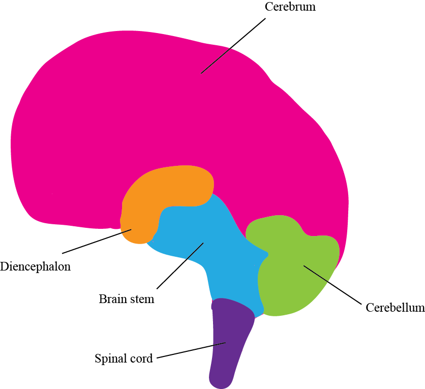
- Spinal cord transfers information from the body to the brain and vice versa.
- Cerebellum processes sensory information; also participates in movement, balance, and coordination.
- Diencephalon includes the thalamus and hypothalamus and regulates homeostatic functions (e.g., body temperature, hunger).
- Cerebrum controls intelligence, learning, perception, and emotion.
- Brain stem controls vital (often involuntary) functions such as breathing, heart rate, and blood pressure.
List the parts of the brain primarily involved in addiction.
Question 1.8
q6Bx9g0AChhHJA0PrVaAbHsYzp//0MU+M3wN2lDcSawSrp8KSDDinVdvypmearLyZ7I2AepbDRRd6EbkkpWlQzooMnp2QUlS2kKA7imtkTM=Question 1.9
tnKoalJHXHEwltwXV2zdnuIFqznLoSFr61A8uMFT45UIHtvRs7omMIZnCKP/6P87nssh+BuceR04kcN/vbfuV4MSZXrouh3CCcJdh4QEnzbAYk0HtYkyhlYuyz8ElP3yQuestion 1.10
oxTBlGjfsB9HDkh8vWTxpIUBfI5MbT0SoIKby/BBhzELcDQqUjIHMVhECC+P9v5ON4nxqUZcHDl/MP4dd63wZ4m2F7aMmVBXcEK5Ya5ZiV4YrkVfYBP7iBK/A/5ZUX+9B91SjmJtyx5xN/EaL2VFYCl8wO82XGak6Tn7XDCc6o+2vGCA5qH5GdlNLkw=Review Questions
Use the following scenario to answer questions 1 and 2.
As you sit studying in a quiet corner of the library, a friend decides to play a joke. He sneaks up behind you and yells “Boo!” in your ear. The following events take place:
A. Your ears receive the stimulus of the loud yell and send a signal to your spinal cord, which sends the signal to your brain.
B. Your brain registers that you are in danger and gets your body ready to freeze, fight, or flee.
C. Your brain sends nerve signals along your spinal cord and out to your adrenal glands to secrete adrenaline.
D. Your adrenal glands receive the brain signal and respond by secreting adrenaline.
Question 1.11
Wzh4lWEy951XVNY6hdGcz/BKLJrm9KvrNnP7uFHsTuVOxGh3xDIQ2eOK7Z6Myx6i3x6CzUqBN3W87lHuW7oWPEi65PbnF+gVEBj7nYZ6HVIOc8fYP778BrTxYwSvPISwJkerfNMSd3Ud7BG3XryMssOkZN8dyga030rqZhQj2QiS4DsAH+KUcg==Question 1.12
bsEbQH5wpDGFnEOtZNRn76+yB90GyMTt6F/1DMVTUw++WPIvMlsnSqwx5yleRRvYcuuipG7kS4NCPpblkaOLUnDzXh4H0Hb7RSl9FdYjAWIAqSIWJFAIDwidNX/FJUbk76gXVruGk7esMJ/wUDnKbTOockfGVBqOePlel22AbhJ0reRIvWVQ1A==Question 1.13
cLQqhVH6rKdqm7A0gxaA1Uz9/XJqrKf8c75FIewtni8vQ0H/oCZr8orgBqoJIysKvdvn8rFwuIfB4mpyMAqcpU2XTInYsZCEePbFxeHjFklLWjzzx0waQsrwIjheCXHnOXREz7DCBQcN8nELrdu+s4HYDd2fklQF7QHmrrzmmOa7F0X/kgG7Mw==Question 1.14
8Bztzdf045Nl838CM4Brsw71VTdegWVRdHOgD+3/l6/mMwxbNhxyBVFzk6fY9J4ujnezPMGL3tk/TwSon3OQR3xcWgEO5sJnJGLPVPs/PbHAGWfakVWQ23CiaFlBXOuF6WZ3Iu1PmVU8lW4rNc9u3LmBR2AH+kGyMslOcFU97AUbvarnAMDrnA0V1iQ9+NKtZUFXlw/1S3g=1.3 Driving Question 2:
Driving Question 2
How do cells in the nervous system transmit signals?
Why should you care?
As is generally the case, we need to understand neuron structure if we are to understand neuron function. The two together help us understand at least the basic problems involved in such conditions and diseases as multiple sclerosis, Parkinson’s disease, and Alzheimer’s disease.
Proper chemical communication between neurons and between neurons, muscles, and glands controls everything from your fight-or-flight response to your ability to move your arms and legs, from memory to the regulation of mood. Many animal venoms work by interfering with this chemical communication process; understanding how it works is also key to developing better treatments for a number of conditions, including Parkinson’s disease and clinical depression.
What should you know?
To fully answer this Driving Question, you should be able to:
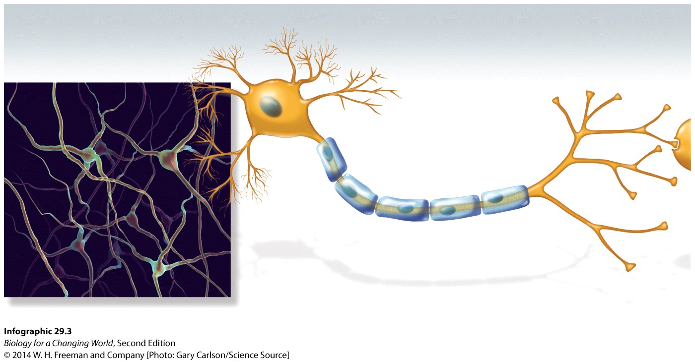
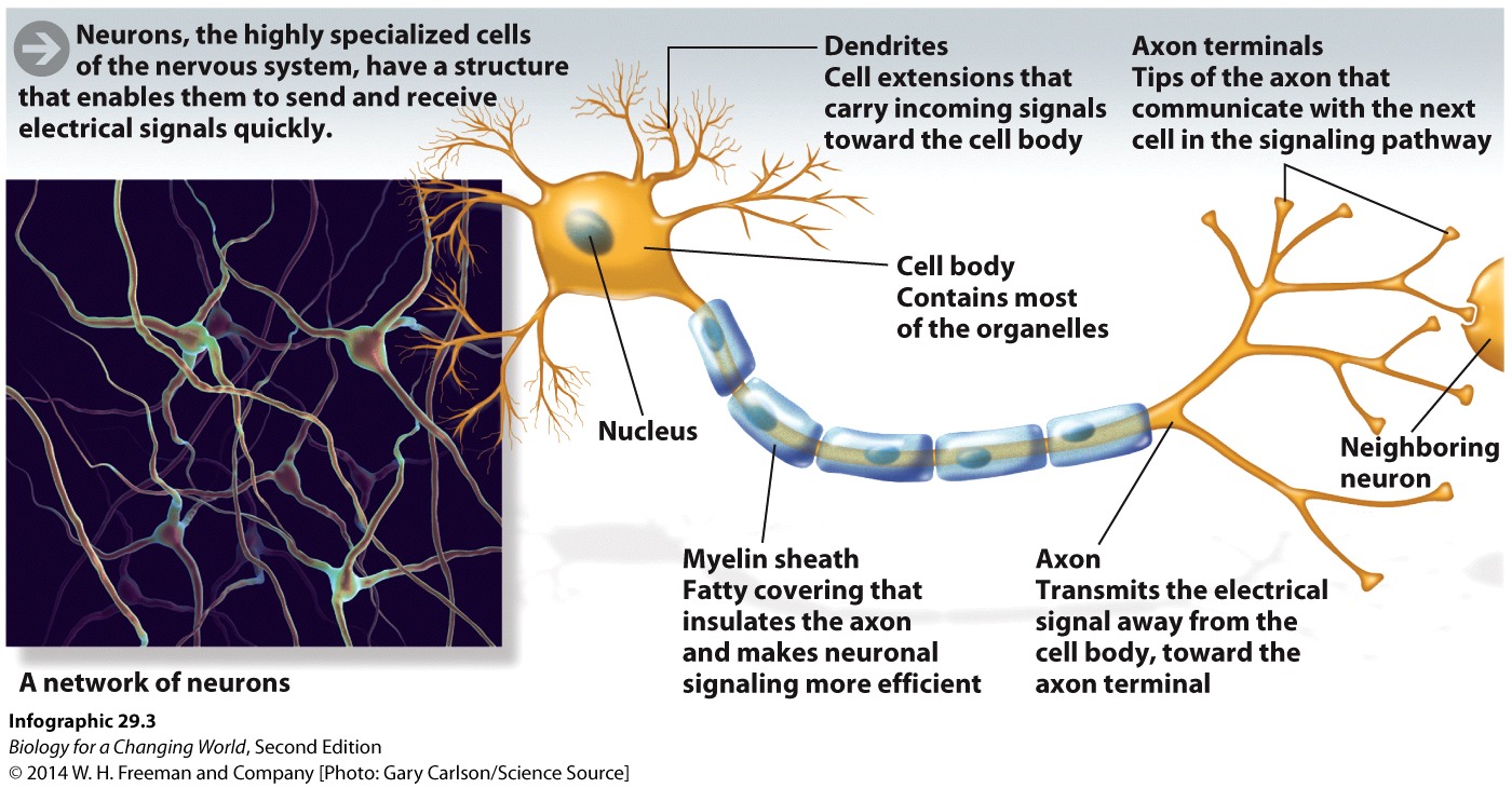
- Identify the parts of a neuron and explain their functions.
- Describe the process by which an action potential is generated.
- Describe the process by which an action potential travels down a neuron.
- Diagram a nerve synapse and explain the function of each of its components.
- Outline transmission of an action potential either between two neurons or between a neuron and another cell, such as a muscle or gland.
Infographic Focus
The Infographics most pertinent to the Driving Question are 29.3, 29.4, and 29.6.
Question Test Your Vocabulary
Choose the correct term for each of the following definitions:
| Term | Definition |
|---|---|
| KIRso7KTL7HikLe8osR8M5kc1RZGby7vKPhx2wmOZa7TwLX+SZHEYDyRw48L+nyonYZsR7iPo9a6AUWxCtPWJlcgc3OgKs+apYf1Msb5eonvJ+eU9w8KxIGSnJpEgcB60RcBOQ8/ijyBDOOg/dufhvJNJ5TNbUfjP5uVePmPrG9ECvePrpWHpZIhs+s= | A fatty substance that insulates the axons of neurons and facilitates rapid conduction of action potentials. |
| UCY+VxVbtsP5vntoIIKOA10fJ5+n/n8JjcnlNihAID4FZT1W1uKHU2v/fP3phNhzx0PdlBhqfR0gwcJOuXgKccO6SVNPiU2xXEwNfbYMjwpQwm1s4PtE25EudYPV+yCxWT1PK410gHfZvqQWtPg9ZN1rOOv4nokPzEFm6zvRXU+uZ8FdLausKi5qSC8= | Branched extensions from the cell body of a neuron, which receive incoming information. |
| h4Nv7bbQxL6ClIB9CBNn41dmkd7YzrkeopHuri69oei2/P5SWS7NVnzFB6NLFPB0V7JoVAKdO+t4/c3jRnGE8WAj6mhD/KjDsiIHPIVse4N/bAX1oGZdy9SMyWbkynxaoL+Dmrt/gdClfDKoH5etSCIiPsoVJBZoJZfUEOGLynmYMvRcyX9IX1NAimY= | The site of transmission of a signal between a neuron and another cell; the synapse includes the axon terminal of the signaling neuron, the space between the cells, and receptors on the receiving cell. |
| jbnuQH9MYa+IKiELebUNNzuJRJpi8QAlNYfhdTckSbrfXvcbyUniyl3vtZrPpvUclz7BsvgxwRLyPKnXLm3wkK63MKq5ERqj46043HBm3MnsNwATw+NWRk1Ja2jnhWD8QyZPC+TQEeoLamP/UXxQKljKXPuBwpV2cD2gtmvCVJKg+d97YpO108cjYVU= | Specialized cells of the nervous system that generate electrical signals in the form of action potentials. |
| yZ4O/UxwUGPPk9EwIo4BrSdPyT6XZG3GuH9V+DAseDzmJ8juBMi7HX5UN9fCOdA3OZY5lVjWT8bz88ZVegSQEFEPTeZMm23FAG9yNZUzQaBQjIgMcP1MzlO1sX+9Q41tP4A92xgWw8/UtlSVZBf8NLpwp4+BCVClWqE6FCPHLxmMKBUCI78bxLG4ieY= | The physical space between a neuron and the cell with which it is communicating. |
| pcTh8QZh3gc9FAV9H/Q7IBO6KgkoAjHNrfXDmkXLQrXWMjILNGyF8DE3mfqqLeJvIsp49VQIELxLSmXKoGsf5xNG0Y/nrO4FpVUagnK8k+qt09cAK7EpkNe+lZuRoUEoa+shkQHwt5IMfAd6XQUlb3jRjtr5CL47ZUj1yYMCWXv3nZ+tcp8/zV5TXo4= | The part of a neuron that contains most of the cell’s organelles, including the nucleus. |
| Sicbk4dBllBv5mplQRfgAnADGo4J99LLwRCmd+YDgXCKvz7KklSn3hub6vOZJPtxu9qYoJy29xzypiH0dV6PSrAVnubX31kshek7wTGWGEn8znyglq93G0sHS+xY6TRM0pT4cjYGu6l110aF7/Hgfy11gL/r1MGvPwfERBRHMToq6jNF98W84deeo8k= | A chemical signaling molecule released by a neuron to transmit a signal to a neighboring cell. |
| ieh2EtDUtBFR6TkCfA/g8aNn8YOw1+rlA/IgicsKLf7Rl5Pn90XJsJaYJvbdfwYqjdb9d+UyirLLEaYYK89O/0I1SY0CgjJ5R/yLsmP/GFISdqhBNR85YUv4qiOmjtrFTET1y6qr+EcL1cG4rf8GxHRL62yhMuaS53Pd9vhXlrGXfVXNq32PBjXVTnk= | The long extension of a neuron that conducts action potentials away from the cell body toward the axon terminal. |
| MG3dckk5QR8jcGCamjGXlQtLnEUszv81i+b4z2nIKrjH8Rlo90Gl9L40O9ouTz5fXkysurEzyiPbHkZdIni1KtQGvGOZjM92bNkm1KnjYwuFJkQDEzcT+r4FnQsolNzjgjn/0wyjoBWeA0zilIivbVYa/FYA95uoIcglDTcTBJ8isyREi+IYq68ekbQ= | Supporting cells of the nervous system. |
| 9r040RXXNdtVtLdjd/CcsVFv7ielpxjAJNN2+jBmnAsjrxuhucaAcPNvL3IYTrDh6wkVR7U7FD3D4aOGZPuGeyKNTMx9HdSUBP0A6bozkuHNPAYm1MI64Fz/2RGTZmdAz/SXN58OGqL5O3TER79xz8fJ9uUixRGLXH02qMOdX41V/AJPwHZDYlFB/i4= | The tip of an axon, which communicates with the next cell in the pathway. |
| DUh49YJaIazL02o5alk8TXcxm3e7va4xXIZZajdaPcJKJj8RkAMSpHQjsP2tsOS7gK4piLmBRP+Fyki4XDP8EB53dMYNv4ZQ4N7x21F4VwVC+Im7OkxCeh27Zik3mczZC/FtOG71OBigdnYlYxK9b7LQRqEfhbDN6Yrr2bc/LpQZ3gNnWdANum6tPaw= | An electrical signal within a neuron caused by ions moving across the cell membrane. |
| uE2j4VTEP6R7MsiPXHB4mzf40bTAAY6+o218T+HslZA/8pqhtlBpxbM7RqhLi/GkTv+yj7lDAOuTokdu+HqxErSHfQ7sN01gemfXjJJQOrPuQAAZnJ9m/mugRKG76C7jmgH0t05hCmwIAmjfDTjdRGNa3ovtEF4/P3fQmTgg3bKCAnzPfCXsyX4Unv0= | A neurotransmitter that participates in conveying a sense of pleasure in the brain. |
Correctly identify the parts of a neuron and explain their functions.
Question 1.15
Question 1.16
M9bYumXiqXNUDWcdQFcbvtvX78V8DK70NbKarb/9jxBojR/wMs/EJl/Mkw6awErqLgFY658px/BisfiYQ2P3LWB9pMLQIGXzNucleus: the command center of the cell, located in the cell body.
Dendrites: extensions of the cell body that carry incoming signals toward the cell body itself.
Axon: nerve cell process that transmits the signal from the cell body to the axon terminals.
Myelin sheath: fatty covering wrapped around axon; insulates the axon and makes signal transduction more efficient.
Axon terminals: processes where signal exits neuron; tips of axon that communicate to the next cell in the signaling pathway.
Describe the process by which an action potential is generated.
Fill in the blanks with the correct words or phrases:
Question 1.17
At rest, when it is not responding to a stimulus, the inside of a neuron is ZJVrNGhcywSMbZ34kX0wqQ== charged relative to the outside.
Question 1.18
When a neuron responds to a stimulus, ul9RhRLJYIKplntJ1JdzQw== channels open.
Question 1.19
Then the 531PIA9dzwfHKzne7PIvmQ== rush into the cell via its open channels, making the inside of the cell more E0Oln+jfXg6UZU3dNevN/9EfSe1vEB/o than the outside.
Question 1.20
To return to its resting charge, the neuron closes the CF3gwNc9NcI/7ahpm1kAIYspE2Y= and opens its bLDYdbxdEmutwUlJxgNsHpx+kk2keuXh, which allow I2oSuNL1gRUWCzAAWUyL2Q== to leave the cell and return the inside of the neuron to a yU5mBK+8pkwzWnVv charge.
Describe the process by which an action potential travels down a neuron.
Fill in the blanks with the correct words or phrases:
Question 1.21
An action potential travels down a neuron when 531PIA9dzwfHKzne7PIvmQ== from an area that is having an action potential triggers rF6W4ysGwMS/QnO0 channels to open on the membrane next to it.
Question 1.22
9Oin8xCSSG5VpygH acts as an insulator for the axon, preventing the leakage of the electrical charge. This makes the electrical signals travel H1BOyzSr1r3qplnt down the neuron.
Question 1.23
Myelin is produced by P8hAcepRwBRwIz9FqzNKTA==.
Diagram a nerve synapse and explain the function of each of its components.
Question 1.24
7LOlCvKs0MQJrpwZZUoPQWLo8TCHvdlzYKQ6PKIZ1KN/gS8meLbp6j+qY8eZWOASSmwPbS7T+3gV4bzZNbKjjjTPG5v7c5SJE8PdMqnRjLuz5TMk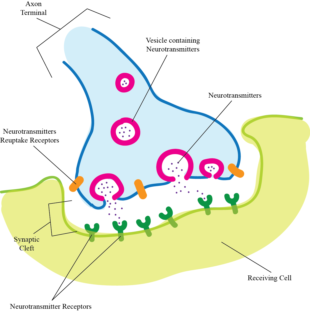
Axon terminal: Where the signal is transmitted to a receiving cell
Vesicles: Packages of neurotransmitters that will fuse with the membrane of the axon terminal and release the neurotransmitters into the synaptic cleft
Neurotransmitter: Signaling molecule
Neurotransmitter receptors: Receptors on the receiving cell that recognize and bind to the neurotransmitter, which in turn generates a signal in the receiving cell
Neurotransmitter reuptake receptors: Receptors on the signaling cell’s axon terminals that remove neurotransmitter from the synaptic cleft and stop the signal
Receiving cell: The target of the signal from the nerve cell’s axon terminals
Outline the transmission of an action potential either between two neurons or between a neuron and another cell, such as a muscle or gland.
Question 1.25
pCS+8vsVmVfymTXodIGcBi+xJz4jx8/UHC/dsDKEFKZiEWR/zgDdD7xQ92H2KZ5er9HEus7P0I03xwnFmcS9qLNzAhn3qLPpBXezkCZuj2/IryhoFFzPx3Kctkljnjt0mfEVZztw0tGi0bLvUiWISBSZWmWavugn/UToFgGLNaWBBq4EfGAU6l7aqHt+EOUo/x+amKL63kzs18fU7rl9iW+CV8RSLsbaR9AzY8jLMJVNm7IsK32NRx7bj0+w0PT51gEZMC/FNqkwTbTtGV9fPbaJU5NfynZYMXFKhS195OMjSBFOReview Questions
Question 1.26
Which part of the neuron can receive signals from surrounding cells? (Choose all that apply.)
| a. sensory neurons | Fd/yhwSeUQ6ZqQj4 |
| b. cell body | uS+xJY+rPntALoOB |
| c. dendrites | uS+xJY+rPntALoOB |
| d. axon | Fd/yhwSeUQ6ZqQj4 |
| e. axon terminal | Fd/yhwSeUQ6ZqQj4 |
Question 1.27
WmVLuLY6AUzs9HDmrtCG8xLTEnXs2QlibQzrXB7nlAJxCX2M8QvZUHaloOJb7EeYM+QKJykgCsUPLjWRiLcOiOjIs2dTlhWbDvVMkdA7QHoLESRZzOyK/qNC0DsrIhmy38RKX+VgTTrO0AMk4HBgCZGg7WcXwqOynAsMW25IfWRLTPu8IXh2G3cMyuJx1l/LzSgK5vhh6J8l70NUCzj0G17xhpi/VClKuPaVySzssk5CqIxG/T/QZS1n7dqyAohRXGuPlku3oIedvEliQuestion 1.28
wreQwKBf7iRTSGzzdDZf5BFlCt2liJkR8PLav7qbt9EkThGJmDXNjn8mFCmG1LoK/ehJnJLRBVXTgkzLTFVMxBsuZSyrA/8mQfW5DWwLQj0jMd2WrAFLVri1RkEmgUgu+J1Umfy0vovXm8cg0fAnMSjytQ37TKbfd6sJfZEF8DE+Wx1edfiCWOBeo2Y7dZ7aVB+MX/zFljz/ByBF0QRSLNbycCnlmn0kvWzwE/jPqZ1B4zvlF/ZC4uPAJQS9tMZcJwbE9YvTm3O92OFZoxSurfC8nUY=Question 1.29
ExctNorIjAq05lCwRlxJaOVs/7ILmZ43hRHjPtltIS5900hf7EUKF0fTdcEn3+Q/9fqxqtR1XFW0rkjiXbqwIce5rYXBU4RSy7+wRoFumGTxrjWJ8iMDTTQ1l+RCj78t1u5DKys7+MIugbTCPp4EKbRlJZ9m367AWnNV8NmnWUtg9J+wQuestion 1.30
M0aEgXC+BZ89T868zglvDvZYL67Cp2ToAuv/wehkMqQQDfuPKe+O4+811JPxUm1NijNPkNkYknkZwrjDVXGAElZl8+Sfdqx1XZD9xuSg+HdPqAYcdQhD40pZd3DcB4ETN7FEFfbndGQypGlxd/HoRhyszj15MQqPwYz3Jspmhx3UiZvemQ7B5WKBYfvL80dONP57RBC9Mhyf22nKOOryeaqOD0bxHV/eXyDSpBz3qBqas4z1ivr5kEnLrExVZ2035SeeioZEZOb0doWyQuestion 1.31
f1RE+xNeVq/zcrGCh5JEAgBJBLlQ47Irkc5HmBG/wFM1rfdKhGY5mABwc62YsU5siKD90JWbClmFfmL0hNCZl2LwdRStCyhUryhUSB9mtMizoyIDsI0HQQhcSA8aMm63pf6m/7u5+i037Ha05UyukzmCVNz5h989P2ZLpxKLFzEigBVlrew1/hvJuJJbnVk3qjiJe2ve3E2B2QzGHtR6pn+0/qpCPu07boA2VcjZs9PAsKhqvl/2hxW1DzYqMlmxoktXArZoGwONLrTTjeoaZsxUIjScOn+FMQ/fA06Zua7QZfMZYAMy4Afc+NZKPMNhQ4nJLIDb/RY=1.4 Driving Question 3:
Driving Question 3
Why are some drugs (and some behaviors) addictive?
Why should you care?
Addiction is a complex process that takes enormous tolls on individuals, families, public health, and more. Scientists are working hard to pinpoint how addictive substances—and even behaviors—affect the brain; their hope is that by doing so they will contribute to the development of more effective ways to help people recover. The good news is that whether the addiction is to nicotine, food, gambling, or illegal drugs, the same areas of the brain and the same underlying mechanisms seem to be involved. The bad news is that the mechanisms are very complex, so much work remains to be done before we understand addiction as well as we need to.
Although details of the individual mechanisms vary, all addictive drugs ultimately affect dopamine signaling the same way: they initially increase the amount of dopamine produced by the neurons of the ventral tegumental area (VTA). This kicks the pleasure centers of our brains into high gear, resulting in feelings of mild to strong euphoria. This simple result isn’t the end of the story, though. Our bodies are the ultimate Goldilocks machines, keeping the levels of critical chemicals in our bodies not too high and not too low, but just right. When a body detects too much dopamine, it responds with mechanisms to reduce it. In the absence of the addictive drug, then, dopamine levels fall below normal. The person starts to feel anxiety, depression, and the urge for more of the drug. To add insult to injury, if a person who uses the drug long enough will need to keep increasing the amount to get the same feeling of pleasure. This is the heart of addiction.
What should you know?
To fully answer this Driving Question, you should be able to:
- Identify the primary chemical (neurotransmitter) involved in addiction, where it is produced, and its role in addiction.
- Discuss the effects of exposure to addictive drugs on the synaptic structure and function of dopamine neurons.
- Explain how these changes in the structure and function of neurotransmitter synapses can lead to long-term addiction.
Infographic Focus
The Infographics most pertinent to the Driving Question are 29.1, 29.5, 29.7, 29.8 and Table 29.1.
Question Test Your Vocabulary
Choose the correct term for each of the following definitions:
| Term | Definition |
|---|---|
| XkMeC2eXDkVILatPFDOpSqvHNERG1ilnTC6uv4csbgr0QE96XVXg1wC3RryGSBFOlAkwbHFyD8I= | The site of transmission of a signal between a neuron and another cell; the synapse includes the axon terminal of the signaling neuron, the space between the cells, and receptors on the receiving cell. |
| Jdi1rTAbr/jGoxWLWvT7pZHwGnUsQRPugzn9N9UwNOcwb3LFlIX7c2PNYHYMyNNWO1DwWncyYJU= | A chemical signaling molecule released by a neuron to transmit a signal to a neighboring cell. |
| Lmf8fGjiw1ppDod8775XjbkY6uqLmgEKbpSPChNFn99qqryud+BWQeDSuqBdyyg7oi85nVmEJPo= | The physical space between a neuron and the cell with which it is communicating. |
| LfR3repmgXfALj8IT+sueOUjbLMqOW2KIa3UtAtyl08qxXFhsOzdABVeIUeI42X3VZzZOktto/c= | A neurotransmitter that participates in conveying a sense of pleasure in the brain. |
Identify the primary chemical (neurotransmitter) involved in addiction, where it is produced, and its role in addiction.
Question 1.32
IAlz3MBBuIk03AbJjugK+KR3VX8sv2aJYAkVG/WA9BFvGiQaiQ/RG88xdrTXRpr6gj25pzGgfOYSbl+VCDiHJA==Question 1.33
B+XnJWoYj33aiHyz/bHdSyuuGYHOcqjxy5LMqU16zg6oyrA8jmK2Jg==Question 1.34
WiYYnO78iDgNqQSrgdIjHhRbsGrhlESat7knjEXpI9hp/D2rn8w313obTXE=Discuss the effects of exposure to addictive drugs on the synaptic structure and function of dopamine neurons.
In the following scenarios, discuss the levels of dopamine and dopamine receptors in the synaptic cleft with respect to levels found during normal functioning. (i.e., greater than or less than levels found during normal functioning). Also, in one or two sentences, describe the effect of raised or lowered dopamine levels on the person who is using the drug.
Question 1.35
yf+KMgh9239ChDLnm5q+7jUKNGuoHp5r+iVFOAVs/WaDHNVWvL7wQNs92HnXALCrL7bVXLSJdzxLaaf2YGIU7mRrOlhUQ7j0pKnVvsItda5xluIxrM0o+/NdDZUPuZlKLw2IzLpJ/faAnONIRoVcx8o6L4c=Question 1.36
QZZYaa1RkkkzRYzPV7qw9M5d4y2J56Ixo3oXdeWbJg7T8qFHHsl/5nT2BV/QM3kTaidNMbb2j+ba9kUnQuestion 1.37
Anwnu9/XISrbH7hFxGA10tgYD0KB1JEL/Xm3NYP+D0WgyXWD1r3h20baeDX1uhQ3zk68AMuPD0zQZLKYExplain how these changes in the structure and function of neurotransmitter synapses can lead to long-term addiction.
Question 1.38
EopoUi9PAP+u1i7xhubO9j4QLw6HUrOeBMaJvwQil9/zhPC+BAG37aB17nA3ldltUAjXTeRKWhEUE0FpBzH84kCyR5M8rvxTCZqiu6+5VDlNEBERT2rGYVnqWdx915jbcYVx3zUTzuq5Ra/6gsMOwNzabNva3iaSGuT5TPPEipO0GOgoE8NdSDMKkuGdRVNddDkUQu/c4W6zZdn5ZdO7r/20L4GmZQMF3TeWPMrR/8F9nGAeu5H2pGQt8iRL2cvBvlRpwvdc0Sz+mfTSukShpd1xzLSz6yH84U8D9ksFp743KZQSfj8Q6UbMKbKtuyt1TFxl1LuvXzT4PyBebZW3N+37Km5+LOA7QGL4Yh9Jod4ASjUG91UMrbilYWDJn5RCeH6M2jPrq+p2b/1S+KLJiv7z1i3QirhETceLiwEcRX6xfjYg1lgjYrguuB4=Review Questions
Question 1.39
At which stage(s) of addiction are dopamine levels in the synaptic cleft the highest? (Mark "yes" for all that apply.)
| a. before addiction | Fd/yhwSeUQ6ZqQj4 |
| b. right after first drug use | Fd/yhwSeUQ6ZqQj4 |
| c. after prolonged drug use | uS+xJY+rPntALoOB |
| d. after the cessation of drug use | Fd/yhwSeUQ6ZqQj4 |
Question 1.40
At which stage(s) of addiction are dopamine receptor levels in the synaptic cleft the lowest? (Mark "yes" for all that apply.)
| a. before addiction | Fd/yhwSeUQ6ZqQj4 |
| b. right after first drug use | Fd/yhwSeUQ6ZqQj4 |
| c. after prolonged drug use | uS+xJY+rPntALoOB |
| d. after the cessation of drug use | uS+xJY+rPntALoOB |