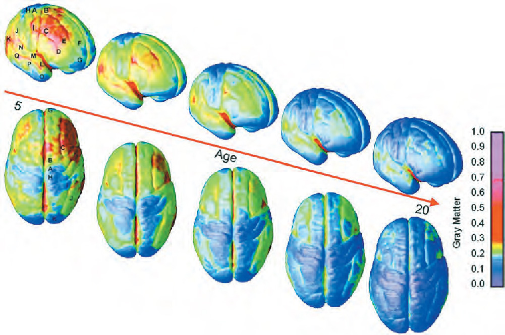
FIGURE 3.9 Brain maturation These views of the right side and top of the brain of 5- to 20-year-olds illustrate maturation over the surface of the cortex. The averaged MRI images come from participants whose brains were scanned repeatedly at 2-year intervals. The bluer the image, the more mature that part of the cortex is (i.e., the more gray matter has been replaced with white matter). Notice that the parts of the cortex associated with more basic functions (i.e., the sensory and motor areas toward the back) mature earlier than the areas involved in higher functions (i.e., attention, executive functioning). Notice particularly that the frontal areas, involved in executive functioning, approach maturity only in early adulthood. (From Gogtay et al., 2004)