Chapter 1. Central Nervous System
Introduction
The nervous system has three broad activities.
- Its sensory systems permit it to sense events occurring inside and outside the body.
- It interprets, integrates and may store this sensory information (in the form of memories), then it decides on an appropriate response, if any.
- It sends commands to muscles and glands (this is called the motor function of the nervous system).
The nervous system is divided into the peripheral nervous system and the central nervous system. The central nervous system is divided into the spinal cord and the brain.
The spinal cord contains motor and sensory neurons, most of them connected with the limbs and trunk by means of the spinal nerves. The spinal cord contains interneurons. It contains the large ascending tracts of nerve fibers that transmit sensory information up the spinal cord to the brain. Additionally, it contains large descending tracts of nerve fibers that transmit motor commands down the spinal cord from the brain to the muscles. The spinal cord can also execute certain simple behavioral activities without involving the brain (its neural networks contain a program for walking, for example).
The spinal cord is actually a continuation of the brain stem. It exits the cranial cavity through the foramen magnum at the base of the skull and extends into the vertebral canal of the vertebral column.
The brain consists of the brain stem, the cerebellum, the diencephalon and the cerebral hemispheres.
The Meninges
The brain and spinal cord lie within the cranial cavity and the vertebral canal, respectively. The brain and spinal cord are covered by three membranes that make up the meninges. The outermost membrane is the dura mater (means “tough mother”); the middle is the arachnoid mater (“spider-like mother”), and the innermost is the pia mater (“delicate mother”).
The dura mater is composed of fibrous connective tissue containing blood vessels and nerves. It often extends between the lobes of the brain, forming partial partitions. In some areas, it splits into two layers, enclosing the dural sinuses.
In the spinal cord, between the dura mater and the wall of the vertebral canal is the epiduralspace. It is filled with fat, connective tissue and blood vessels. The epidural space inferior to the second lumbar vertebra is the site for the injection of anesthetics, such as a saddle block in childbirth.
The arachnoid mater is a thin, spider’s web-like membrane lacking blood vessels. Beneath it lies the subarachnoid space that contains cerebrospinal fluid.
The pia mater is thin, and contains nerves and blood vessels. It follows the contours of the brain and spinal cord quite closely.
From the cranial cavity, the dura mater extends into the vertebral canal and ends as a blind sac just below the end of the spinal cord.
Clinical Application. A condition known as meningitis can be caused by bacteria or viruses. The meninges (usually the arachnoid mater or pia mater) become inflamed. If not treated promptly, death, mental retardation, paralysis, or loss of sight and hearing can occur.
Clinical Application. A severe blow to the head (e.g., a bicycle or motorcycle accident) can rupture meningeal blood vessels. If the blood released from the rupture pools beneath the dura mater, a subdural hematoma occurs. Because the blood has no place to go, its accumulation compresses the softer underlying brain tissue. If the increase in pressure is not relieved, brain damage or death can occur.

Ventricles
The cerebral hemispheres and brain stem are hollow, and contain four interconnected cavities called ventricles (see Lecture notes). The ventricles are continuous with the central canal of the spinal cord, and contain cerebrospinal fluid.
- The first is the right lateral ventricle.
- The second is the left lateral ventricle.
- The third ventricle is small. It connects with the lateral ventricles through interventricular foramina.
- The fourth ventricle is connected at its anterior end to the third ventricle via the aqueduct of Sylvius (cerebral aqueduct), and is continuous with the central canal of the spinal cord at its posterior end. The fourth ventricle has openings in its roof that permit cerebrospinal fluid to pass into the subarachnoid space.

Cerebrospinal Fluid (CSF)
A. Production of Cerebrospinal Fluid
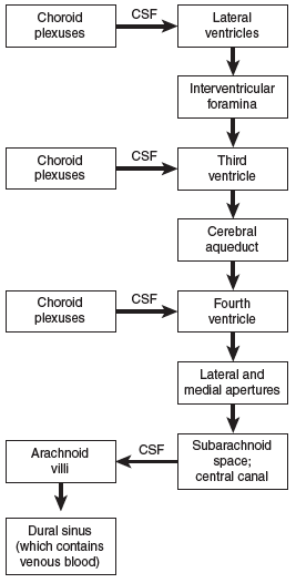
Most of the cerebrospinal fluid (about 140 ml) is found in the four ventricles, where it is formed as a plasma ultrafiltrate from networks of capillaries called the choroid plexuses. The rate of production is about 600–700 ml per day. The choroid plexus consists mainly of capillary networks surrounded by epithelial-like ependymal cells of the neuroglia.
Cerebrospinal fluid flows from the lateral ventricles through the interventricular foramina into the third ventricle. From the third ventricle it flows into the fourth ventricle through the cerebral aqueduct. From the fourth ventricle, cerebrospinal fluid flows into the central canal of the spinal cord and also out of the fourth ventricle into the subarachnoid space. Within the subarachnoid space, cerebrospinal fluid flows over the surface of the pia mater covering the brain and spinal cord.
B. Absorption of Cerebrospinal Fluid
Cerebrospinal fl uid is returned to the blood via arachnoid granulations (= arachnoid villi). These are fi nger-like processes that project into the lumen of the dural sinuses, which are fi lled with venous blood.
C. Functions of the Cerebrospinal Fluid
Because it is in equilibrium with the extracellular fl uid of the brain, the cerebrospinal fl uid is important in maintaining a constant external environment for the neurons and neuroglia. The cerebrospinal fl uid provides a mechanical cushion that protects the brain from impact with the bones of the skull when the head moves. By its buoyant action, the cerebrospinal fl uid allows the brain to fl oat, reducing its effective weight from 1,400 grams in air to less than 50 grams in the head. Finally, the cerebrospinal fl uid serves to remove waste substances from the brain and transfer them to the blood, and may act to distribute some peptide hormones and nutrients.
The Brain
The brain contains hundreds of billions of neurons that communicate with each other and with other parts of the central nervous system through numerous nerve fi bers. The brain serves as a center for receiving sensory information. It also issues commands for movement. Other regions of the brain are also involved in memory and reasoning.

The Brain Stem
The brain stem consists of the medulla, pons, and midbrain. The brain stem contains many nerve fiber tracts and masses of gray matter called nuclei.
- The medulla oblongata, like the spinal cord, consists of a layer of white matter that surrounds a mass of gray matter. The white matter of the medulla consists of ascending and descending tracts that serve as the communication link between the spinal cord and the brain. On the lateral surfaces of the medulla are oval projections called the olives. The olivary nuclei connect with the cerebellum. Other nuclei within the medulla function as control centers for many vital processes. Examples are the cardiac center, which regulates the heart rate, the vasomotor center, which causes the dilation or the constriction of blood vessels, and the respiratory center, which regulates rate, rhythm, and depth of breathing. Other nuclei are involved in certain nonvital reflexes such as coughing, sneezing, swallowing, and vomiting.
- The pons appears as a bulge on the underside of the brain stem and separates the midbrain from the medulla. Through it run the nerve fibers of the middle cerebellar peduncles. Also present are the nuclei for several of the cranial nerves that relay sensory impulses from peripheral nerves to higher brain centers, and nuclei that are involved in the regulation of the rate and depth of breathing.
- The midbrain is located just above the pons. It contains many myelinated nerve fibers that connect the brain stem and spinal cord with higher parts of the brain. The cerebral peduncles are two prominent bundles of nerve fibers on the underside of the midbrain. They contain the corticospinal tracts and the certain sensory fibers. On the upper surface of the midbrain are four rounded eminences (two on each side of the midline) collectively called the corpora quadrigemina. The superior colliculi contain nuclei responsible for movements of the eyes and of the head and neck in response to visual stimuli. The inferior colliculi contain nuclei responsible for movement of the head and neck in response to auditory stimuli.
The Cerebellum
The cerebellum is important for coordinated smooth movement and posture. Like the cerebrum, it consists of two hemispheres and is made up of an outer layer of gray matter, called the cerebellar cortex, and an inner layer of white matter. Like the cerebrum, there are deep masses of gray matter called the fastigial, interposed, and dentate nuclei.
The inferior, middle and superior cerebellar peduncles are nerve tracts that permit communication between the cerebellum and other parts of the brain.
The cerebellum is involved in the execution of smooth, coordinated voluntary movements. It regulates the rate, range, force, direction, and timing of movements by controlling the activities of agonist, antagonist, and synergist muscles.
In this respect, the cerebellum appears to be a device that compares intention with actual movements. When the actual movement deviates from that which has been planned, the cerebellum issues compensatory commands via the motor systems. The cerebellum is involved in posture, balance, and equilibrium. It also has a role in learning motor skills.
The Diencephalon
The diencephalon is associated with the optic tracts, optic chiasm, infundibulum, pituitary gland, mamillary bodies, and the pineal gland. However, the two major structures are the thalamus and the hypothalamus.
- The thalamus is the major relay and processing station for all information passing to the cerebral cortex. The information may be sensory in nature; it may be from the cerebellum and basal ganglia.
- The hypothalamus contains many nuclei, as well as neuronal receptor cells that monitor the osmotic pressure of body fl uids and the body temperature. The hypothalamus is important in homeostasis, such as maintaining the body’s water balance. The hypothalamus also controls the autonomic nervous system, directs the release of hormones from the pituitary gland, and is involved in many of the emotional and motivational behaviors associated with the limbic system.

The Cerebrum
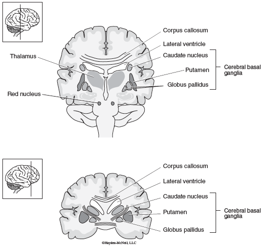
The cerebrum develops from the anterior portion of the forebrain, and consists of left and right cerebral hemispheres. The surface layer of the cerebrum is composed of gray matter (neurons), and is called the cerebral cortex. Other areas of gray matter (neurons) are found in masses located deep within the cerebral hemispheres. They make up the cerebral basal ganglia: the putamen, caudate nucleus, and the globus pallidus (other components of the basal ganglia are found in the midbrain, and consist of the substantia nigra and the subthalamic nucleus).
The cerebral cortex overlies the cerebral white matter, composed of billions of myelinated nerve fi bers that conduct nerve impulses into and out of the cerebral cortex, and also between different areas of the cerebral cortex. The corpus callosum is a huge band of these fi bers that connects the two cerebral hemispheres.
During human embryonic development, the surface area of the cerebral cortex increases enormously, in order to accommodate the increasing numbers of neurons. Consequently, it becomes highly convoluted and folded. The folds are called gyri (plural of gyrus). Gyri are separated from each other by grooves called sulci (plural of sulcus). Very deep grooves are called fissures.
The longitudinal fissure separates the right and left cerebral hemispheres. The lateralfissure separates the temporal from the frontal lobe. An extension of the dura mater called the falx cerebri dips down into this fissure.
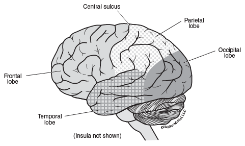
Anatomically, the cerebral cortex is divisible into five lobes, mostly named after the skull bones that overlie them.
- Frontal lobe—contains the precentral gyrus, which represents the primary motor area.
- Parietal lobe—separated from the frontal lobe by the central groove or sulcus. The postcentral gyrus represents the somatosensory area.
- Temporal lobe—separated from the frontal lobe by the lateral fissure.
- Occipital lobe
- Insula (Latin for “island”)—lies deep within the lateral fi ssure under the parietal, frontal and temporal lobes. It cannot be seen in an external view of the brain unless the temporal lobe is pulled out and away from the rest of the brain. It is delineated by the circular sulcus.
The Cerebral Cortex—Functional Areas
The cerebral cortex has areas that have different functions. Large regions of the cerebral cortex are committed to movement (the motor areas) and sensation (the sensory areas). Association areas are involved in our highest intellectual activities and often provide the link between sensation and action. The association areas integrate diverse information, often from many different forms of sensation (sensory modalities) and from other cortical areas.
1. Motor Areas
The motor area contains primary and higher-order areas. Primary motor areas execute voluntary movements, while higher-order motor areas are involved in the planning of movements. The major primary motor area is located in the precentral gyrus of the frontal lobe. Different parts of this area control movements in different parts of the body, which are roughly mapped on the surface of the cerebral cortex.
The motor speech area, or Broca’s area, is in the frontal lobe (usually on the left side). Neurons in this region control the complex movements of mouth, tongue, larynx, and breathing that make speech possible. Broca discovered this area in the 1860s by noting that patients with damage to the left frontal lobe were unable to speak (i.e., they suffered from Broca’s aphasia).
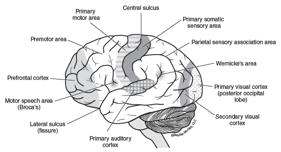

Wernicke’s area is a complex area involved in language reception and comprehension. It is found in the temporal lobe, close to the primary auditory area. It processes both verbal and visual commands. A bundle of fi bers called the arcuate fasciculus connects Wernicke’s area with Broca’s area. A lesion in Wernicke’s area does not impair a patient’s ability to speak. Such patients often speak fl uently, but the words often convey little meaning and they cannot convey the ideas they have in their minds.
The frontal eye fi eld is superior to Broca’s area, and is involved in gaze control. Another eye field area is found in the occipital lobes, and seems to be involved in tracking movements of the eyes, as when a spectator watches the ball in a tennis match.
2. Sensory Areas
Primary sensory areas receive information from sensory receptors, while higher order sensory areas process more complex aspects of the sensation, and analyze and integrate information that they receive from the primary sensory areas.
The primary somatic sensory area is located in the parietal lobe, in the postcentral gyrus (B in the diagram). The body surface is mapped roughly on the surface of this gyrus. Damage to this area can lead to loss of fi ne localization of stimuli, loss of the ability to judge weights, shapes, and textures.
The primary visual area is located on the medial surface of the occipital lobe, and the primary auditory area is located in the superior part of the temporal lobe near the lateral fi ssure. Primary areas for taste and smell are found in the parietal and temporal lobes, respectively.
3. Association Areas
Association areas integrate diverse information, often for purposeful action. In consequence they often provide the link between sensation and action. Association areas are found in regions of the parietal, temporal, occipital and frontal lobes. One part of the parietal lobe is known to be important in our awareness of our bodies and where all the parts are located in relation to our surroundings. People with damage to this region may fail to recognize parts of their body as belonging to them. For example, such a person might wake up startled, believing that someone had put a fake leg into bed with them. It was really their own leg they were looking at.
During mammalian evolution the prefrontal association cortex in the frontal lobe expanded dramatically. In human evolution, the development of a distinctively high forehead seems to be associated with the need to accommodate the increasing size of the prefrontal cortex. The prefrontal cortex seems to be involved in the highest intellectual functions that include thinking and problem-solving. It appears to play an important role in personality and emotional drive: it has extensive linkages with the limbic system via the thalamus.
Other Components of the Cerebrum
1. The Limbic System
The prefrontal association area is often considered part of the limbic system. The limbic system is a loose term for a group of gyri and associated structures (not necessarily cortical) that roughly encircles the corpus callosum and the diencephalon. In turn, the limbic system is encircled by the temporal, occipital, parietal, and frontal lobes.
Functions of the limbic system include self-preservation (feeding, fight, flight), reproduction (mating, care of offspring), emotions, goal-related behavior, motivation, sensations of reward, pleasure and punishment, and memory. The limbic system provides a link between the conscious functions of the cerebral cortex and the autonomic nervous system and endocrine system via the hypothalamus.
Much of the output of the limbic system goes to the hypothalamus.
2. Basal Ganglia
The cerebral basal ganglia are masses of gray matter lying deep within the cerebral hemispheres. They are the caudate nucleus, the putamen, and the globus pallidus. Two midbrain structures are also included with the basal ganglia: the substantia nigra and the subthalamic nucleus. Neurons in the basal ganglia receive major input from the cerebral cortex. The output of the basal ganglia goes via the thalamus back to the cerebral cortex. One of the best understood functions of the basal ganglia is related to voluntary movements.
Clinical Application. Different parts of the basal ganglia facilitate movement, while other parts suppress them. This becomes very apparent when studying two well-known disorders of the basal ganglia—Parkinson’s disease and Huntington’s chorea. Refer to lecture notes for more information.
Memory
Memory can be divided into procedural or “knowing how,” immediate or working memory, and declarative or “knowing that.” Declarative memory is divided into short term (recent) memory and long term memory.
- Procedural memory or “knowing how” memory—is concerned with the learning of motor skills. Knowing how to type, play the piano, or ride a bicycle are examples. Some procedural memories appear to involve the cerebellum.
- Immediate or working memory—working memory is a store that holds important information in the mind for brief periods of time. Working memory is temporary and its capacity seems to be limited. It. seems to be associated with “inner speech,” helping us to express our ideas in a meaningful way. Working memory seems to involve many brain areas, but particularly the prefrontal cortex.
- Declarative memory or “knowing that” memory—The kind of memory that most people think of as memory holds the names of things and people, occurrences, events, and other pieces of facts for periods of time ranging from days to a lifetime. It is known as declarative memory. Declarative memory is divisible into two types. Short-term or recent memory concerns events that occurred days to weeks before. Long-term memories can last years or a lifetime. The hippocampus is a region of the limbic system that seems to be involved in declarative memory. Removal of the hippocampus in humans erases memories going over the few months before surgery. Long-term memory, from many years ago, however, remains intact. Further, there is loss of the ability to form new long-term memories. It is, therefore, believed that declarative memories are first stored in the hippocampus as short-term memories. After time, there is a reshuffling and reorganization of the information and it is placed into more stable long-term memory banks in other brain locations.
There seems to be general agreement that learning and memory are associated with long-lasting, use-dependent changes in the “strength” of synapses.When synapses in the hippocampus are repeatedly stimulated, they are “strengthened”. That is, it becomes easier to excite the postsynaptic (receiving) neurons. In lecture 13, we described this process as long-term potentiation (LTP).
There is also evidence for dendritic growth associated with memory formation.
Question 1.1 On the diagram of the brain below, label the following anatomic areas
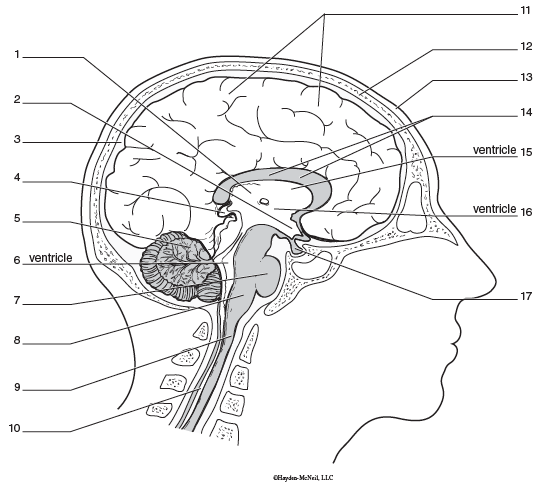
| scalp | +e5m8Wi5evA= |
|---|---|
| skull | DDH6Tw1RFEk= |
| thalamus | 0VV1JcqyBrI= |
| hypothalamus | XvVM00l89Is= |
| pineal | 0VV1JcqyBrI= |
| cerebrum | cVy0Xs6Cud4= |
| cerebellum | DYU2tVvtzEQ= |
| pituitary | ITC7oswgWJE= |
| pons | I0N5oPvoADQ= |
| medulla oblongata | Pz2PEfhsNWI= |
| lateral ventricle | U1syIiEJmco= |
| 3rd ventricle | zhw5AiG32jY= |
| 4th ventricle | L6bSXEGJIC8= |
| corpus callosum | a+gNtjpaMI8= |
| spinal cord | Pz2PEfhsNWI= |
List the function of the parts of the brain listed below.
Question 1.2 List the function of the parts of the brain listed below.
dgDc2g/ASD2C+dfT rTER13+aILjGmPDOMVctuXDM1i4= bbwZzPlb1EG63Rwj QzQfhBjq12gVWkpefg4COw== stggnLaBPB5kn1MXm+RbrQ== 9qYRrhGMUI0kcLml pcvnvdb9UVIjZI8g8IKmvA== a5WS7XCziyTm+abnpJcxpVBDQ4bVzWid 8qpaFVc7Dq6wSsKs4Q8L51kcywZ+d9yt bofhqdQ2oy0= 6vacpLy7IOpHTmiw tr8lc/65PVHwG0xuosqdkQ== WqSHxwDJOA5pNjPgIM06CUeMmUY= I6uCQR2kjdA1AtSxTdUPHMxe9AsdegiL fO4EDSlwpjpSY9KFn7YyMAqo/dY=1.1 Activity Completed!
Activity results are being submitted...