Chapter 2. SKIN, MUSCLES, AND BONES
I. OVERVIEW
This week, we will build on our knowledge of tissues and focus on three important organs and organ systems: skin, muscles, and bones. In most cases, an organ is made from multiple tissues. For each of the organ systems we will be examining (this week and subsequent weeks), we will be looking at the “gross anatomy” or macroscopic anatomy, as well as the microscopic anatomy. At each level, we will be looking for how form fits function.
II. SKIN AND THE INTEGUMENTARY SYSTEM
The largest organ in the body is the skin. As an organ, it is made up of several different kinds of tissue. It is the primary external epithelial covering. The central function of the skin, as well as the structures that develop from the skin, such as hair or scales, is protection. The skin provides protection from bacteria, desiccation, and to an extent, from predators. In addition, in mammals, the skin also helps in thermoregulation and can be important in communication. Although the skin of different vertebrates may look fairly different (compare the skin of an elephant with that of a frog!), the basic structure is similar among species.
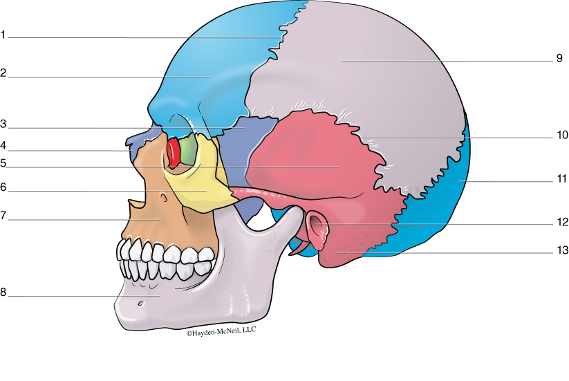
In mammals, the outermost layer is the epidermis (Fig. 2.1). The basal layer of the epidermis is a single layer of epithelium comprised of mitotically active cells that are continually dividing. In terrestrial animals, the layers closer to the surface have keratin, a proteinaceous material that makes the layer very tough and helps to keep inner layers from desiccating or drying out. The outermost layer, called the stratum corneum, is made up of dead, cornified cells. Mammals continually shed this layer. Much of the dust in our houses is made up of these dead cells.
Below the epidermis is the dermis, a relatively thick and dense layer made up of fibrous connective tissue and collagen. Collagen is tough and flexible, both important properties for the outer layer of an organism. Within the dermis are blood vessels (made of epithelial tissue), sensory organs (also epithelial tissue), glands such as the sebaceous and sweat glands (epithelial tissue), hair follicles, muscles associated with hair follicles (muscle tissue), and nerves.
PROCEDURE
From the side table, get a microscope slide showing a cross section of human skin. Again, work with a partner, but every person should have a microscope. It is important that everyone see at least two slides. Find as many structures as you can and make a labeled sketch of the cross section. Identify what kind of tissue makes up the epidermis, dermis, and subcutaneous layer. Use the figure as a guide; your drawing must be based on what you see and not be copied from the figure. Be able to identify examples of each of the four categories of tissue (i.e., epithelial, connective, muscle, and nervous).
Next, examine and sketch the slides of skin from an amphibian. How is this similar to mammalian skin? How is it different?
III. MUSCLES
Last week we examined skeletal muscle and talked briefly about the different kinds of muscle tissue. This week, we will learn to differentiate between skeletal, smooth, and cardiac muscle tissue when viewed microscopically. For each, you should know where that kind of muscle tissue is found and what makes it distinctive. Of course, you should also be able to distinguish muscle tissue from the other categories of tissue. In lecture, you will discuss the mechanisms of muscle contraction.
Muscle tissue is comprised of elongate, spindle-shaped cells that are contractile. Each muscle cell is called a muscle fiber, and a muscle is made of many cells or fibers. Each fiber or cell is made of many contractile units called myofibrils.
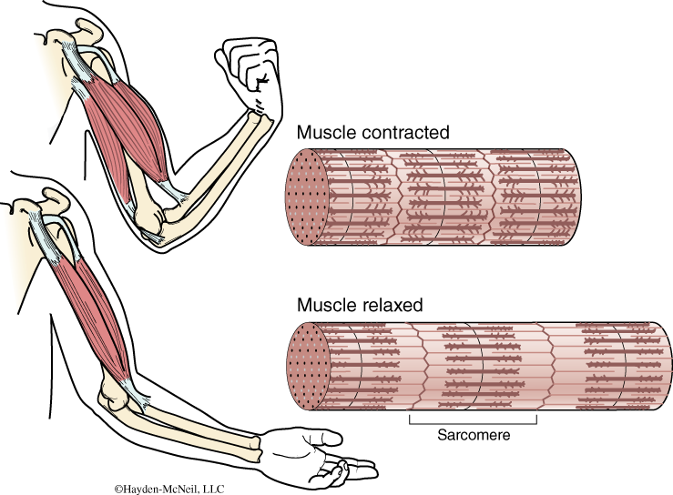
Skeletal muscle is muscle that is attached to the skeleton and is under conscious control. It is sometimes called striated muscle. It is distinguished by its striations or banding and by the multinucleated muscle fibers. Fig. 2.2 shows a bicep, an example of skeletal muscle. It also shows how the muscle looks when it is contracted and when it is relaxed.
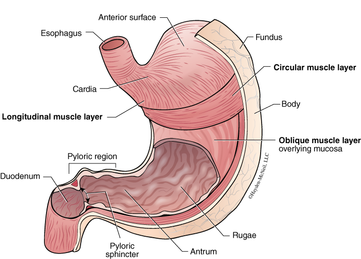
Smooth muscle is found in the walls of internal organs and is not under conscious control. It also lacks the distinctive striations that are seen in skeletal muscle, and it is single nucleated. Many organs, such as the stomach shown in Fig. 2.3, have several layers of muscles that are oriented in different planes. Since any one muscle can only contract or lengthen, it takes multiple muscles to make something like a stomach contract and churn.
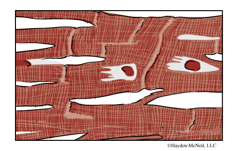
The heart is primarily made of cardiac muscle, and this muscle has some special properties. The fibers of cardiac muscle are branched, and there are connections between the muscle fibers called intercalated disks (light bands seen in Fig. 2.4). Both the branching and the disks help the muscle to contract as a single unit. It is also single nucleated and, like smooth muscle, it is not under conscious control.
PROCEDURE
Get examples of skeletal, smooth, and cardiac muscle slides and make sketches of each. In your labeled sketches, make notes about what makes the muscle type distinct. List examples of where each of the types of muscle are found. Ask yourself: “How can I recognize this as skeletal muscle or cardiac muscle or smooth muscle?”
IV. BONES AND THE SKELETAL SYSTEM
There are three kinds of skeletal systems found in the animal kingdom: hydrostatic skeletons, exoskeletons, and endoskeletons. In general, the skeletal system, no matter what kind, provides both protection and also structure to which muscles can attach or gives rigidity against which muscles can pull. A hydrostatic skeleton is seen in many worms, where a fluid-filled cavity provides a somewhat rigid structure. Arthropods have an exoskeleton; their outermost covering clearly provides protection and a place for attachment of muscles. Vertebrates and echinoderms have an endoskeleton. With endoskeletons, the skeleton is internal, but is still very important for protection as key areas of the body (e.g., thoracic cavity and brain) are enclosed in a hard, bony case. Animals with exoskeletons have to molt in order to grow. They shed their exoskeleton just before a growth spurt. Endoskeletons can grow continuously with the animal.
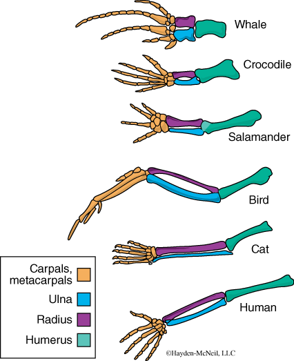
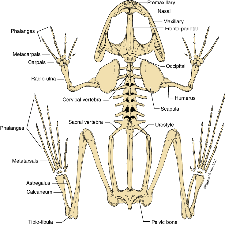
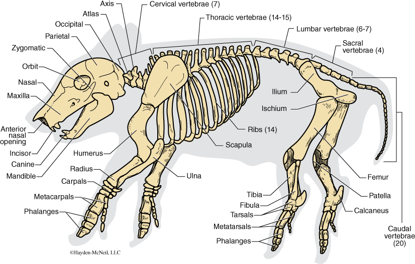
As bones are hard and decompose more slowly than other tissues, they have provided a very rich and interesting fossil record. In addition, vertebrate bones provide a very good example of homologies in comparative studies. Homologies are similarities due to common ancestry. When you look at a series of front limbs from different vertebrates, you can see a basic similarity in the bones (Fig. 2.5; compare also Figs. 2.9, 2.10, and 2.11). In this lab we will focus first on the cross section of bone. We will then learn the elements of the human appendicular and axial skeleton. Finally, we will compare parts of the human skeleton with parts from the frog skeleton and the pig skeleton.
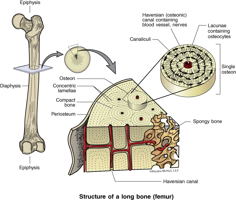
Bone is a type of connective tissue and much of it is a matrix that is acellular. However, there are cells within bones, which can be seen, for example, if you take a cross section of a long bone such as a femur (Fig. 2.6). The outermost layer of such a bone is the periosteum, a layer of connective tissue that is continuous with ligaments and tendons. The periosteum is a growth layer and can increase the diameter of a bone. The main shaft of a bone is the diaphysis, while each end is called an epiphysis. In children, there is a growth center or cartilage between the epiphysis and diaphysis called the metaphysis. The outer shell of bone is called compact bone, which is very dense. Individual bone cells (osteocytes) are held within an opening called a lacuna. There are tiny channels connecting the osteocytes called canaliculi. A Haversian canal in the middle of each osteon provides routes for blood vessels and nerves. To the inside of the dense bone is the less dense (and less strong) spongy bone. In addition, most bones have a central marrow cavity where red blood cells are produced.
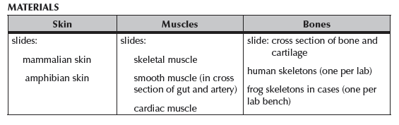
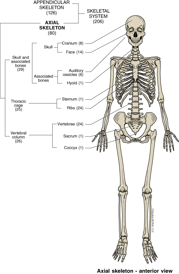
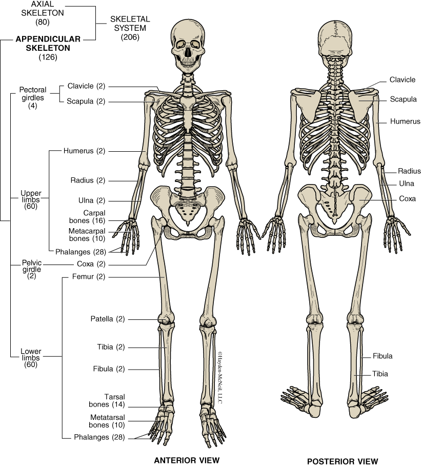
In vertebrates, the skeletal system is divided into two parts: the axial skeleton and the appendicular skeleton. The axial skeleton is the skeleton along the central axis of the body which includes the skull, vertebral column, and the rib cage. The appendicular skeleton is comprised of the bones that form the limbs: the leg bones including the pelvis, and the arm bones including the shoulder girdle. We will learn the elements of both the axial skeleton and the appendicular skeleton.
PROCEDURE
Each team of students will spend approximately 8 minutes at each of 7 stations. Each station has directions and specific bones to look at.
Bone Station 1: Axial Skeleton. Learn all bones labeled in Fig. 2.7 and be able to find each on the skeleton.
Bone Station 1: Appendicular Skeleton. Learn all bones labeled in Fig. 2.8 and be able to find each on the skeleton.
Bone Station 2: Femur
This is a detailed study of one bone. Note that each ridge and valley is named and has a function. Be able to distinguish right femur and left femur, anterior and posterior aspects.
Be able to find and identify:
Diaphysis
Head
Neck
Linea aspera
Greater trochanter
Lesser trochanter
Medial condyle
Lateral condyle
Adductor tubercle
Bone Station 3: Forelimbs
Be able to identify:
Radius
Ulna
Carpals
Metacarpals
Phalanges
Compare homologies in Fig. 2.5 with the frog skeleton (Fig. 2.13).
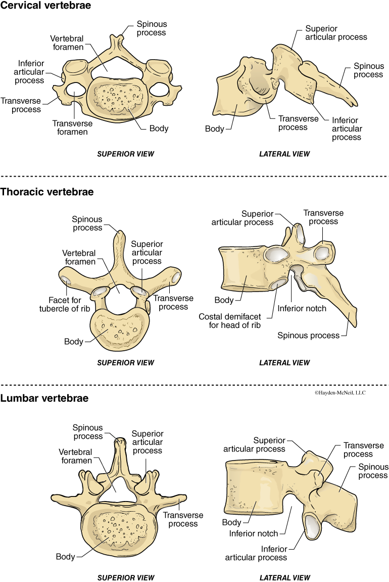
Bone Station 4: Vertebrae
On a vertebral column be able to identify numbers and kinds of each vertebrae.
12 thoracic vertebrae
5 lumbar vertebrae
5 sacral vertebrae (fused)
3–5 coccyxal vertebrae (fused)
Be able to identify each type when it is disarticulated.
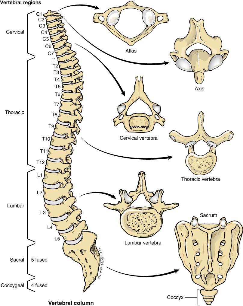
For thoracic vertebrae, identify:
Superior articular process
Transverse process
Vertebral foramen
Spinous process
Inferior articular process
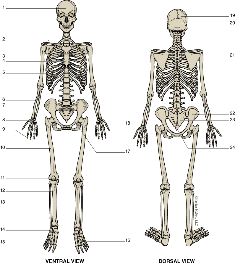
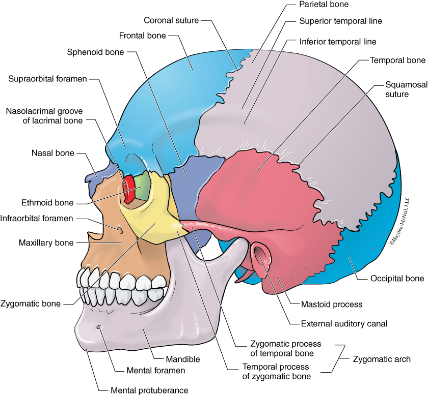
Bone Station 5: Cranium and Face. Be able to find and identify on a skull all of the terms listed in Fig. 2.12.
Bone Station 6: Frog and Pig Skeleton. On the frog skeleton, find and identify:
Radio-ulna
Femur
Tibio-fibula
Where is the calcaneum in the pig skeleton?
What is the shape of the humerus in the frog skeleton?
Find 9 differences in the frog skeleton compared with the human skeleton (other than size!).
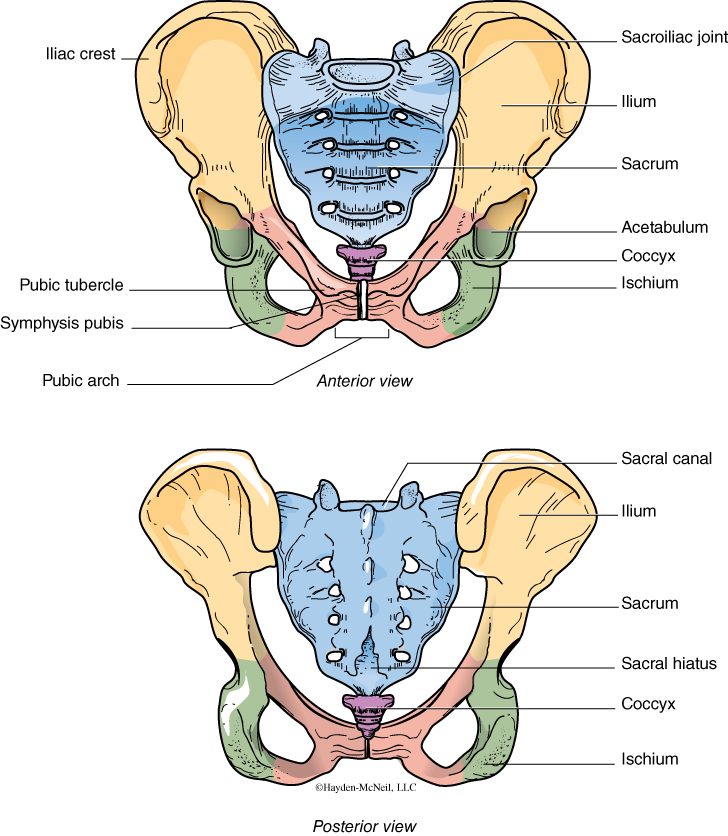
Bone Station 7: Pelvis. Be able to identify:
Iliac crest
Sacrum
Sacroiliac joint
Ischium
Pubic bone
Pubic arch
Pubic symphysis
Coccyx
V. NOTES TO INSTRUCTORS AND MATERIALS FOR LAB
This lab can be fairly equally divided into two parts. You can start with introducing the integumentary system and muscles, and have students look at and sketch examples of each in the first half of the lab. In the second half of the lab, you can introduce the skeletal system and involve students in learning both the appendicular and axial skeleton. The more students can use lab time to actively learn the material, the better they will know it. You must insist that lab time be used to learn!
At the end of class, it is the TA’s responsibility to be sure that students return slides to the correct tray and that the lab is clean and ready for the next group. Nobody should leave until the lab is clean, organized, and ready for the next group.