Chapter 6. NERVOUS AND ENDOCRINE SYSTEMS
I. OVERVIEW
All living organisms can respond to environmental stimuli, but animals have taken this response to a very high level indeed. There are two systems that are part of this response: the nervous system and the endocrine system. Although all animals except sponges have a nervous tissue, we will focus primarily on the vertebrate system by examining nerve cells, the brain (using sheep as our model), and the spinal cord. Your text makes the point that the nervous system is the principle regulatory system and that it regulates both behavior and physiological processes. The endocrine system is also regulatory. While the nervous system acts quickly (and briefly) in regulation, the endocrine system acts more slowly and has longer lasting regulation. The nervous system is involved with reception of stimuli (both external and internal), integration of the information from those stimuli and then transmission to effectors that respond to the information. In the Biology Department at the University of Mississippi, Dr. Lainy Day is a neuroethologist who works on neural mechanisms underlying natural behaviors. She offers Introductory Neuroscience and Functional Neuroanatomy. Another faculty member, Dr. Brad Jones, studies the development and differentiation of glial cells that surround nerves. Dr. Jones teaches Developmental Biology, as well as Cell and Molecular Biology. You can see some of his work on the posters on the first floor of Shoemaker Hall.
In vertebrates, the nervous system is broadly divided into the Central Nervous System (CNS) consisting of the brain and spinal cord, and the Peripheral Nervous System (PNS), consisting of motor neurons, sensory neurons, cranial nerves, and all nerves outside of the CNS. In this lab we will first review nerve cells. Next, we will examine the protective coverings of the fetal pig brain, a sheep brain, a model of a human brain, and the spinal cord. We will then move on to examine the endocrine system, both from a gross anatomy perspective and a functional perspective.
II. NERVE CELLS AND GLIAL CELLS
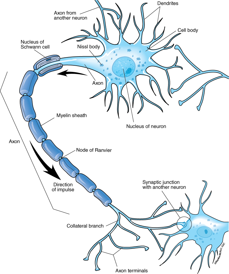
Nerve cells comprise their own tissue type and are among the most unusual cells in the body. A typical nerve cell or neuron has many processes (axons and dendrites), which may be connected to thousands of other cells (Fig. 6.1). Although very small in diameter, the projections of the nerve cells can be many feet in length. For example, in a giraffe, the axon may be 6 feet long, traveling the entire length of the neck. The length of some of these processes is part of what makes recovery from a severed spinal cord so difficult. Typically there is a single long axon and many short dendrites extending out from the cell body (Fig. 6.1). The axon conducts nerve impulses (or action potentials) away from the cell body, while dendrites conduct nerve impulses toward the cell body (note the attachment of axons from other cells to the dendrites of the cell indicated below). The axons of some neurons are surrounded by a specialized cell called Schwann cells (Fig. 6.1). These cells provide a myelin sheath, which protects the axon. There are regularly spaced gaps in the sheath called nodes of Ranvier. The Schwann cells (and the nodes) allow for much faster conduction of the nerve impulse along the axon. The action potential, or nervous impulse, jumps from node to node by what is called saltatory conduction. In lecture, you will study the details of an action potential.
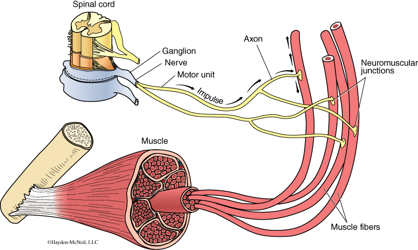
Neurons are sometimes classified by their function. Sensory neurons (or afferent neurons) conduct signals from sense organs to the central nervous system. Motor neurons (efferent neurons) conduct signals from the CNS to muscles or glands, as is shown in Fig. 6.2. Interneurons connect neurons to other neurons within the CNS.
Closely associated with nerve cells are glial cells, or cells that support and nourish the neurons. It is now known that there are many kinds of glial cells, and their functions are quite varied, including the following: provide physical support for neurons, maintain extracellular fluids, function in signaling, serve as stem cells for the development of new neurons, secrete growth factors, and produce the cerebrospinal fluid. The Schwann cells are a specific kind of glial cell.
A nerve is a bundle of axons, sometimes thousands, that is surrounded by connective tissue (Fig. 6.2 shows a nerve exiting the spinal cord and connecting with a muscle cell via neuromuscular junctions). A ganglion in a nerve is where the cell bodies of many neurons are concentrated. Nerves also connect to organs and glands as well as muscles.
PROCEDURE
Examine a prepared microscope slide showing nerve cells. Find and sketch a cell body, as well as axons and dendrites. Be able to differentiate nervous tissue from muscle, epithelial, and connective tissue.
III. THE BRAIN
As the complete dissection of the brain of a fetal pig is difficult (the brain tissue is very soft and fragile, partly because of the developmental stage of the brain in the fetal pig), we will use the fetal pig to examine the protective layers of the brain, and then examine brains from adult sheep and models of the human brain.
The Central Nervous System (CNS) in vertebrates consists of the brain and the spinal cord. The Peripheral Nervous System (PNS) is comprised of all the nerves and receptors outside of the brain and spinal cord, including the sensory organs, cranial nerves, and spinal nerves.The CNS is the integrating center for the vertebrate nervous system; it receives and interprets sensory information and initiates reponses to the stimuli. The PNS can be functionally subdivided into the sensory or afferent division, which carries impulses toward the CNS, and the motor or efferent division that carries signals away from the CNS to effectors (muscles or glands). The motor division can be subdivided into two parts: the somatic nervous system (or voluntary nervous system, which controls the skeletal muscles) and the autonomic nervous system, also called the involuntary system. The autonomic system regulates the smooth muscle (for example, in the digestive system) and glands.
Recall that in our very first lab we looked at the early development of animals. The ectoderm is the outer layer of the zygote, and it becomes both the skin and the nervous system. In vertebrates, the ectoderm develops into the neural tube that runs along the dorsal side of the developing embryo (see Lab 1). During development, the neural tube differentiates into the brain and spinal cord. The anterior end becomes the brain while the posterior portion becomes the spinal cord. The brain and the spinal cord are continuous. As the brain develops, three bulges appear in all groups of vertebrates. The bulges are called the forebrain, midbrain, and hindbrain. While all classes of vertebrates have these three areas, in each vertebrate class, a different area becomes the largest or predominant part in the adult animal. It is thus possible to see that these areas of the brain are homologous in the different classes of vertebrates (Figs. 6.3 and 6.4). That is, each section of the brain has a common developmental origin even though in the adult animal, the brains end up looking quite different. In the Comparative Anatomy class, you will do an extensive comparison of the different brains of vertebrates.
We will trace what happens to the forebrain, midbrain, and hindbrain in the development of the animal (summarized in Fig 6.4). The hindbrain differentiates into the metencephalon and the myelencephalon. The metencephalon becomes the cerebellum, a portion of the brain that looks like a cauliflower, and the pons. The cerebellum coordinates muscle activity, as well as muscle tone, posture, and equilibrium. The pons is a mass of fibers that connects the spinal cord to the brain and helps regulate respiration. The most posterior part of the brain, the myelencephalon, gives rise to the medulla (or medulla oblongata), the part of the brain that is continuous with the spinal cord. The medulla is the center of regulation for respiration, heartbeat, and blood pressure. The brain stem, the portion that looks like a stem, is made of medulla, pons, and midbrain.
The midbrain, the middle bulge that is seen early in development, differentiates into the mesencephalon, which is the main association area. In fishes and amphibians, the midbrain is prominent. In mammals, the midbrain is relatively small and consists of the superior and inferior colliculi, centers for visual reflexes (such as pupil dilation) and auditory reflexes. In diagrams, this is often just labeled as “midbrain.”
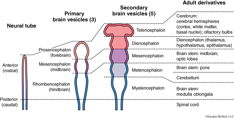
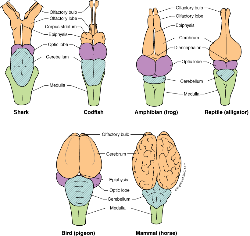
The forebrain becomes the telencephalon and diencephalon, the anterior-most portion of the brain, and it in turn develops into the cerebrum and olfactory bulbs. In humans, the cerebrum is very large and prominent. In several vertebrate groups such as many of the fishes, the olfactory bulbs are large and well developed, which clearly shows that olfaction (the sense of smell) is very important to fishes. In humans and other mammals, the olfactory bulbs are much smaller than in fishes. The forebrain also differentiates into the diencephalon, a part that becomes the thalamus and hypothalamus. The thalamus is a relay center for motor and sensory messages. The hypothalamus is a regulating center for the peripheral nervous system, and it links the nervous and endocrine systems.
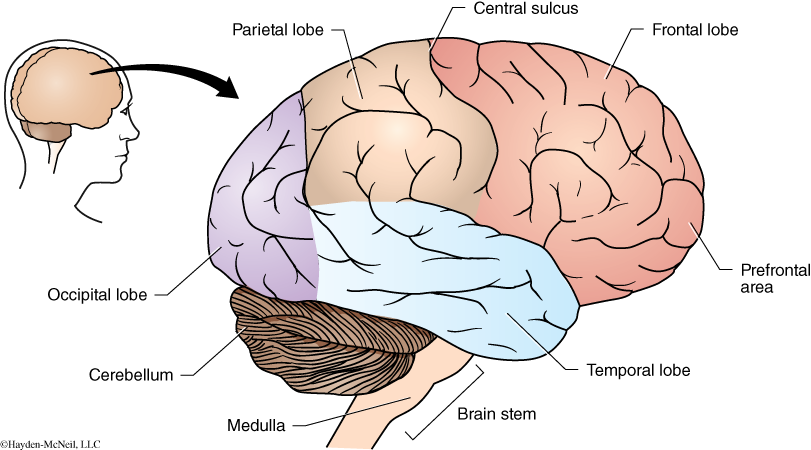
Much of the cerebrum is white matter, or is comprised of myelinated axons. The outer portion of the cerebrum is the cerebral cortex and, at least in mammals, is made of gray matter, or nonmyelinated axons. Externally, the most visible aspect of the brain is the many wrinkles or folds. These wrinkles and folds, called sulci (sing. sulcus), greatly increase the surface area of the brain. The human brain is comprised of as many as 30–50 billion neurons and glial cells (with 20–40% variation between individuals). According to one source, a single cubic millimeter of human brain tissue has more than 1 million neurons. If each neuron is connected to 10 others (a conservative estimate), the neurons in the cubic millimeter would have 10 million connections.
In humans, the cerebrum is divided into regions or lobes (Fig. 6.5): The frontal lobe is the center for judgement, creativity, problem solving, and planning. It is a region you should be using extensively in preparation for this class. The parietal lobes process (and integrate) sensory information and have language functions. The temporal lobes are important in hearing, memory, and language. You engage both of these lobes when you listen to lectures about the brain. The occipital lobe is the visual processing center. Examining pictures of the brain or looking at a brain would engage the occipital lobe.
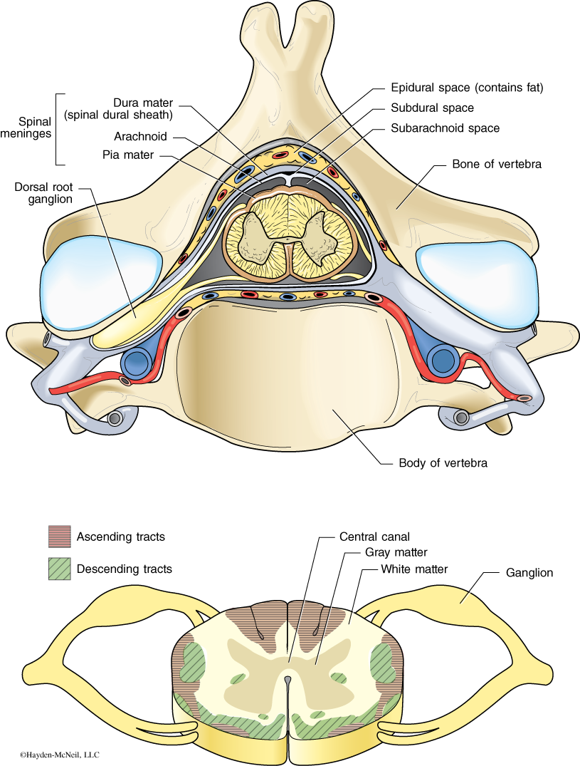
The importance of the brain is reflected in the many layers of protection that it has. It is entirely enclosed in the bony case called the cranium. In addition, there are several layers of connective tissue that provide protection. The outermost layer of connective tissue is the dura mater, which is tough and leathery. The middle layer is called the arachnoid mater, and beneath this is the pia mater. In between the arachnoid and pia mater is the subarachnoid space, which is filled with cerebrospinal fluid. These three layers, as well as the cerebrospinal fluid, also protect the brain as well as the spinal cord.
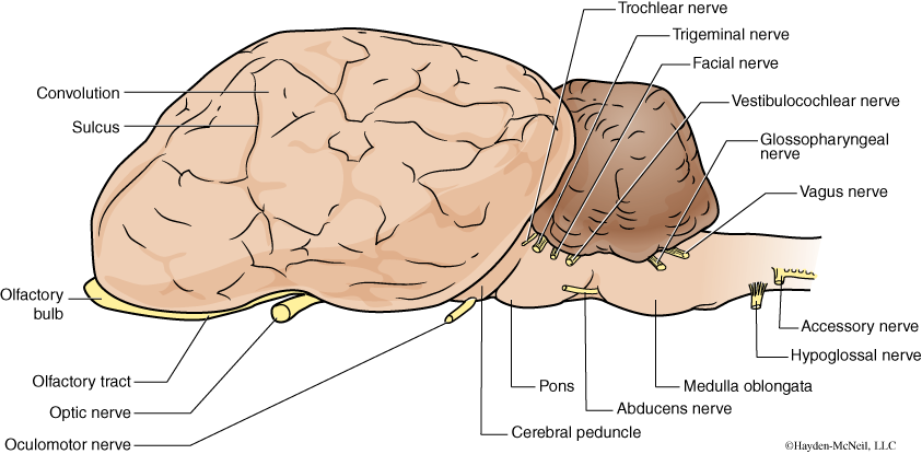
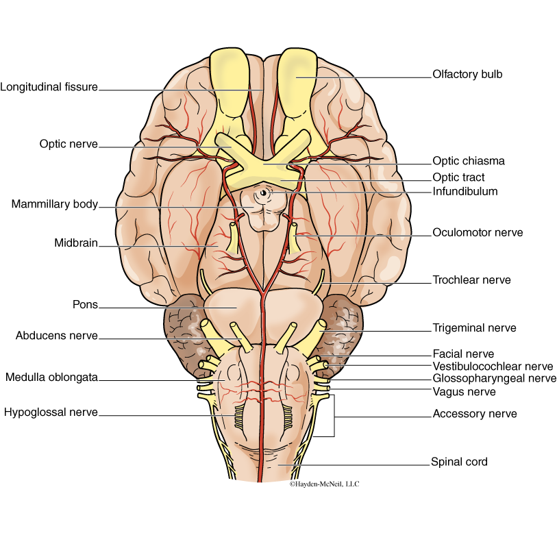
The brain has 12 pairs of cranial nerves that exit the brain. One example of a cranial nerve that we should be able to see is the optic nerve (see Figs. 6.6, 6.7, and 6.8). In the Comparative Anatomy class, you will learn the cranial nerves.
PROCEDURE
Examine the skull of the fetal pig. Carefully open the top of the brain by shaving off layers of the skull with your scalpel to find the three layers of connective tissue beneath the skull. Find the dura mater, the arachnoid, and the pia mater. To look at the brain in detail, we will use sheep brains and models of the human brain.
Next, get one of the sheep brains. There should be enough brains for there to be one per 2 or 3 students. As with all materials, please handle these gently. It is important to look at more than one brain. Using this diagram and additional diagrams provided in class, find and identify the structures listed below. Start with a lateral view and move to a ventral view, and finally, move to a midsagittal view. Label your sketch from each view.
For each structure you label you should be able to say if it developed from the hindbrain, the midbrain, or the forebrain. Look for the cranial nerves. However, they are often difficult to find in brains that have been removed from the skull.
Know All Regions, Their Developmental Origins and Their Functions
Lateral view of sheep brain. Find, sketch, and label these structures:
Medulla oblongata
Cerebellum
Cerebrum
Midbrain
Olfactory bulb
Brain and spinal cord
Ventral view of sheep brain. Find, sketch, and label these structures:
Medulla oblongata
Midbrain
Cerebellum
Cerebrum
Optic tract
Optic chiasma
Optic nerve
Pons
Olfactory bulb
Anterior and posterior ends of the brain
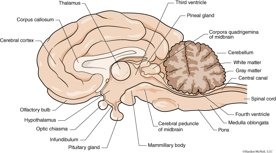
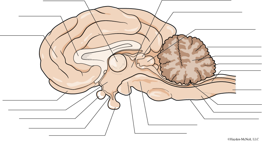
Midsagittal view. Find, sketch, and label these structures:
Medulla oblongata
Cerebellum
Spinal cord
Thalamus
Corpus callosum
Pons
Hypothalamus
On the human brain model, identify these lobes and know their functions:
Parietal
Temporal
Occipital
IV. THE SPINAL CORD
The second part of the CNS, clearly continuous with the brain, is the spinal cord. The spinal cord is enclosed within the vertebral column and extends from the base of the skull to the first lumbar vertebrae, where it branches into a bundle of nerve roots called the cauda equina. The spinal cord is a two-way conduction pathway from the brain to the rest of the body and is important in spinal reflexes. In humans, there are 31 pairs of spinal nerves that arise from the spinal cord and exit from the vertebrae. In a cross section of a spinal cord (Fig. 6.10), it is possible to identify white matter and gray matter. Although they are not visibly distinct, different tracts within the spinal cord have been identified. A tract is a group of axons from a similar source. Ascending tracts as well as descending tracts have been mapped. These are shown in Fig. 6.10 by the different shading.
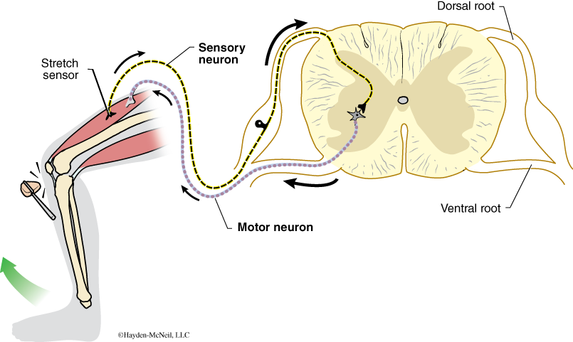
The spinal cord is also important in reflexes, and some reflexes happen entirely within the spinal cord and do not require the brain to moderate the response. For example, if you tap the tendons on the front of the knee, there is a knee-jerk response that is moderated only by the spinal cord (Fig. 6.11). This is an example of a reflex arc.
In humans, good posture is important for the spinal cord. Poor posture can put pressure on spinal nerves, resulting in pain and nerve damage.
PROCEDURE
Examine the prepared slide that shows the cross section of the spinal cord. Be able to recognize white matter and gray matter. Using the diagram provided, identify regions of ascending tracts and regions of descending tracts.
White matter (ascending and descending axons)
Gray matter (motor and association neuron cell bodies)
Pia mater
Central canal
Examine a reflex arc with your lab partner. Have your partner sit on the table with his/her leg resting comfortably. Gently tap the tendons on the front of the knee. Be able to describe the pathway of the response, from sensory detection to movement.
Finally, dissect one or more vertebrae from your fetal pig to find one of the spinal nerves exiting the spinal cord. This can be done by slicing thin layers off of the dorsum of the pig.
V. ENDOCRINE ORGANS
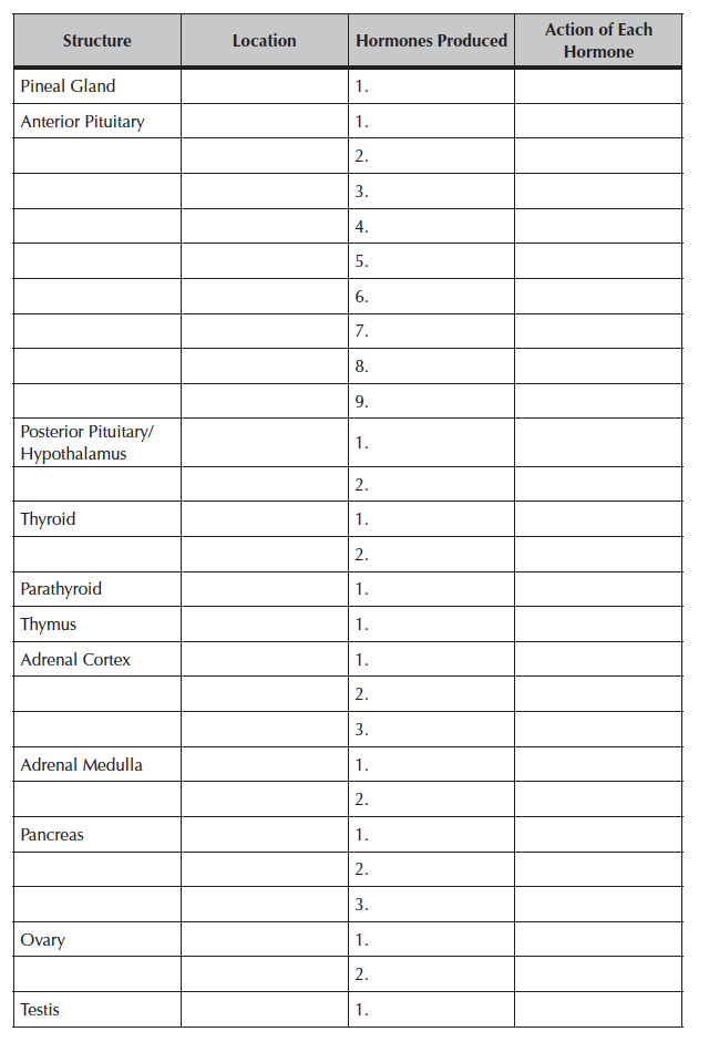
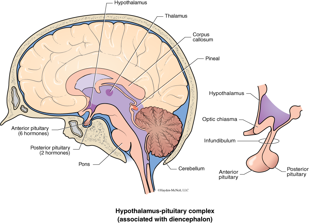
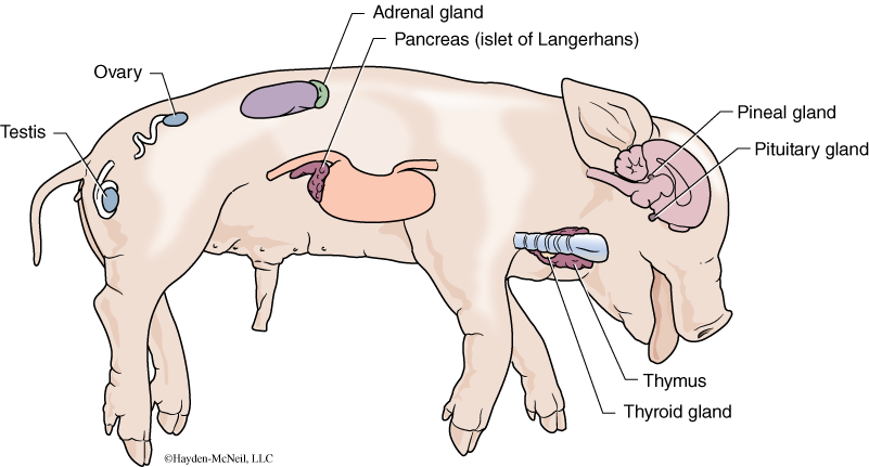
The endocrine system is a diverse collection of cells, tissues, and organs that together produce and secrete hormones or chemical messages. Generally, a hormone is produced by one gland, carried by the circulatory system and, once it encounters its target organ, has a very specific effect on that organ. In addition to specific endocrine glands, like the thyroid, there are many different tissues that produce chemical messages. The list of glands or organs known to have an endocrine function is growing. In addition to the usual endocrine glands (listed below), the heart, kidney, fat cells, and even the skeletal system have an endocrine function. Many hormones have complex feedback loops to control their relative volumes and effects. Endocrine glands have no ducts; they secrete their products directly into capillaries. In lab today, we will focus on the glands listed below. You need to know where these glands are in the fetal pig and their functions. Fig. 6.12 shows the location of the pineal gland, hypothalamus, and pituitary gland in the human brain. Fig. 6.13 shows the location of the endocrine organs in the fetal pig.
The pineal gland is found within the brain and produces melatonin.
The pituitary gland, located at the base of the brain, has two distinct regions: an anterior and posterior region. Each region secretes several hormones. The anterior pituitary secretes growth hormone (GH), prolactin, follicle-stimulating hormone (FSH), luteinizing hormone (LH), thyroid-stimulating hormone (TSH), and adrenocorticotropic hormone (ACTH). The posterior pituitary secretes oxytocin and antidiuretic hormone (ADH).
The thyroid gland is very important in regulating metabolism. It produces thyroxine and calcitonin. Thyroxine influences the metabolic rate of cells and affects growth and development. Calcitonin stimulates osteoblasts to lay down bone tissue which, as a result, lowers the level of calcium in the blood. The thyroid is located in the neck under the thymus.
The thymus is much larger in a fetus than in the adult animal. It probably was very evident in our first day of dissection, but may have been removed for us to see the trachea. It produces thymosin, a hormone that stimulates the development of the immune system. Also, it is the location for maturation of T-cells or T-lymphocytes of the immune system.
Adrenal glands are located near the anterior medial edge of the kidney. Like the kidneys, they have an outer layer called the cortex and an inner layer called the medulla. The outer layer of the adrenals secretes aldosterone, a hormone important in water balance. Stress hormones such as cortisol are produced in the middle layer. The stress hormones have a variety of effects, including changing blood pressure and blood sugars, as well as causing immunosuppression. The inner layer of the cortex produces androgens (e.g., testosterone). The medulla of the adrenal gland produces the neurotransmitters epinephrine and norepinephrine. These hormones produce the “fight or flight response.” Curiously, the portion of the adrenal gland that produces neurotransmitters has a neural origin.
The pancreas, previously discussed in the context of the digestive system, also has a role in the endocrine system. Throughout the pancreas are small bits of endocrine tissue called islets of Langerhans. These cells secrete insulin, glucagon, and somatostatin into the capillaries of the pancreas. Insulin causes liver and muscle cells to take up glucose from the blood and convert it to glycogen for temporary storage. Glucagon has the opposite effect; it stimulates the conversion of glycogen to glucose when blood glucose levels are low. Somatostatin is important in regulating both insulin and glucagon.
The ovary produces both estrogen and progesterone. Estrogens are present in both males and females, but have a higher level in females than males. They are involved in the development of secondary sexual characteristics and are important in the thickening of the endometrium of the uterus in the menstrual cycle. There is a much higher level of progesterone after ovulation.
The testis functions in the endocrine system by producing the hormone testosterone. Specialized cells called interstitial cells produce and secrete the testosterone. Curiously one of the functions of testosterone may be to protect the developing sperm from the immune response of the individual making the sperm. That is, sperm may not be recognized as “self” and need some protection! Testosterone is also important in energy level, immune function, and libido. It is also produced (in small amounts) by the ovaries and adrenal glands in females.
PROCEDURE
Get out microscopes and look at slides to review microanatomy of an ovary and a testis. We will also look at a cross section of the pancreas. Make labeled sketches of each of these.
Locate each of these endocrine glands on your fetal pig. Be able to describe what hormones are produced and their effects. Using your text and materials available in lab, make a table that summarizes each gland, the hormones produced, and the action of the hormones.
Adrenal gland
Pituitary gland
Pancreas
Thyroid gland
Ovary
Thymus
Testis
VI. NOTES TO INSTRUCTORS AND MATERIALS FOR LAB
To TAs: This tends to be a fun and interesting lab. It works well to divide into sections and to give the introduction and information for each section before students start work.
Nerve cells. Be sure students understand the differences between nerve cells and glial cells. They should know the parts of a nerve cell and be able to distinguish nervous tissue from the other types of tissue.
Brain. Students should know the regions, developmental origin, and functions of the different parts of the brain. They should examine the protective layers of the brain in the fetal pig. The sheep brains and model brains must be handled gently. It is important to be clear that students are responsible for everything in the lab manual. Help students use the lab time efficiently.
Spinal cord.
Endocrine organs.
MATERIALS
Fetal pig (students use their own dissecting tools)
Sheep brains (1 per 2 students)
Human brain model
Prepared slide of spinal cord
Medical reflex hammer
SUMMARY OF ENDOCRINE GLANDS, HORMONES, AND ACTIONS
Use your lab manual (pages 90–93) and Chapter 41 in your textbook to complete this table.