Chapter 7. BACTERIA AND PROTISTA
NOTE TO STUDENTS
In some semesters, we will start the laboratory sequence with the chapters and lab exercises on diversity. This makes for a logical sequence from what we cover in the first semester class to where we finish the semester examining evolution. Diversity is the product of evolution. That is, the processes that we talked about last semester has given rise to the amazing variety of organisms with whom we share the earth. In this first lab we will start with the prokaryotes and some of the eukaryotes.
I. OVERVIEW: DIVERSITY AND COMPARISON OF PROKARYOTES AND EUKARYOTES
One of the most exciting areas of biology right now is the study of phylogenetics or the study of relationships of different groups. In the last 20 years, there has been a revolution in our understanding of relationships, due partly to the ability to compare sequences of RNA and DNA from a variety of groups or organisms. The unity of life is clear. Closely related organisms share a wealth of characteristics in addition to their RNA and DNA sequences. In this lab, we will start a four-lab sequence in which we will examine some of the diversity of life on earth. As there are millions of species of organisms, we will be looking at some of the larger groupings in order to make sense of this diversity.
The largest grouping of organisms (the most inclusive group) is called a domain. There are three domains and each domain is comprised of many kingdoms. Since the 1930s, it has been recognized that there are very large differences between the cells of bacteria and the cells of animals, plants, and fungi. Bacteria have been called prokaryotes as they lack a nucleus and other membrane-bound organelles (the term prokaryote means “before (pro) the kernel (karyote),” or before the nucleus). Cells from Protista, fungi, plants, and animals are more complex, have one or more nuclei, and have other membrane bound organelles. These organisms have been called eukaryotes as they are made from eukaryotic cells. The term eukaryote means “true kernel” and that refers to the nucleus.
In 1977, Carl Woese, by looking at the sequences of the small subunit of 16S ribosomal RNA, discovered there were two large groupings of prokaryotes and one group of eukaryotes. He subsequently suggested the division of all living organisms into three domains based on this. The two large groups of prokaryotes differ not only in the RNA sequences but also in their mechanism of transcription and translation. The two groups of prokaryotes now recognized are the Bacteria and the Archaea. The Archaea are more similar to eukaryotes than they are to the Bacteria (see Fig. 7.1).
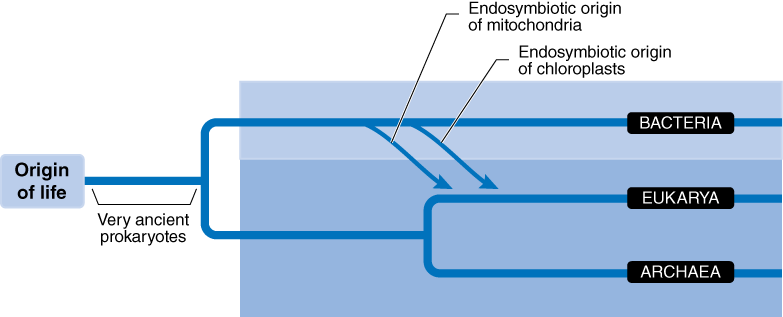
In this class, we will be looking at the Domain Eukarya in the greatest detail. The Domain Eukarya is subdivided into clades, or groups.
II. DIVERSITY: DOMAIN BACTERIA AND DOMAIN ARCHAEA
Although both Bacteria and Archaea are prokaryotes, they are very different from each other. Members of the Domain Archaea are sometimes called the “extremeophiles.” They are often found in areas that are extremely hot (as in the hot sulfur springs of Yellow Stone National Park), extremely salty (as in the halophiles found in the Great Salt Lake or the Dead Sea) or even in extremely cold areas such as the oceans near Antarctica. Archaea are the source of a very important compound used by molecular biologists called Taq polymerase, a polymerase that was isolated from Thermus aquaticus (thus the abbreviation “Taq”), an Archaean bacteria from hot springs. Because it is adapted for high temperatures, it can be used for DNA work such as PCR or amplification of DNA and it can function to repair DNA at temperatures that inactivate other enzymes.
The Domain Bacteria includes most of the bacteria that we tend to be more familiar with. Indeed, we are very familiar with this domain. An individual human may be made up of 100 trillion human cells. Curiously, we may have as many as 1000 trillion bacterial cells living associated with us! That is, there may be more bacterial cells than human cells in each one of us. We tend to think of bacteria as mostly being harmful because they can be deadly; however, the vast majority are more helpful than harmful. Bacteria are important as decomposers, they recycle nitrogen, phosphorus, and sulfur, they are very important in nutrient cycling and energy flow in most environments, and yes, some are pathogenic.
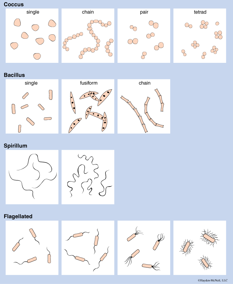
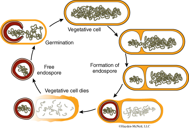
Bacterial cells are small, typically 1–5 microns long (which is why there can be so many bacterial cells than human cells in a human body). One of the ways to group bacteria is by their shape as is indicated in Fig. 7.2. Most bacteria reproduce by binary fission and many can form endospores, which are cells that can be dormant during very harsh conditions (Fig. 7.3).
Although they lack nuclei and other membrane-bound organelles, they do have ribosomes, a cell wall, and a nuclear area where there is a circular ring of DNA. A second way to categorize and identify bacteria is by stain. Important differences in the cell wall allow different groups to stain very differently. For example, a commonly used staining procedure is called the Gram stain. Species that are Gram + (Gram positive) typically stain a violet color when stained the crystal violet. Gram – (Gram negative) bacteria do not retain the stain when they are rinsed with ethanol. Gram + bacteria have a peptidoglycan in their cell walls, and it is this layer that retains the stain. We will have a Gram + and Gram – demo in lab this week.
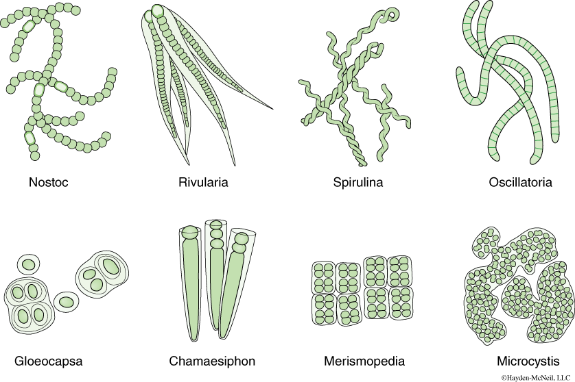
An important group of bacteria that are are photosynthetic that most people have seen (but not necessarily recognized) are the cyanobacteria (Fig. 7.4). These are Gram-negative, photosynthetic bacteria. Although they are unicellular, in several species the cells will be loosely attached to each other via a gelatinous covering or will grow in filaments. In either case, although individual cells are micoscopic, they are often visible macroscopcally (as green algae or scum) and when conditions are suitable, they are often the culprits in algal blooms. Many cyanobacteria can withstand drying out, sometimes for long periods. In many cases, the cells may not be remarkable, but there are specialized cells that allow the cyanobacteria to fix atmospheric nitrogen. A cyanobacterium may have been the ancestor of the chloroplasts, photosynthetic organelles found in plants. Use your text to explore the evidence for this.
PROCEDURE
Know the basis of the three domains and some of the organisms in each domain. Make a chart in your notebook summarizing this information.
Examine the prepared slide “Bacterial types.” If you use the oil immersion lens, it must be used correctly. In your labeled sketch, be sure to include the magnification. Examine the demonstration of Gram-negative and Gram-positive bacteria. You should understand the basis of the Gram stain.
Examine the examples of cyanobacteria available in the lab and look for the specialized cells. Oscillatoria are able to move by oscillation, reproduce by fragmentation. Anabaena often have heterocysts (important in fixing nitrogen). Gleocapsa occur singly or in groups of up to eight individuals. Nostoc are made of filaments of round cells and can form gelatinous balls with hundreds of filaments.
Note that for the exam, you will need to recognize each of the organisms we will be looking at in lab. You should look at them with the questions: How will I recognize this? What makes it distinctive?
III. DIVERSITY: PROTISTA
DOMAIN EUKARYA, PROTISTA
We start our study of the eukaryotes with the Protista. Unlike the other clades or groups that are clearly monophyletic, the Protista is more a grouping of convenience and it is not monophyletic. That is, its members may not be each other’s closest relatives. Anything that is not animal plant or fungus has historically been put in with the protists. It is large—there may be as many as 60 major taxa covered by the term Protista. (You may be happy that we will not try to cover 60 groups this week in lab!)
Plants, animals, and fungi all evolved from a protistan-like ancestor. And indeed, once it is better known, the Protista are likely to be many different kingdoms because there is such a huge variety. Because they are so diverse, it is very difficult to point to one or a few characteristics that unite all of the Protista, except for the fact that all are eukaryotes. Many (but not all) are single celled. Some are heterotrophic, some are autotrophic, and some are either or both. Some are colonial, many are solitary, and others are truly multicellular with specialization of structures.
The protistans are ecologically important in a variety of ways. The algae (or phytoplankton) in the ocean are very important photosynthesizers and may be the most important source of atmospheric oxygen in the world. Some estimates are that 70–80% of the atmospheric oxygen comes from marine algae. The phytoplankton are eaten by zooplankton (more Protista!), who in turn are eaten by larger predators. Thus the Protista are also at the base of the marine food web.
Many protistans are unicellular and are able to perform all of life’s functions within the one cell. Many others are multicellular and can be colonial, or grouping of cells, or very large with specialized structures, as we see in the giant marine kelp or brown algae. Some form colonies and provide us with a model of how multicelluarity evolved.
Many have some form of locomotion, including cilia, flagella, and pseudopodia. The means of locomotion can be helpful in recognizing different groups. Some are autotrophs, some are heterotrophs and some have the very impressive ability to be either. Some form long-term associations with other organisms. For example, lichens are an association between fungi and certain species of either green algae or cyanobacteria. Although this has long been considered an mutualism (with both members benefiting), it may be a type of parasitism. There are some algae that live in the tissues of coral animals. This association is being studied by UM Professor Dr. Tamar Goulet. The ability to sequence the genes of the different organisms has given Dr. Goulet a wealth of information about the history and biology of the associations. Another important association is between termites and Protista.
Protists were the first eukaryotic cells to evolve and thus provide the critical link between the prokaryotes, which have been on the planet for 3 billion years, to the multicellular organisms such as ourselves, who are relative latecomers to the planet (the oldest eukaryotic cells in the fossil record are from 750 million years to 1.5 billion years ago; the first multicellular organisms are present in the fossil record from around 600 million years ago). You should be familiar with this term and the ideas of endosymbiosis.
In this lab we will look at representatives of the main groups. We will be looking at a total of nine clades. Note that the red algae and green algae are in the same clade as the land plants and will be covered with the plants. The choanoflagellates are in the same clade as the fungi and animals. The clades presented here all share close similarity with each other based on the nucleotide sequences of their ribosomal RNA.
Here is the taxonomy that you are responsible for in this class. For each of these, you need to be able to recognize the examples that will be available in lab. These groups are called clades because the ribosomal RNA show that they are monophyletic groups. The names in parentheses for each group are the common names (or, in some cases the genus name, recall that names for genera are capitalized and italicized) for the different groups.
Taxonomy
Clade Stramenopiles (brown algae, diatoms)
Clade Plantae (red algae, chlorophytes, land plants—will be covered in Lab 8)
Clade Excavates (Giardia, Trichonympha, Trypanosomes, Euglena)
Clade Rhizaria (forams and radiolaria)
Clade Unikonts
Clade Amoebozoans (Amoeba and slime molds)
Clade Alveolates (ciliates, dinoflagellates, and apicomplexans); Clade Stramenopiles ((brown algae, diatoms)
These two groups are considered together because even though their morphology is quite different. This is a large and diverse group whose monophyly is still not well established. They are generally photosynthetic (and their chloroplasts are similar) and have cellulose in their cell wall.
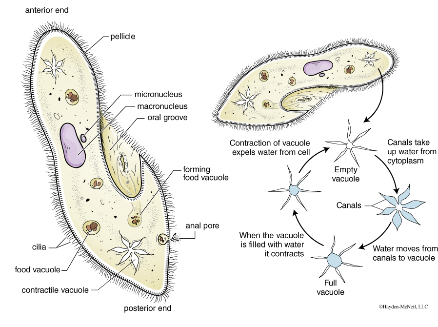
The Clade Alveolates have alveoli or sacs below their plasma membrane. They include ciliates (like Paramecia), dinoflagellates, and apicomplexans and are perhaps the best known of the Protista. One of the best known of these is the ciliate Paramecium (Fig.7.5). It is the “lab rat” for the Protista and is seen in many classes. The Paramecia and other ciliates are covered with short cilia that they use for locomotion. They are able to move with some precision, going both forward and backward. They can back themselves out of a tight place. They are found in freshwater and can occur in huge numbers when conditions are right. They are heterotrophic and feed on decaying material. Contractile vacules help them to osmoregulate and it is not difficult to see them filling and emptying excess fluid from their vacuoles. Curiously, they have two kinds of nuclei: micronuclei that are important in reproduction and macronuclei important in regulating cell metabolism and growth. They have a curious form of sexual behavior called conjugation, which involves an exchange of genetic material without reproducing.
A second group of the alveolates are the dinoflagellates, found in both freshwater and in saltwater. In the oceans, they are a significant part of the marine phytoplankton. Dinoflagellates typically have two flagella: one flagellum lies that in a longitudinal groove and one that lies in the transverse groove (see Fig. 7.6). Most are photosynthetic and while some are free-living (and are bioluminescent), others are endosymbionts in the tissues of corals, jellyfish, and some large clams. Some of the free-living dinoflagellates can cause algal blooms called red tides. The endosymbionts are called zooxanthellae, and Dr. Tamar Goulet in the Biology Department studies the endosymbionts and their hosts.
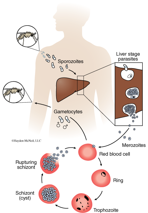
Apicomplexa are a group in the Clade Alveolates that are entirely parasitic. One disease caused by an apicomplexan is malaria, a debilitating disease that affects nearly half a billion people in the world. The symptoms of malaria are fever and chills that occur in waves every 24–48 hours. The fever occurs when red blood cells that have the parasite burst and release the parasites to enter more blood cells. Malaria is a disease that has a growing resistance to treatments and is a serious concern for most tropical countries (Fig. 7.7).
PROCEDURE: CLADE ALVEOLATES
Make a wet mount from the live Paramecia culture. How large are the they? Get one individual to watch and look for the filling and emptying of the contractile vacuole. In a short paragraph, describe their movement.
In the prepared slide of Paramecium, find the micronucelus and macronucelus. Be able to describe conjugation.
For comparison, we have prepared slides of a dinoflagellate and of the Plasmodium. You need to be familiar with the life cycle of the Plasmodium, and with the disease malaria.
The Clade Stramenopiles include the brown algae, diatoms and water molds. Stramenopiles have two unequal flagella, one of which has a row of tubular hairs. The water molds are a filamentous group used to be classified with the fungi. However, their cell walls are made from cellulose (as other protists) and not chitin (as with the fungi). A species of water mold caused the potato blight or famine in Ireland. The potato blight of Ireland in 1846–1847 was significant in the deaths of over a million people and was also a significant factor in the immigration of millions of Irish to North America.
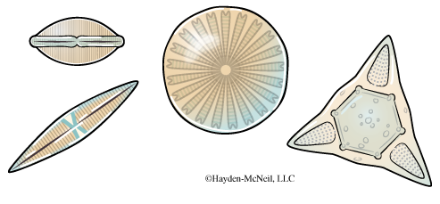
Diatoms are mostly unicellular, and are generally made from two shells containing silica that fit together. Because the silica preserves well, the diatoms are very important in the fossil record. Diatomaceous earth is rock that is formed from billions of shells. Fig. 7.8 shows some examples of diatoms.
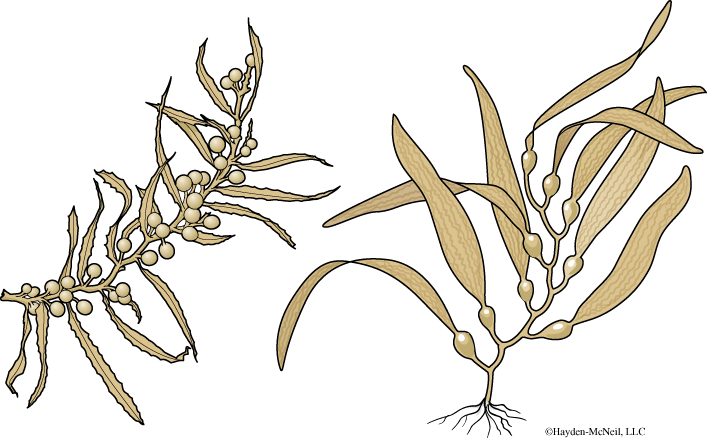
A very different example of the stramenopiles is the very large brown algae such as the kelps. Many kelps have specialized regions, such as “blades” or leaf-like structures, stipes, (similar to stems) and holdfasts, similar to roots (See Fig. 7.9). Brown algae is the source for algin, a thickening agent used in ice cream and many other foods. Kelps are important in providing habitat for a host of other marine animals. Sargassum is mostly found in the Sargasso Sea, an area of the Atlantic with a large area of floating brown algae in the North Atlantic. It is an important area for migrating turtles and eels.
PROCEDURE: CLADE STRAMENOPILES
We will have prepared slides of diatoms for you to examine and sketch. In addition, there will be examples of brown algae for you to examine and sketch.
Clade Plantae
Red algae, green algae, land plants. This group will be covered in the next chapter.
Clade Excavates
(Giardia, Trichonympha, Trypanosomes, Euglena)
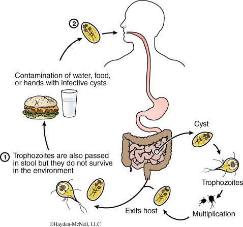
The excavates include several groups that lack mitochondria. At one point they were considered to be basal in the evolution of eukaryotes, but there is growing evidence that they evolved from organisms that at one time had mitochondria. The genus Giardia is an intestinal parasite that infects people and causes severe diarrhea and dehydration (Fig. 7.10). It causes what is sometimes called Backpackers’ diarrhea, as it can be found in seemly clear mountain streams. Giardia have two haploid nuclei. Most other eukaryotes have a diploid nucleus. Perhaps the ancestor of this group reflects the transition between the haploid prokaryotes and the mostly diploid eukaryotes!
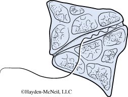
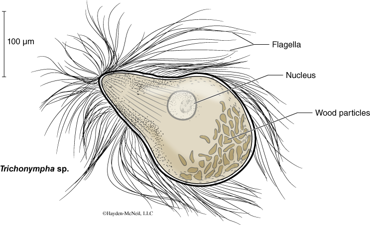
A second important group is the Trichonympha, a group of protists that live symbiotically in the guts of termites and cockroaches (Fig. 7.11). Trichonympha have numerous long flagella (and look a bit like rock stars!). Termites rely on the Trichonympha to digest the cellulose in the wood that they eat; the Trichonympha in turn rely on endosymbiotic bacteria to digest the cellulose.
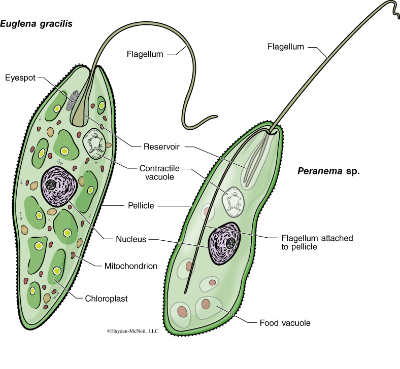
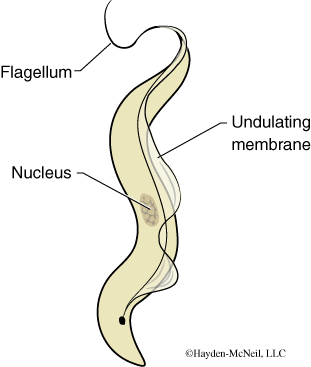
Euglenoids and trypanosomes are protists that have a disk-shaped cristae in their mitochondria. Cristae are inward projections on the inner membrane of mitochondria. These protists move by flagella. One well-known example that we will look at in lab this week is Euglena. Euglena are very small, can reproduce only by mitosis, and are autotrophic when light is available (note the green color of the culture!) and heterotrophic when light is not available. They are found in freshwater and can contribute to algal blooms. In addition to the above, they have a very flexible outer covering, called the pellicle (Fig. 7.12).
Trypanosomes are parasitic and cause serious diseases. For example, African sleeping sickness is caused by a trypanosome and is spread by the bite of a tsetse fly. They also are very small and can be seen in the blood of people with the infection. We will have a prepared slide for you to examine. Trypanosomes have a remarkable ability to evade the host’s immune system, and that is an area of active research (Fig. 7.13).
PROCEDURE: CLADE EXCAVATES
Examine the demonstration slide of Giardia. Next, at least one member of each table should dissect the hindgut of a termite and make a wet mount using insect ringers to look for the Trichonympha. You should sketch and label (and be able to recognize!) both the Giardia and Trichonympha.
Make a wet mount from the live Euglena culture. How large are the Euglena? What magnification do you need to see any details? Next, examine the prepared slide of Trypanosoma. Note that they are mixed in with the red blood cells. Sketch, label, and be able to identify each of these.
Clade Rhizaria (forams and radiolaria)
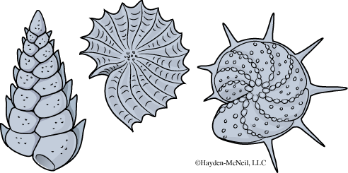
The foraminiferans are microscopic and include cells that are Amoeba-like but are enclosed in a carbonate shell. The shell has minute openings, through which cytopasmic projections can be extended. They are predators, and the tiny pseudopodia of the organism are extended through those holes and capture food floating by. Allthough individually tiny, together they can form massive deposits. The famous White Cliffs of Dover in England contain fossil foraminifera shells. Because of the shell, they are important microfossils (Fig. 7.14).
PROCEDURE: CLADE RHIZARIA
Examine the prepared slides of the foraminifera. Make a sketch and be sure to indicate size.
Clade Unikonts; Clade Amoebozoans (Amoeba and slime molds)
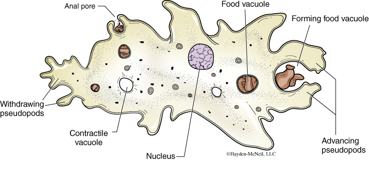
Amoebas are well-known and fascinating organisms. They are a single cell, and can be “huge” for a cell (such as the giant amoeba, which can be more than half a millimeter long!). They are also remarkable for their ability to change size and shape. Indeed, they move by pseudopodia, or flexible extensions of their cytoplasm. They are predators and scavengers, and some, such as the Entamoeba histolytica, are parasites in humans (Fig. 7.15).
Another very curious group within this clade is the plasmodial slime mold, or Physarum polycephalum. This is a multinucleate mass of streaming cytoplasm that is macroscopic and not uncommon in gardens. It creeps along and ingests bacteria, molds, and any decaying oraganic matter. Note that this is another example where the common name is “mold,” but this organism is a protist.
PROCEDURE: CLADE AMOEBOZOA
We will examine live Amoeba in lab today. Typically they are located in the bottom of the jars and can be gotten by a gentle use of a pipette. There may be a demo example, made with a depression slide. We will also have a live example of a plasmodial slime mold and look for cytoplasmic streams. Look also at the pictures in the lab.
Clade Unikonts; Clade Opisthokonts (fungi, choanoflagellates, and animals—will be covered in more detail in Lab 9)
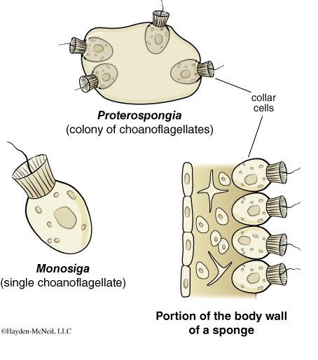
The final clade of Protista that we will consider is the Clade Opisthokonts. This is another very large and very diverse clade that includes both the fungi and the animals. The opisthokonts are characterized by its representative having at some point in their lives, single posterior flagellum in a motile cell, (the name “opistho” means backward, kont refers to flagellum). In animals, this characteristic is seen in sperm cells. In some fungi, this characteristic is seen in some motile spores. This group is also united by similarity in the ribosomal RNA sequences. In this lab, we will look at a choanoflagellate. The choanoflagellate is a free-living marine group, but it is of interest because very similar cells are seen in the most primitive animals, the sponges (Fig. 7.16). Choanoflagellates have a collar that surrounds their flagellum. This collar helps to capture microscopic debris that the flagellate can then feed on.
PROCEDURE: CLADE OPISTHOKONTS
Examine the prepared slide of the choanoflagellate.
IV. MATERIALS FOR LAB CHECKLIST FOR STUDENTS AND TAs
NOTE TO TAs
Bacteria: 15-minute introduction; 30–45 minutes to complete this section
Protista: 10–15-minute introduction; 1 hour, 45 minutes to complete this section
I. OVERVIEW: DIVERSITY AND COMPARISON OF PROKARYOTES AND EUKARYOTES; BASIS OF THREE DOMAINS
Review overview of prokaryotic cells and domains.
Know the basis of the three domains and some of the organisms in each domain.
II. DIVERSITY: DOMAIN BACTERIA AND DOMAIN ARCHAEA
PROCEDURE
Examine prepared slide “Bacterial types,” which has all three morphological types of bacteria (coccus, spirillum, and bacillus or rod). Use oil immersion lenses correctly. Oil immersion is to be used only with the 100× lens and oil must be cleaned from the lens with lens cleaner and tissue when you are done. Examine the demonstration slide of Gram-positive and Gram-negative bacteria. You should understand the basis of how the Gram stain works. Examine the SEM pictures available in the lab manual.
Examine cyanobacteria. Make wet mounts and make sketches of each of the following species. (Be careful to not mix the cultures!) Look for heterocycts and akinetes. If you use oil immersion lenses do so correctly.
Anabaena
Gloecapsa
Nostoc
III. DIVERSITY: PROTISTA
Notes to TAs: Students need to keep careful sketches of each of the groups covered in class and should know the formal clade name as well as the genus or informal name. In their sketches, they should pay particular attention to the magnification, as that gives them an indication of the size of the organism. For students to succeed in this lab, they must come to class already knowing many of the names. And, students need to believe that we really expect them to know these groups!
Note to students: You should know the formal and common names for each of these groups as well as what is in your lab book about each. How does each move? How does each get food? Your TA will have a list of the sketches you need to do for this lab.
You should make a summary table that includes clade, common name, movement, and reproduction.