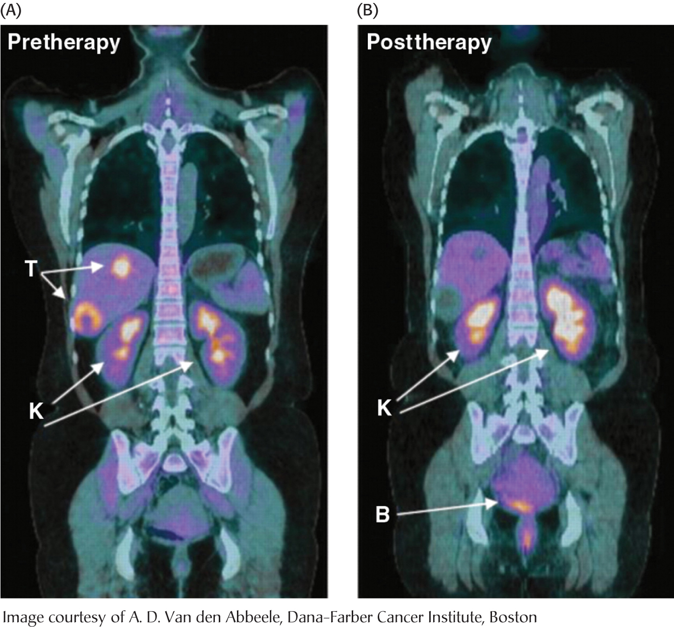
Figure 16.17 Tumors can be visualized with 2-18F-2-D-deoxyglucose (FDG) and positron emission tomography. (A) A nonmetabolizable glucose analog (FDG) infused into a patient and detected by a combination of positron emission and computer-aided tomography reveals the presence of a malignant tumor (T). (B) After 4 weeks of treatment with a tyrosine kinase inhibitor, the tumor shows no uptake of FDG, indicating decreased metabolism. Excess FDG, which is excreted in the urine, also visualizes the kidney (K) and bladder (B).