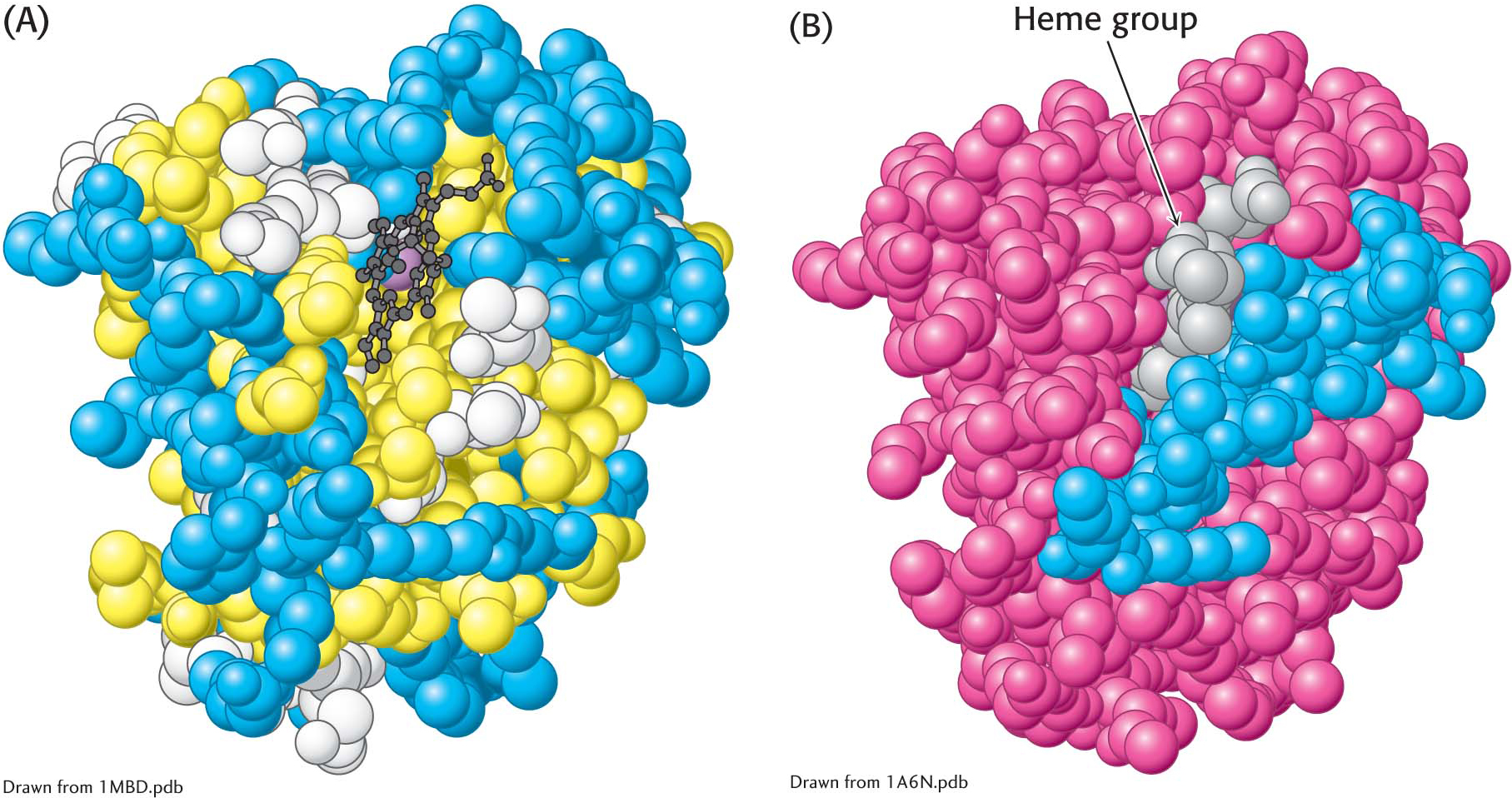
Figure 4.26  The distribution of amino acids in myoglobin. (A) A space-filling model of myoglobin, with hydrophobic amino acids shown in yellow, charged amino acids shown in blue, and others shown in white. Notice that the surface of the molecule has many charged amino acids, as well as some hydrophobic amino acids. (B) In this cross-sectional view, notice that mostly hydrophobic amino acids are found on the inside of the structure, whereas the charged amino acids are found on the protein surface.