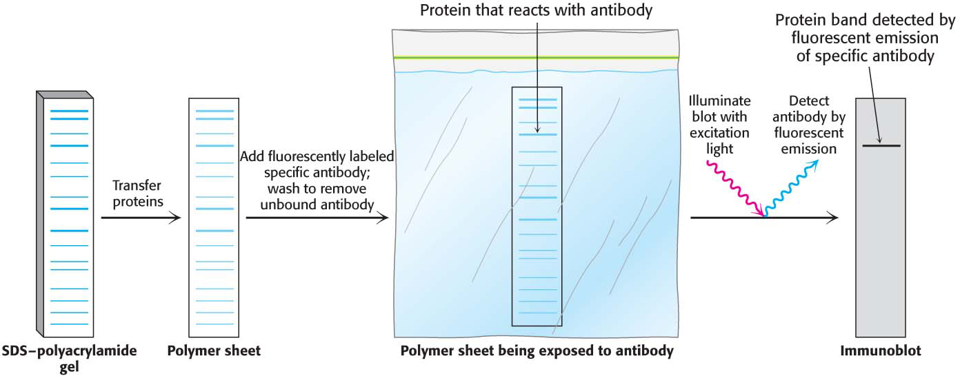
Figure 5.23 Western blotting. Proteins on an SDS–polyacrylamide gel are transferred to a polymer sheet and stained with fluorescent antibody. The fluorescent antibody is excited by light, and the band corresponding to the protein to which the antibody binds is visualized with an appropriate detector.