21.3 Cellular Respiration is Regulated by the Need for ATP

✓ 4 Identify the ultimate determinant of the rate of cellular respiration.

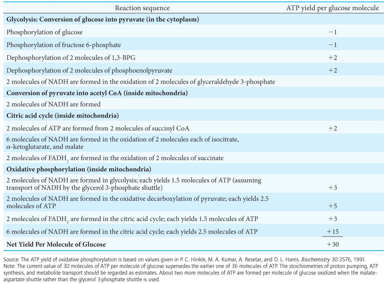
We have observed many times that most catabolic pathways are regulated in some fashion by the ATP concentration. Because ATP is the end product of cellular respiration, the ATP needs of the cell are the ultimate determinant of the rate of respiratory pathways and their components. Before we explore the nature of this regulation, let us calculate the ATP yield from the conversion of glucose to CO2 and H2O.

The Complete Oxidation of Glucose Yields About 30 Molecules of ATP

We can estimate how many molecules of ATP are formed when glucose is completely oxidized to CO2. We say “estimate” because, in contrast with the ATP yield of glycolysis and the citric acid cycle (which yield 4 molecules of ATP per molecule of glucose and 1 molecule of ATP per molecule of pyruvate, respectively), the stoichiometries of proton pumping, ATP synthesis, and metabolite-transport processes need not be an integer or even have fixed values. As stated earlier, the best current estimates for the number of protons pumped out of the matrix by NADH-Q oxidoreductase, Q-cytochrome c oxidoreductase, and cytochrome c oxidase per electron pair are four, four, and two, respectively. The synthesis of a molecule of ATP is driven by the flow of about three protons through ATP synthase. An additional proton is consumed in transporting ATP from the matrix to the cytoplasm. Hence, about 2.5 molecules of cytoplasmic ATP are generated as a result of the flow of a pair of electrons from NADH to O2. For electrons that enter at the level of Q-cytochrome c oxidoreductase, such as those from the oxidation of succinate or cytoplasmic NADH transferred by the glycerol-phosphate shuttle, the yield is about 1.5 molecules of ATP per electron pair. Hence, as tallied in Table 21.1, about 30 molecules of ATP are formed when glucose is completely oxidized to CO2. Most of the ATP, 26 of 30 molecules formed, is generated by oxidative phosphorylation. Recall that the anaerobic metabolism of glucose yields only 2 molecules of ATP. The efficiency of cellular respiration is manifested in the observation that one of the effects of endurance exercise, a practice that calls for much ATP for an extended period of time, is to increase the number of mitochondria and blood vessels in muscle and therefore increase the extent of ATP generation by oxidative phosphorylation.

394

Table 21.1 ATP yield from the complete oxidation of glucose

395

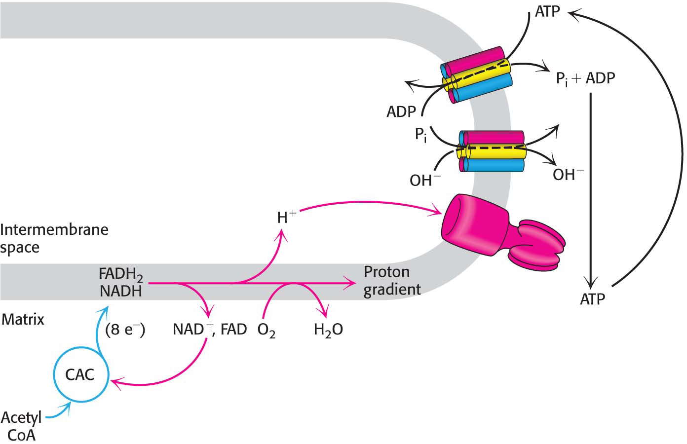
The Rate of Oxidative Phosphorylation Is Determined by the Need for ATP

Figure 21.16: Respiratory control. Electrons are transferred to O2 only if ADP is concomitantly phosphorylated to ATP.

How is the rate of the electron-transport chain controlled? Under most physiological conditions, electron transport is tightly coupled to phosphorylation. Electrons do not usually flow through the electron-transport chain to O2 unless ADP is simultaneously phosphorylated to ATP. When ADP concentration rises, as would be the case in active muscle that is continuously consuming ATP, the rate of oxidative phosphorylation increases to meet the ATP needs of the cell. The regulation of the rate of oxidative phosphorylation by the ADP level is called respiratory control or acceptor control. Experiments on isolated mitochondria demonstrate the importance of ADP level (Figure 21.16). The rate of oxygen consumption by mitochondria increases markedly when ADP is added and then returns to its initial value when the added ADP has been converted into ATP.

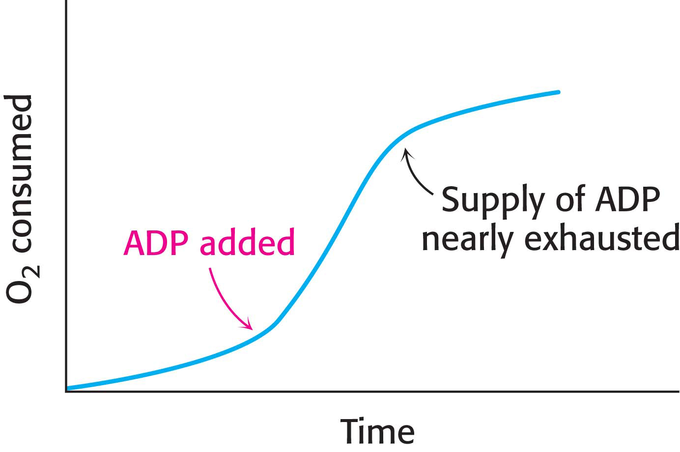
As discussed in Chapter 19, the level of ADP likewise indirectly affects the rate of the citric acid cycle. At low concentrations of ADP, as in a resting muscle, NADH and FADH2 produced by the citric acid cycle are not oxidized back to NAD+ and FAD by the electron-transport chain. The citric acid cycle slows because there is less NAD+ and FAD to feed the cycle. As the ADP level rises and oxidative phosphorylation speeds up, NADH and FADH2 are oxidized, and the citric acid cycle becomes more active. Electrons do not flow from fuel molecules to O2 unless ATP needs to be synthesized. We see here another example of the regulatory significance of the energy charge (Figure 21.17).

Figure 21.17: The energy charge regulates the use of fuels. The synthesis of ATP from ADP and Pi controls the flow of electrons from NADH and FADH2 to oxygen. The availability of NAD+ and FAD in turn control the rate of the citric acid cycle (CAC).

!clinic! CLINICAL INSIGHT: ATP Synthase Can Be Regulated

Mitochondria contain an evolutionarily conserved protein, inhibitory factor 1 (IF1), that specifically inhibits the potential hydrolytic activity of the F0F1 ATP synthase. What is the function of IF1? Consider a circumstance where tissues may be deprived of oxygen (ischemia). Without oxygen as the electron acceptor, the electron transport chain will be unable to generate the proton-motive force. The ATP in the mitochondria would be hydrolyzed by the synthase, working in reverse (problem 22). The role of IF1 is to prevent the wasteful hydrolysis of ATP by inhibiting the hydrolytic activity of the synthase.

396

IF1 is over-expressed in many types of cancer. This over-expression plays a role in the induction of the Warburg effect, the switch from oxidative phosphorylation to aerobic glycolysis as the principle means for ATP synthesis.

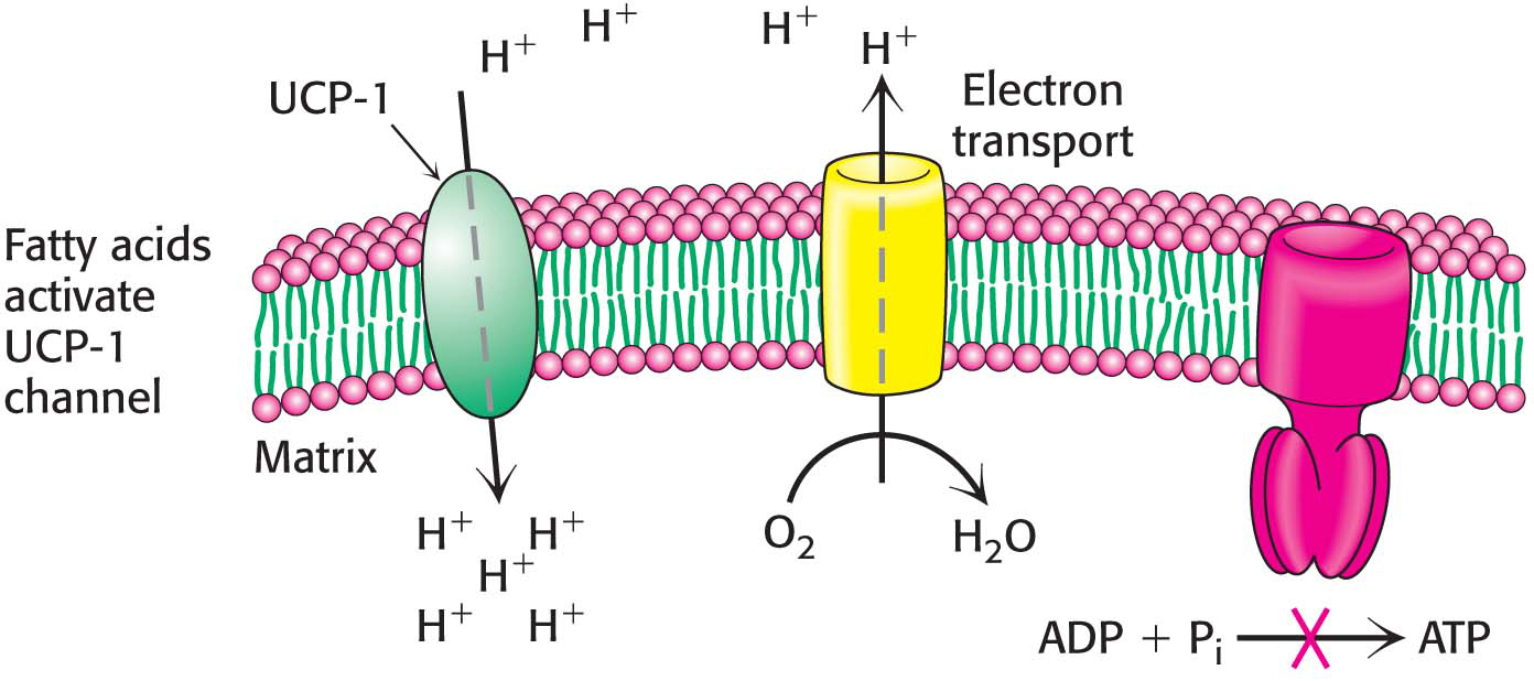
!bio! BIOLOGICAL INSIGHT: Regulated Uncoupling Leads to the Generation of Heat

Some organisms possess the ability to uncouple oxidative phosphorylation from ATP synthesis to generate heat. Such uncoupling is a means to maintain body temperature in hibernating animals, in some newborn animals (including human beings), and in mammals adapted to cold. In animals, brown fat or brown adipose tissue (BAT) is specialized tissue for this process of nonshivering thermogenesis. In contrast, white adipose tissue (WAT), which constitutes the bulk of adipose tissue, plays no role in thermogenesis but serves as an energy source.

Brown adipose tissue is very rich in mitochondria, often called brown-fat mitochondria. The inner mitochondrial membrane of these mitochondria contains a large amount of uncoupling protein 1 (UCP-1), also called thermogenin. UCP-1 transports protons from the intermembrane space to the matrix with the assistance of fatty acids. In essence, UCP-1 generates heat by short-circuiting the mitochondrial proton battery. The energy of the proton gradient, normally captured as ATP, is released as heat as the protons flow through UCP-1 to the mitochondrial matrix (Figure 21.18). This dissipative proton pathway is activated when the core body temperature begins to fall. In response to a temperature drop, a adrenergic hormones stimulate the release of free fatty acids from triacylglycerols stored in cytoplasmic lipid droplets. Long chain fatty acids bind to the cytoplasmic face of UCP-1, and the carboxyl group binds a proton. This causes a structural change in UCP-1 so that the protonated carboxyl now faces the proton-poor environment of the matrix, and the proton is released. Proton release resets UCP-1 to the initial state.

Figure 21.18: The action of an uncoupling protein. Uncoupling protein 1 (UCP-1) generates heat by permitting the influx of protons into the mitochondria without the synthesis of ATP.

397

We can witness the effects of a lack of nonshivering thermogenesis by examining pig behavior. Pigs are unusual mammals in that they have large litters and are the only ungulates (hoofed animals) that build nests for birth (Figure 21.19). These behavioral characteristics appear to be the result of a biochemical deficiency. Pigs lack UCP-1 and, hence, brown fat. Piglets must rely on other means of thermogenesis. Nesting, large litter size, and shivering are means by which pigs compensate for a lack of brown fat.

Figure 21.19: Nesting piglets. A wild boar, a member of the pig family, is shown with her nesting piglets.

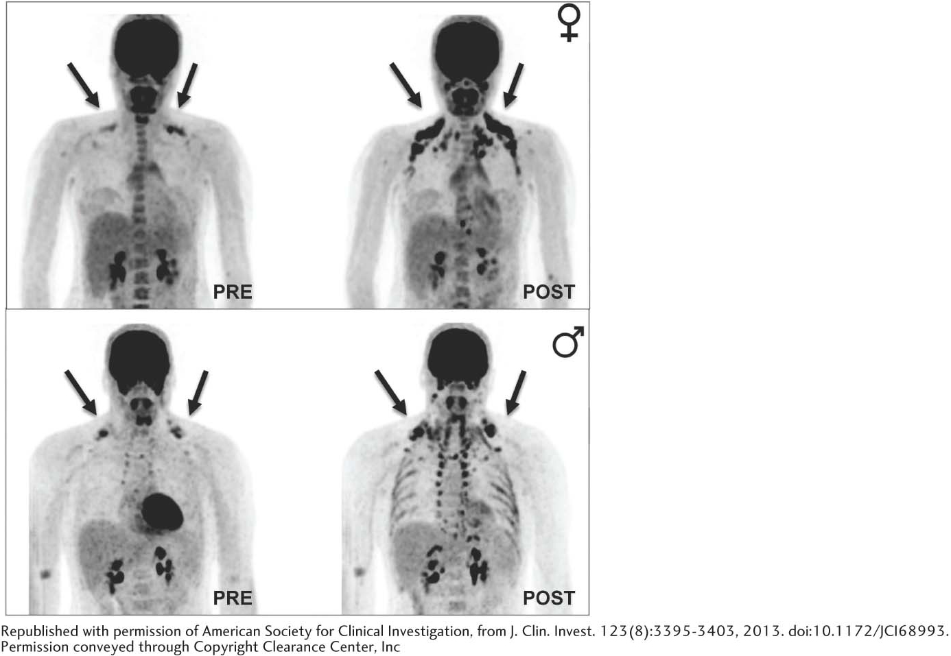
Until recently, adult humans were believed to lack brown fat tissue. However, new studies have established that adults, women especially, have brown adipose tissue on the neck and upper chest regions that is activated by cold (Figure 21.20). Obesity leads to a decrease in brown adipose tissue.

Figure 21.20: Brown adipose tissue is revealed on exposure to cold. The results of PET–CT (positron emission and computerized tomography) scanning show the uptake and distribution of 18F-fluorodeoxyglucose (18F-FDG), a nonmetabolizable glucose analog, in adipose tissue. The patterns of 18F-FDG uptake in the same subject are dramatically different under thermoneutral conditions (left) and after exposure to cold (right).

398

!clinic! CLINICAL INSIGHT: Oxidative Phosphorylation Can Be Inhibited at Many Stages

Many potent and lethal poisons exert their effects by inhibiting oxidative phosphorylation at one of a number of different locations.

Inhibition of the electron-transport chain The four complexes of the electron-transport chain can be inhibited by different compounds, blocking electron transfer downstream and thereby shutting down oxidative phosphorylation. NADH-Q oxidoreductase (Complex I) is inhibited by rotenone, which is used as a fish and insect poison, and amytal, a barbiturate sedative. Inhibitors of Complex I prevent the utilization of NADH as a substrate (Figure 21.21). Rotenone exposure, along with a genetic predisposition, has been implicated in the development of Parkinson disease, a condition characterized by resting tremor, slowness of movement, inability to initiate movement, rigidity, and postural instability. Inhibition of Complex I does not impair electron flow from FADH2, because these electrons enter through QH2, beyond the block. Q-cytochrome c oxidoreductase (Complex III) is inhibited by antimycin A, an antibiotic isolated from Streptomyces that is used as a fish poison. Furthermore, electron flow in cytochrome c oxidase(Complex IV) can be blocked by cyanide (CN), azide (N3), and carbon monoxide (CO). Cyanide and azide react with the ferric form (Fe3+) of heme a3, whereas carbon monoxide inhibits the ferrous form (Fe2+). Inhibition of the electron-transport chain also inhibits ATP synthesis because the proton-motive force can no longer be generated.

Figure 21.21: Sites of action of some inhibitors of electron transport.

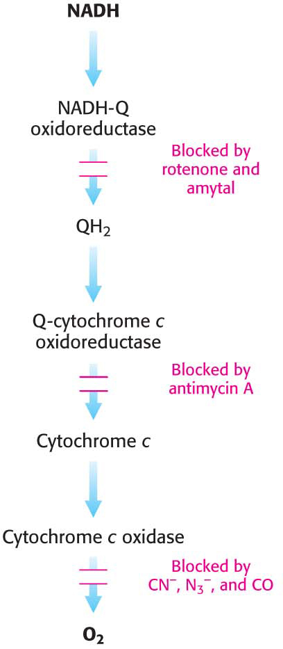
Inhibition of ATP synthase Oligomycin, an antibiotic used as an antifungal agent, and dicyclohexylcarbodiimide (DCC), used in peptide synthesis in the laboratory, prevent the influx of protons through ATP synthase by binding to the carboxylate group of the c subunits required for proton binding. Modification of only one c subunit by DCC is sufficient to inhibit the rotation of the entire c ring and hence ATP synthesis. If actively respiring mitochondria are exposed to an inhibitor of ATP synthase, the electron-transport chain ceases to operate. This observation clearly illustrates that electron transport and ATP synthesis are normally tightly coupled.

399

Uncoupling electron transport from ATP synthesis The tight coupling of electron transport and phosphorylation in mitochondria can be uncoupled by 2,4-dinitrophenol (DNP) and certain other acidic aromatic compounds. These substances carry protons across the inner mitochondrial membrane, down their concentration gradients. In the presence of these uncouplers, electron transport from NADH to O2 proceeds in a normal fashion, but ATP is not formed by mitochondrial ATP synthase, because the proton-motive force across the inner mitochondrial membrane is continuously dissipated. This loss of respiratory control leads to increased oxygen consumption and the oxidation of NADH. Indeed, in the accidental ingestion of uncouplers, large amounts of metabolic fuels are consumed, but no energy is captured as ATP. Rather, energy is released as heat. DNP is the active ingredient in some herbicides and fungicides. Remarkably, some people consume DNP as a weight-loss drug, despite the fact that the U.S. Food and Drug Administration (FDA) banned its use in 1938. There are also reports that Soviet soldiers were given DNP to keep them warm during the long Russian winters. Chemical uncouplers are nonphysiological, unregulated counterparts of uncoupling proteins.

Drugs are being sought that would function as mild uncouplers, uncouplers not as potentially-lethal as DNP, for use in treatment of obesity and related pathologies. Xanthohumol, a prenylated chalcone found in hops and beer, shows promise in this regard. A chalcone is an aromatic ketone. Xanthohumol also scavenges free radicals and is used for treatment of certain types of cancers.

Inhibition of ATP export ATP-ADP translocase is specifically inhibited by very low concentrations of atractyloside (a plant glycoside) or bongkrekic acid (an antibiotic found in fermented coconut contaminated by the bacterium Burkholderia gladioli). Atractyloside binds to the translocase when its nucleotide site faces the cytoplasm, whereas bongkrekic acid binds when this site faces the mitochondrial matrix. Oxidative phosphorylation stops soon after either inhibitor is added, showing that ATP-ADP translocase is essential for maintaining adequate amounts of ADP to accept the energy associated with the proton-motive force.

!clinic! CLINICAL INSIGHT: Mitochondrial Diseases Are Being Discovered in Increasing Numbers

The number of diseases that can be attributed to mitochondrial mutations is steadily growing in step with our growing understanding of the biochemistry and genetics of mitochondria. Mitochondrial diseases are estimated to affect from 10 to 15 per 100,000 people, roughly equivalent to the prevalence of the muscular dystrophies. The first mitochondrial disease to be understood was Leber hereditary optic neuropathy (LHON), a form of blindness that strikes in midlife as a result of mutations in Complex I. Some of these mutations impair NADH utilization, whereas others block electron transfer to Q. Mutations in Complex I are the most frequent cause of mitochondrial diseases. The accumulation of mutations in mitochondrial genes in a span of several decades may contribute to aging, degenerative disorders, and cancer.

A human egg harbors several hundred thousand molecules of mitochondrial DNA, whereas a sperm contributes only a few hundred and thus has little effect on the mitochondrial genotype. Because the maternally-inherited mitochondria are present in large numbers and not all of the mitochondria may be affected, the pathologies of mitochondrial mutants can be quite complex. Even within a single family carrying an identical mutation, chance fluctuations in the percentage of mitochondria with the mutation lead to large variations in the nature and severity of the symptoms of the pathological condition as well as the time of onset. As the percentage of defective mitochondria increases, energy-generating capacity diminishes until, at some threshold, the cell can no longer function properly. Defects in cellular respiration are doubly dangerous. Not only does energy transduction decrease, but also the likelihood that reactive oxygen species will be generated increases. Organs that are highly dependent on oxidative phosphorylation, such as the nervous system, the retina and the heart, are most vulnerable to mutations in mitochondrial DNA.

400

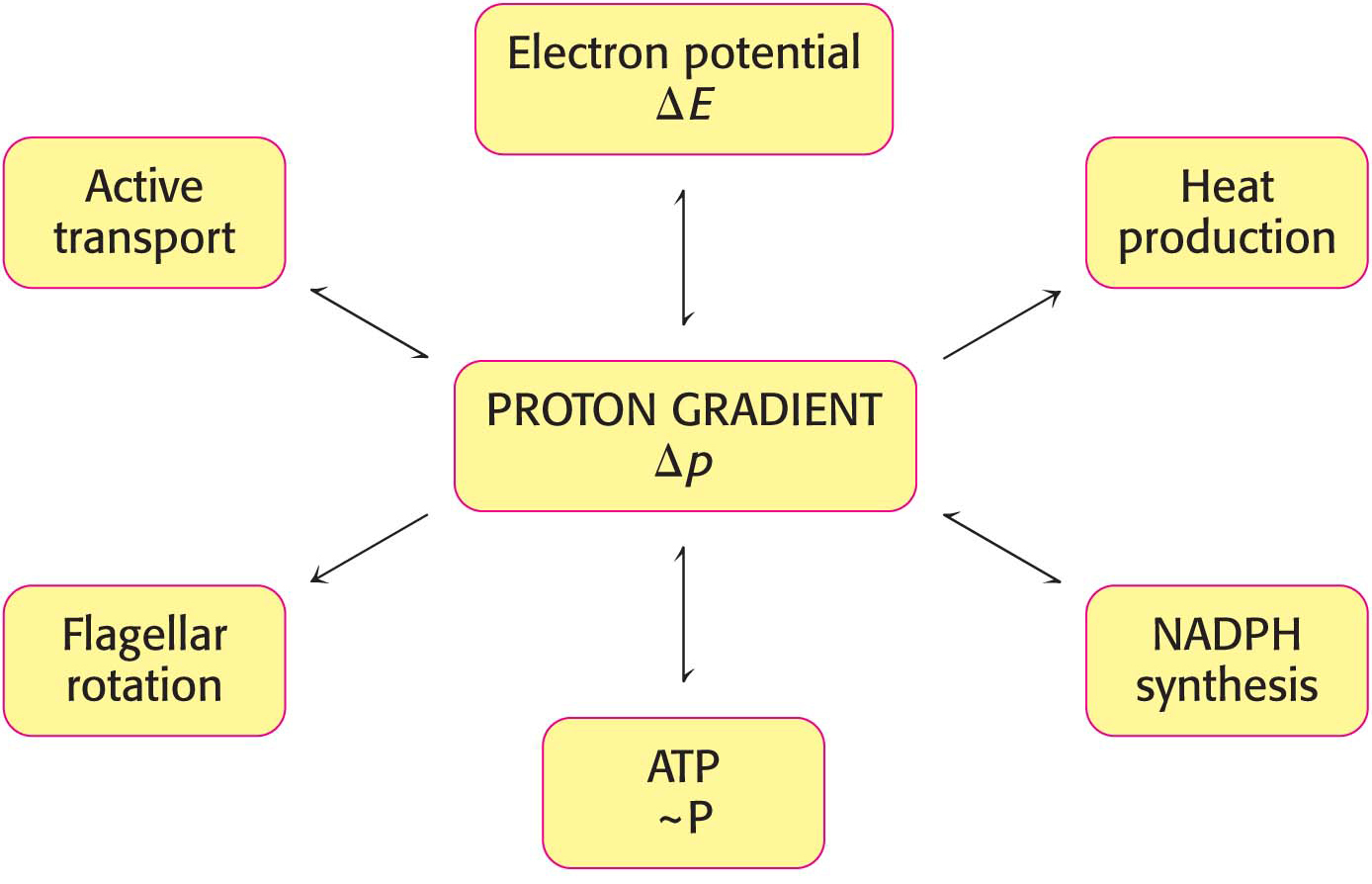
Power Transmission by Proton Gradients Is a Central Motif of Bioenergetics

The main concept presented in this chapter is that mitochondrial electron transfer and ATP synthesis are linked by a transmembrane proton gradient. ATP synthesis in bacteria and chloroplasts also is driven by proton gradients. In fact, proton gradients power a variety of energy-requiring processes such as the active transport of calcium ions by mitochondria, the entry of some amino acids and sugars into bacteria, the rotation of bacterial flagella, and the transfer of electrons from NADP+ to NADPH. As we have already seen, proton gradients can also be used to generate heat. It is evident that proton gradients are a central interconvertible currency of free energy in biological systems (Figure 21.22). Peter Mitchell noted that the proton-motive force is a marvelously simple and effective store of free energy because it requires only a thin, closed lipid membrane between two aqueous phases.

Figure 21.22: The proton gradient is an interconvertible form of free energy.