32.3 The Purine Ring Is Assembled on Ribose Phosphate

Figure 32.4: De novo pathway for purine nucleotide synthesis. The origins of the atoms in the purine ring structure are indicated.

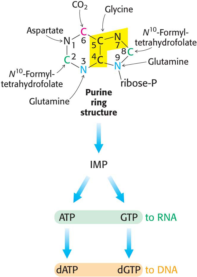
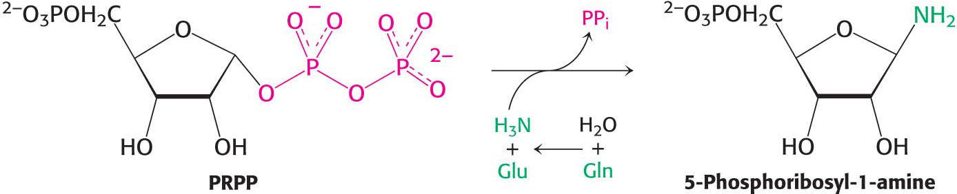
Purines, like pyrimidines, are synthesized de novo, beginning with simple starting materials (Figure 32.4). In contrast with pyrimidine synthesis, the first step in purine assembly begins with the attachment to ribose. De novo purine biosynthesis, like pyrimidine biosynthesis, requires PRPP, but, for purines, PRPP provides the foundation on which the bases are constructed step by step. The initial step is the displacement of the pyrophosphate of PRPP by ammonia, rather than by a preassembled base, to produce 5-phosphoribosyl-1-amine, with the amine in the β configuration. Glutamine again provides the ammonia. Glutamine phosphoribosyl amidotransferase catalyzes this reaction, which is the committed step in purine synthesis.

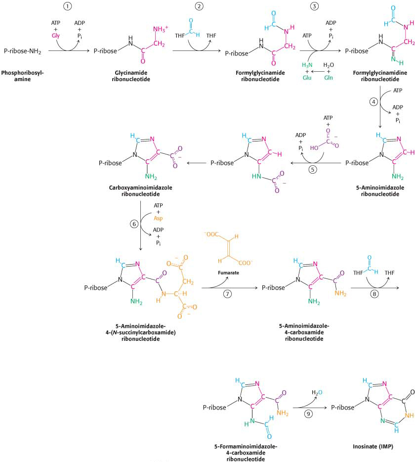
Nine additional steps are required to assemble the purine ring. De novo purine biosynthesis proceeds by successive steps of activation by phosphorylation followed by displacement, as shown in (Figure 32.5). The final product is the nucleotide inosine monophosphate (IMP, or inosinate). The amino acids glycine, glutamine, and aspartate are required precursors. The synthesis of purines depends on tetrahydrofolate, a prominent carrier of activated one-carbon units.

Figure 32.5: De novo purine biosynthesis. (1) Glycine is coupled to the amino group of phosphoribosylamine. (2) N10-Formyltetrahydrofolate (THF) transfers a formyl group to the amino group of the glycine residue. (3) The inner amide group is phosphorylated and then converted into an amidine by the addition of ammonia derived from glutamine. (4) An intramolecular coupling reaction forms a five-membered imidazole ring. (5) Bicarbonate adds first to the exocyclic amino group and then to a carbon atom of the imidazole ring. (6) The imidazole carboxylate is phosphorylated, and the phosphoryl group is displaced by the amino group of aspartate. (7) Fumarate leaves, followed by (8) the addition of a second formyl group from N10-formyl tetrahydrofolate. (9) Cyclization completes the synthesis of inosinate, a purine nucleotide. Abbreviation: P stands for phosphoryl group.

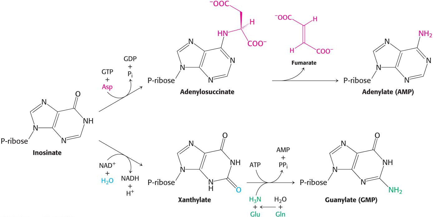
AMP and GMP Are Formed from IMP

DID YOU KNOW?

In the ring form of ribose sugars, the β configuration means that the group attached at C-1 is on the same side of the ring as the —CH2OH group. In the α configuration, the group attached to C-1 and —CH2OH are on opposite sides of the ring.

Inosinate, although a component of some RNA molecules, serves primarily as a precursor to the other purines. Inosinate is at the base of a branched pathway that leads to both AMP and GMP (Figure 32.6). Adenylate is synthesized from inosinate by the substitution of an amino group for the carbonyl oxygen atom at C-6. The addition of aspartate, followed by the departure of fumarate, contributes the amino group. GTP, rather than ATP, is required for the synthesis of the adenylosuccinate intermediate from inosinate and aspartate.

Figure 32.6: Generating AMP and GMP. Inosinate is the precursor of AMP and GMP. AMP is formed by the addition of aspartate followed by the release of fumarate. GMP is generated by the addition of water, dehydrogenation by NAD+, and the replacement of the carbonyl oxygen atom by —NH2 derived by the hydrolysis of glutamine.

!quickquiz! QUICK QUIZ 2

Identify the sources of all of the atoms in a purine ring.

Guanosine monophosphate (GMP, or guanylate) is synthesized by the oxidation of inosinate to xanthine monophosphate (XMP, or xanthylate), followed by the incorporation of an amino group at C-2. NAD+ is the hydrogen acceptor in the oxidation of inosinate. The carbonyl group of xanthylate is activated by the transfer of an AMP group from ATP. Ammonia, generated by the hydrolysis of glutamine, then displaces the AMP group to form guanylate, in a reaction catalyzed by GMP synthetase. Note that the synthesis of adenylate requires GTP, whereas the synthesis of guanylate requires ATP, a contrast that, as we will see, enables the flux down each branch to be balanced.

591

592

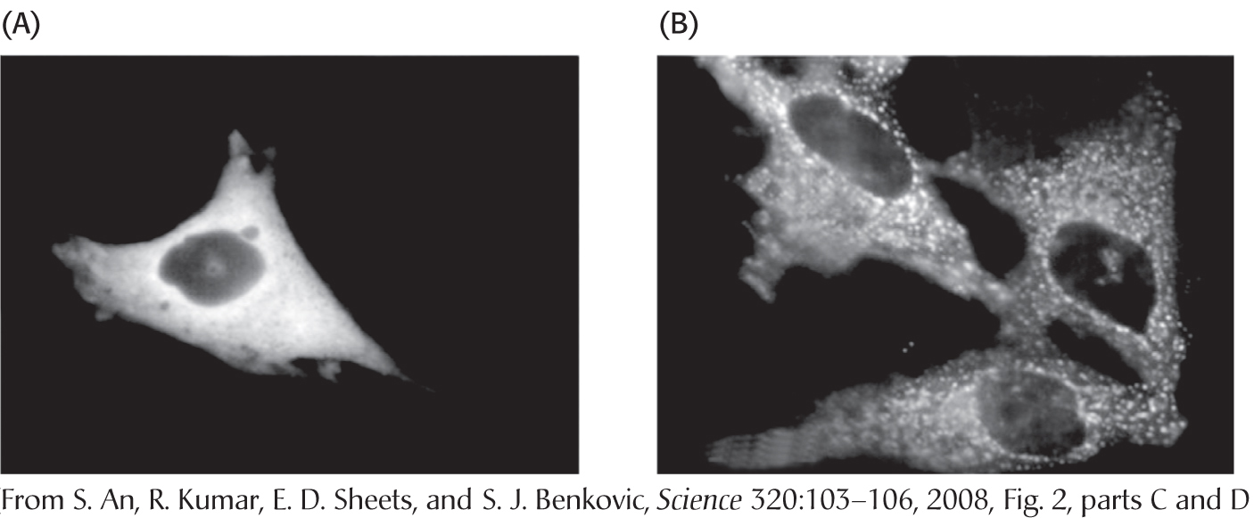
!bio! BIOLOGICAL INSIGHT: Enzymes of the Purine-Synthesis Pathway Are Associated with One Another in Vivo

Biochemists believe that the enzymes of many metabolic pathways, such as glycolysis and the citric acid cycle, are physically associated with one another. Such associations would increase the efficiency of pathways by facilitating the movement of the product of one enzyme to the active site of the next enzyme in the pathway. The evidence for such associations comes primarily from experiments in which one component of a pathway, carefully isolated from the cell, is found to be bound to other components of the pathway. However, these observations raise the question, do enzymes associate with one another in vivo or do they spuriously associate during the isolation procedure? Recent in vivo evidence shows that the enzymes of the purine-synthesis pathway associate with one another when purine synthesis is required. Various enzymes of the pathway were fused with green fluorescent protein (GFP), which renders the enzymes visible with a fluorescent microscope, and transfected into cells. When cells were grown in the presence of purine, GFP was spread diffusely throughout the cytoplasm (Figure 32.7A). When the cells were switched to growth media lacking purines, purine synthesis began and the enzymes became associated with one another, forming complexes dubbed purinosomes (Figure 32.7B). The experiments were repeated with other enzymes of the purine-synthesis pathway, and the results were the same: purine synthesis takes place when the enzymes form the purinosomes. What causes complex formation? Although the results are not yet established, it appears that several G-protein coupled receptors, including those responding to epinephrine as well as ATP and ADP (purinergic receptors), instigate complex formation, while human creatine kinase II (hCK2), responding to the presence of purines, causes disassembly of the purinosome.

Figure 32.7: A Formation of purinosomes. A gene construct encoding a fusion protein consisting of formylglycinamidine synthase and green fluorescent protein (GFP) was transfected into Hela cells (a human cell line) and expressed in them. (A) In the presence of purines (the absence of purine synthesis), the GFP was seen as a diffuse stain throughout the cytoplasm. (B) When the cells were placed in a purine-free medium, purinosomes formed, seen as cytoplasmic granules, and purine synthesis took place.

593

Bases Can Be Recycled by Salvage Pathways

Purine nucleotides, like pyrimidine nucleotides, can also be synthesized by salvaging and recycling intact purines released by the hydrolytic degradation of nucleic acids and nucleotides. Such salvage pathways skip most of the energy-requiring steps of de novo nucleotide synthesis, thereby saving substantial amounts of ATP.

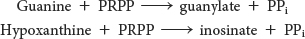
Two salvage enzymes with different specificities recover purine bases. Adenine phosphoribosyltransferase catalyzes the formation of adenylate,

whereas hypoxanthine-guanine phosphoribosyltransferase (HGPRT) catalyzes the formation of guanylate as well as inosinate, which, you will recall, is a precursor of guanylate and adenylate:

Purine salvage pathways are especially noteworthy in light of the amazing consequences of their absence.