33.5 RNA Can Adopt Elaborate Structures
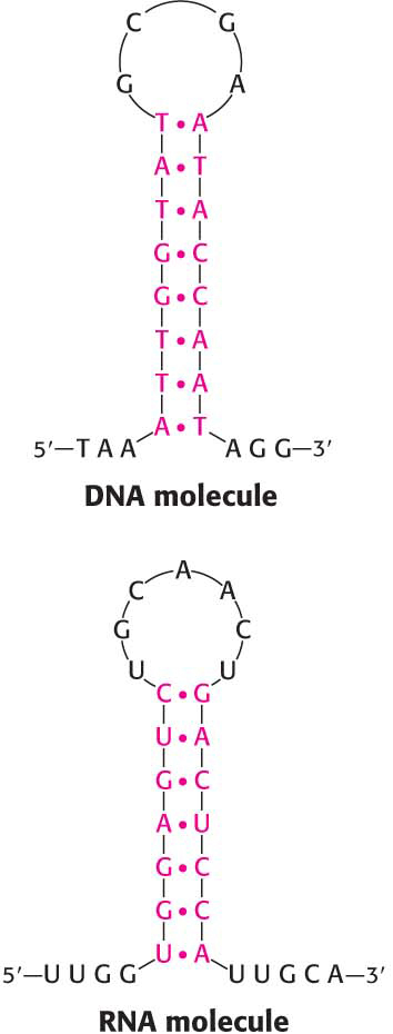
Figure 33.30: Stem-loop structures. A stem-loop structure can be formed from single-stranded DNA or from RNA.
As we have seen, DNA is usually not a simple helix but is compacted into a more complex structure. Even single-stranded nucleic acids, most commonly RNA, often fold back on themselves to form well-defined and often complex structures. These complex structures allow RNA to perform a host of functions that the double-stranded DNA molecule cannot. Indeed, the complexity of some RNA molecules rivals that of proteins, and these RNA molecules perform a number of functions that had formerly been thought to be the exclusive role of proteins. For instance, one of the RNA components of ribosomes—a large complex of RNAs and proteins on which proteins are synthesized—is the actual catalyst for protein synthesis (Chapter 40).
The simplest and most common structural motif formed is a stem-loop, created when two complementary sequences within a single strand come together to form a double-helical structure (Figure 33.30). In many cases, these double helices are made up entirely of Watson–Crick base pairs. In other cases, however, the structures include mismatched base pairs or unmatched bases that bulge out from the helix. Such mismatches destabilize the local structure but introduce deviations from the standard double-helical structure that can be important for higher-order folding and for function.
Single-stranded nucleic acids can adopt structures more complex than simple stem-loops through the interaction of more widely separated bases. Often, three or more bases interact to stabilize these structures. In such cases, hydrogen-bond donors and acceptors that do not participate in Watson–Crick base pairs form hydrogen bonds in nonstandard pairings (Figure 33.31). Metal ions such as magnesium ion (Mg2+) often assist in the stabilization of these more elaborate structures.
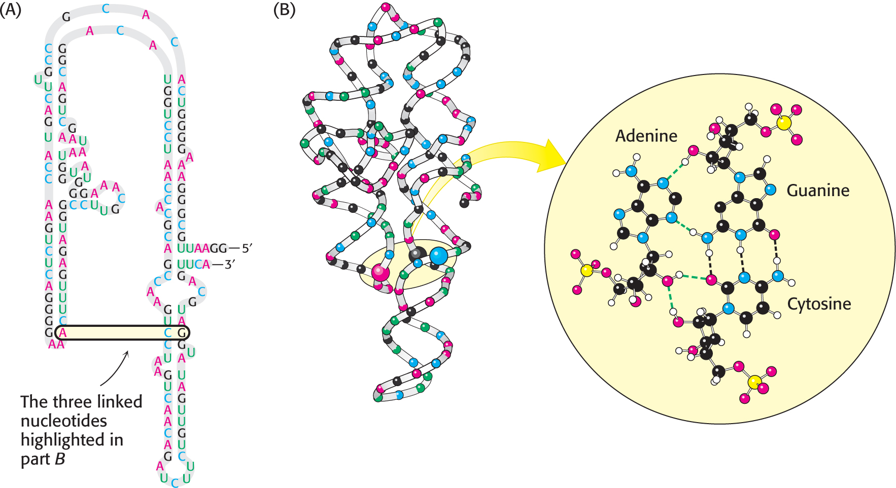
Figure 33.31: The complex structure of an RNA molecule. A single-stranded RNA molecule can fold back on itself to form a complex structure. (A) The nucleotide sequence showing Watson–Crick base pairs and other nonstandard base pairings in stem-loop structures. (B) The three-dimensional structure and one important long-range interaction between three bases. In the three-dimensional structure to the left, cytidine nucleotides are shown in blue, adenosine in red, guanosine in black, and uridine in green. In the blown-up view, hydrogen bonds within the Watson–Crick base pair are shown as dashed black lines; additional hydrogen bonds are shown as dashed green lines.