Chapter 2. DIVERSITY III—ANIMALS
Learning Objectives
Lab 11
false
false
General Purpose
Conceptual
- Be able to compare and contrast the major groups within the kingdom Animalia.
- Be able to describe the major evolutionary trends in structures within the kingdom Animalia.
Exercise 1. Phylum Porifera
PROCEDURE
- Examine the demonstration specimens of porifera.
- Make a sketch of one or more of the specimens in your laboratory notebook.
- Look at the prepared slide of the cross section of porifera using the compound microscope. Make a sketch of the specimen in your laboratory notebook.
- Answer the following questions in your laboratory notebook: Are definite tissue layers present? If so, how many?
Exercise 2. Phylum Cnidaria
PROCEDURE
- Place several drops of freshwater pond or culture water in a depression slide.
- Use a dropper to obtain a living hydra from the culture available, and place the hydra in the water on the slide.
- Using a compound microscope, observe the hydra structure and make a sketch of the specimen in your laboratory notebook.
- Look at the prepared slide of hydra sections using the compound microscope. Make a sketch of the specimen in your laboratory notebook. Note and label the central cavity, called a gastrovascular cavity. Food is digested in this water-filled cavity and is then directly absorbed into the tissue layers of the body wall.
- Answer the following questions in your laboratory notebook: Are definite tissue layers present? If so, how many?
Exercise 3. Phylum Platyhelminthes
PROCEDURE
- Using your stereoscopic microscope, observe a live planarian specimen and make a sketch of the specimen in your laboratory notebook.
- Add a very small piece of fresh liver to the water near the planarian. The planarian may approach the food and begin to feed by extending a long tubular pharynx out of the mouth, a circular opening on the ventral side of the body.
- Make a sketch of your observations in your laboratory notebook.
- Using the compound microscope, observe a prepared slide of a planarian cross section. Make a sketch of the specimen in your laboratory notebook.
- Answer the following questions in your laboratory notebook: Compared with other phyla, what differences are seen? Are definite tissue layers present? If so, how many?
Exercise 4. Phylum Nematoda
PROCEDURE
- Observe the several species of nematoda and make a sketch of one or more of the specimens in your laboratory notebook.
- View the diagram of the life cycle of a representative parasitic nematode. Answer the following question in your laboratory notebook: How many hosts does the nematode have during its life cycle?
- Using the medium power on the compound microscope, observe a prepared slide of an Ascaris sp. cross section. Make a sketch of the specimen in your laboratory notebook.
- Answer the following questions in your laboratory notebook: Compared with other phyla, what differences are seen? Are definite tissue layers present? If so, how many?
Exercise 5. Phylum Annelida
Materials
Dissecting pan
Scissors
Pins
PROCEDURE
- Obtain a preserved earthworm and identify its anterior end by locating the mouth, which is overhung by a flesh dorsal protuberance called the prostomium. The anus at the posterior end has no such protuberance. Also, a swollen glandular band, the clitellum (a structure that secretes a cocoon that holds eggs), is located closer to the mouth than to the anus.
- Using scissors make a mid-dorsal incision along the anterior third of the animal. You can identify the dorsal surface in a couple of ways. The prostomium is dorsal, and the ventral surface of the worm is usually flattened, especially in the region of the clitellum. Cut to the prostomium. Pin the body open in a dissecting pan near the edge. You may need to cut through the septa that divide the body cavity into segments.
- Using a stereoscopic microscope or hand lens, look for the small brain just behind the prostomium on the surface of the digestive tract. Note the two nerves that pass from the brain around the pharynx and meet ventrally. These nerve tracts continue towards the posterior as a ventral nerve cord.
- Look for the large blood vessel on the dorsal wall of the digestive tract. You may be able to see the enlarged lateral blood vessels (hearts) around the anterior portion of the digestive tract.
- Identify (from anterior to posterior) the pharynx, esophagus, crop (a soft, swollen region of the digestive tract), gizzard (smaller and more rigid than the crop), and intestine.
- Make a sketch of the specimen in your laboratory notebook and label all of the structures noted in this procedure.
- Using the compound microscope, observe the prepared slide of a cross section of the earthworm. Make a sketch of the specimen in your laboratory notebook.
- Answer the following questions in your laboratory notebook: Compared with other phyla, what differences are seen? Are definite tissue layers present? If so, how many?
Exercise 6. Summary of Animal Characteristics
PROCEDURE
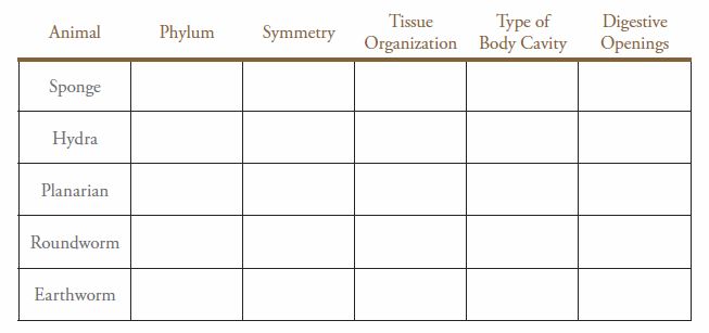
Based on your observations of the animal specimens, make a table in your laboratory notebook of the comparative characteristics. Use Table 11-1 as a template for your table.
Table 11-1. Summary table of animal characteristics.
Post-Lab Quiz
Proceed to the Post-Lab Quiz