1.2 DNA Illustrates the Interplay Between Form and Function
A fundamental biochemical feature common to all cellular organisms is the use of DNA for the storage of genetic information. The discovery that DNA plays this central role was first made in studies of bacteria in the 1940s. This discovery was followed by a compelling proposal for the three-dimensional structure of DNA in 1953, an event that set the stage for many of the advances in biochemistry and many other fields, extending to the present.
The structure of DNA powerfully illustrates a basic principle common to all biological macromolecules: the intimate relation between structure and function. The remarkable properties of this chemical substance allow it to function as a very efficient and robust vehicle for storing information. We start with an examination of the covalent structure of DNA and its extension into three dimensions.
DNA is constructed from four building blocks
DNA is a linear polymer made up of four different types of monomers. It has a fixed backbone from which protrude variable substituents, referred to as bases (Figure 1.4). The backbone is built of repeating sugar–phosphate units. The sugars are molecules of deoxyribose from which DNA receives its name. Each sugar is connected to two phosphate groups through different linkages. Moreover, each sugar is oriented in the same way, and so each DNA strand has directionality, with one end distinguishable from the other. Joined to each deoxyribose is one of four possible bases: adenine (A), cytosine (C), guanine (G), and thymine (T).
These bases are connected to the sugar components in the DNA backbone through the bonds shown in black in Figure 1.4. All four bases are planar but differ significantly in other respects. Thus, each monomer of DNA consists of a sugar–phosphate unit and one of four bases attached to the sugar. These bases can be arranged in any order along a strand of DNA.
Figure 1.4: Covalent structure of DNA. Each unit of the polymeric structure is composed of a sugar (deoxyribose), a phosphate, and a variable base that protrudes from the sugar–phosphate backbone.
Two single strands of DNA combine to form a double helix
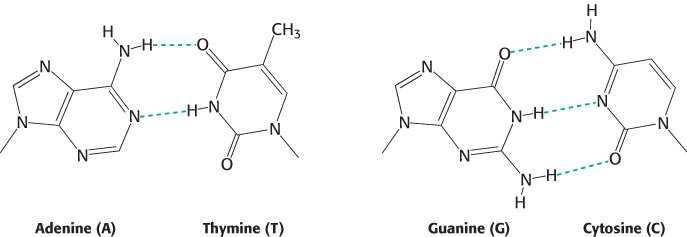
Most DNA molecules consist of not one but two strands (Figure 1.5). In 1953, James Watson and Francis Crick deduced the arrangement of these strands and proposed a three-dimensional structure for DNA molecules. This structure is a double helix composed of two intertwined strands arranged such that the sugar–phosphate backbone lies on the outside and the bases on the inside. The key to this structure is that the bases form specific base pairs (bp) held together by hydrogen bonds (Section 1.3): adenine pairs with thymine (A–T) and guanine pairs with cytosine (G–C), as shown in Figure 1.6. Hydrogen bonds are much weaker than covalent bonds such as the carbon–carbon or carbon–nitrogen bonds that define the structures of the bases themselves. Such weak bonds are crucial to biochemical systems; they are weak enough to be reversibly broken in biochemical processes, yet they are strong enough, particularly when many form simultaneously, to help stabilize specific structures such as the double helix.
Figure 1.5: The double helix. The double-helical structure of DNA proposed by Watson and Crick. The sugar–phosphate backbones of the two chains are shown in red and blue, and the bases are shown in green, purple, orange, and yellow. The two strands are antiparallel, running in opposite directions with respect to the axis of the double helix, as indicated by the arrows.
Figure 1.6: Watson–Crick base pairs. Adenine pairs with thymine (A – T), and guanine with cytosine (G – C). The dashed green lines represent hydrogen bonds.
DNA structure explains heredity and the storage of information
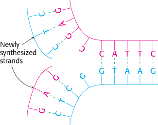
Figure 1.7: DNA replication. If a DNA molecule is separated into two strands, each strand can act as the template for the generation of its partner strand.
The structure proposed by Watson and Crick has two properties of central importance to the role of DNA as the hereditary material. First, the structure is compatible with any sequence of bases. While the bases are distinct in structure, the base pairs have essentially the same shape (Figure 1.6) and thus fit equally well into the center of the double-helical structure of any sequence. Without any constraints, the sequence of bases along a DNA strand can act as an efficient means of storing information. Indeed, the sequence of bases along DNA strands is how genetic information is stored. The DNA sequence determines the sequences of the ribonucleic acid (RNA) and protein molecules that carry out most of the activities within cells.
Second, because of base-pairing, the sequence of bases along one strand completely determines the sequence along the other strand. As Watson and Crick so coyly wrote: “It has not escaped our notice that the specific pairing we have postulated immediately suggests a possible copying mechanism for the genetic material.” Thus, if the DNA double helix is separated into two single strands, each strand can act as a template for the generation of its partner strand through specific base-pair formation (Figure 1.7). The three-dimensional structure of DNA beautifully illustrates the close connection between molecular form and function.