12.3 Phospholipids and Glycolipids Readily Form Bimolecular Sheets in Aqueous Media

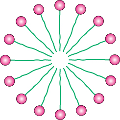
What properties enable phospholipids to form membranes? Membrane formation is a consequence of the amphipathic nature of the molecules.Their polar head groups favor contact with water, whereas their hydrocarbon tails interact with one another in preference to water. How can molecules with these preferences arrange themselves in aqueous solutions? One way is to form a globular structure called a micelle. The polar head groups form the outside surface of the micelle, which is surrounded by water, and the hydrocarbon tails are sequestered inside, interacting with one another (Figure 12.9).

Figure 12.9: Diagram of a section of a micelle. Ionized fatty acids readily form such structures, but most phospholipids do not.

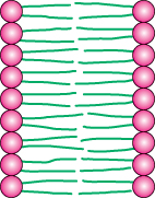
Alternatively, the strongly opposed preferences of the hydrophilic and hydrophobic moieties of membrane lipids can be satisfied by forming a lipid bilayer, composed of two lipid sheets (Figure 12.10). A lipid bilayer is also called a bimolecular sheet. The hydrophobic tails of each individual sheet interact with one another, forming a hydrophobic interior that acts as a permeability barrier. The hydrophilic head groups interact with the aqueous medium on each side of the bilayer. The two opposing sheets are called leaflets.

Figure 12.10: Diagram of a section of a bilayer membrane.

The favored structure for most phospholipids and glycolipids in aqueous media is a bimolecular sheet rather than a micelle. The reason is that the two fatty acid chains of a phospholipid or a glycolipid are too bulky to fit into the interior of a micelle. In contrast, salts of fatty acids (such as sodium palmitate, a constituent of soap) readily form micelles because they contain only one chain. The formation of bilayers instead of micelles by phospholipids is of critical biological importance. A micelle is a limited structure, usually less than 200 Å (20 nm) in diameter. In contrast, a bimolecular sheet can extend to macroscopic dimensions, as much as a millimeter (107 Å, or 106 nm) or more. Phospholipids and related molecules are important membrane constituents because they readily form extensive bimolecular sheets.

Lipid bilayers form spontaneously by a self-assembly process. In other words, the structure of a bimolecular sheet is inherent in the structure of the constituent lipid molecules. The growth of lipid bilayers from phospholipids is rapid and spontaneous in water. Hydrophobic interactions are the major driving force for the formation of lipid bilayers. Recall that hydrophobic interactions also play a dominant role in the stacking of bases in nucleic acids and in the folding of proteins (Sections 1.3 and 2.4). Water molecules are released from the hydrocarbon tails of membrane lipids as these tails become sequestered in the nonpolar interior of the bilayer. Furthermore, van der Waals attractive forces between the hydrocarbon tails favor close packing of the tails. Finally, there are electrostatic and hydrogen-bonding attractions between the polar head groups and water molecules. Thus, lipid bilayers are stabilized by the full array of forces that mediate molecular interactions in biological systems. Because lipid bilayers are held together by many reinforcing, noncovalent interactions (predominantly hydrophobic), they are cooperative structures. These hydrophobic interactions have three significant biological consequences: (1) lipid bilayers have an inherent tendency to be extensive; (2) lipid bilayers will tend to close on themselves so that there are no edges with exposed hydrocarbon chains, and so they form compartments; and (3) lipid bilayers are self-sealing because a hole in a bilayer is energetically unfavorable.

Lipid vesicles can be formed from phospholipids

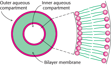
The propensity of phospholipids to form membranes has been used to create an important experimental and clinical tool. Lipid vesicles, or liposomes, are aqueous compartments enclosed by a lipid bilayer (Figure 12.11). These structures can be used to study membrane permeability or to deliver chemicals to cells. Liposomes are formed by suspending a suitable lipid, such as phosphatidylcholine, in an aqueous medium, and then sonicating (i.e., agitating by high-frequency sound waves) to give a dispersion of closed vesicles that are quite uniform in size. Vesicles formed by this method are nearly spherical and have a diameter of about 500 Å (50 nm). Larger vesicles (of the order of 1 μm or 104 Å in diameter) can be prepared by slowly evaporating the organic solvent from a suspension of phospholipid in a mixed-solvent system.

Figure 12.11: Liposome. A liposome, or lipid vesicle, is a small aqueous compartment surrounded by a lipid bilayer.

349

Figure 12.12: Preparation of glycine-containing liposomes. Liposomes containing glycine are formed by the sonication of phospholipids in the presence of glycine. Free glycine is removed by gel filtration.

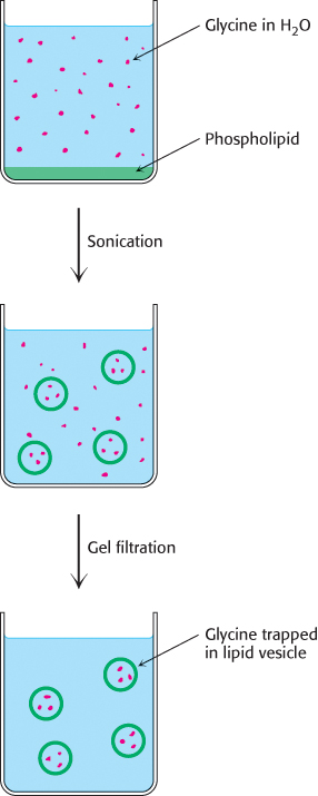
Ions or molecules can be trapped in the aqueous compartments of lipid vesicles by forming the vesicles in the presence of these substances (Figure 12.12). For example, 500-Å-diameter vesicles formed in a 0.1 M glycine solution will trap about 2000 molecules of glycine in each inner aqueous compartment. These glycine-containing vesicles can be separated from the surrounding solution of glycine by dialysis or by gel-filtration chromatography (Section 3.1). The permeability of the bilayer membrane to glycine can then be determined by measuring the rate of efflux of glycine from the inner compartment of the vesicle to the ambient solution. Liposomes can be formed with specific membrane proteins embedded in them by solubilizing the proteins in the presence of detergents and then adding them to the phospholipids from which liposomes will be formed. Protein–liposome complexes provide valuable experimental tools for examining a range of membrane-protein functions.

Therapeutic applications for liposomes are currently under active investigation. For example, liposomes containing drugs or DNA can be injected into patients. These liposomes fuse with the plasma membrane of many kinds of cells, introducing into the cells the molecules that they contain. Drug delivery with liposomes often lessens its toxicity. Less of the drug is distributed to normal tissues because long-circulating liposomes concentrate in regions of increased blood circulation, such as solid tumors and sites of inflammation. Moreover, the selective fusion of lipid vesicles with particular kinds of cells is a promising means of controlling the delivery of drugs to target cells.

Another well-defined synthetic membrane is a planar bilayer membrane. This structure can be formed across a 1-mm hole in a partition between two aqueous compartments by dipping a fine paintbrush into a membrane-forming solution, such as phosphatidylcholine in decane, and stroking the tip of the brush across the hole. The lipid film across the hole thins spontaneously into a lipid bilayer. The electrical conduction properties of this macroscopic bilayer membrane are readily studied by inserting electrodes into each aqueous compartment (Figure 12.13). For example, the permeability of the membrane to ions is determined by measuring the current across the membrane as a function of the applied voltage.

Figure 12.13: Experimental arrangement for the study of a planar bilayer membrane. A bilayer membrane is formed across a 1-mm hole in a septum that separates two aqueous compartments. This arrangement permits measurements of the permeability and electrical conductance of lipid bilayers.

Lipid bilayers are highly impermeable to ions and most polar molecules

Figure 12.14: Permeability coefficients (P) of ions and molecules in a lipid bilayer. The ability of molecules to cross a lipid bilayer spans a wide range of values.

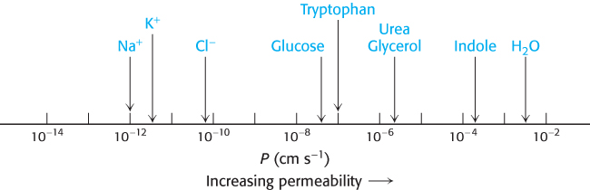
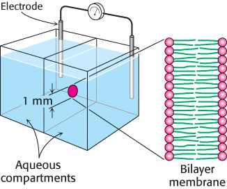
Permeability studies of lipid vesicles and electrical-conductance measurements of planar bilayers have shown that lipid bilayer membranes have a very low permeability for ions and most polar molecules. Water is a conspicuous exception to this generalization; it traverses such membranes relatively easily because of its low molecular weight, high concentration, and lack of a complete charge. The range of measured permeability coefficients is very wide (Figure 12.14). For example, Na+ and K+ traverse these membranes 109 times as slowly as does H2O. Tryptophan, a zwitterion at pH 7, crosses the membrane 103 times as slowly as does indole, a structurally related molecule that lacks ionic groups. In fact, the permeability of small molecules is correlated with their solubility in a nonpolar solvent relative to their solubility in water.This relation suggests that a small molecule might traverse a lipid bilayer membrane in the following way: first, it sheds its solvation shell of water; then, it is dissolved in the hydrocarbon core of the membrane; and, finally, it diffuses through this core to the other side of the membrane, where it becomes resolvated by water. An ion such as Na+ traverses membranes very slowly because the replacement of its coordination shell of polar water molecules by nonpolar interactions with the membrane interior is highly unfavorable energetically.

350